- PDB-12uw: Crystal structure of KRAS(Q61K) bound to GDP and ligand -
+
Open data
ID or keywords:
Loading...
-
Basic information
Entry
Database: PDB / ID: 12uw
Title
Crystal structure of KRAS(Q61K) bound to GDP and ligand
Components
Isoform 2B of GTPase KRas
Keywords
ONCOPROTEIN / KRAS / GTPase / GDP-bound
Function / homology
Function and homology information
response to mineralocorticoid / GMP binding / LRR domain binding / response to isolation stress / response to gravity / myoblast proliferation / cardiac muscle cell proliferation / Signaling by RAS GAP mutants / Signaling by RAS GTPase mutants / Activation of RAS in B cells ...response to mineralocorticoid / GMP binding / LRR domain binding / response to isolation stress / response to gravity / myoblast proliferation / cardiac muscle cell proliferation / Signaling by RAS GAP mutants / Signaling by RAS GTPase mutants / Activation of RAS in B cells / RAS signaling downstream of NF1 loss-of-function variants / RUNX3 regulates p14-ARF / SOS-mediated signalling / Activated NTRK3 signals through RAS / Activated NTRK2 signals through RAS / SHC1 events in ERBB4 signaling / Signalling to RAS / SHC-related events triggered by IGF1R / Activated NTRK2 signals through FRS2 and FRS3 / Estrogen-stimulated signaling through PRKCZ / SHC-mediated cascade:FGFR3 / MET activates RAS signaling / positive regulation of Ras protein signal transduction / SHC-mediated cascade:FGFR2 / SHC-mediated cascade:FGFR4 / Signaling by PDGFRA transmembrane, juxtamembrane and kinase domain mutants / Signaling by PDGFRA extracellular domain mutants / PTK6 Regulates RHO GTPases, RAS GTPase and MAP kinases / Erythropoietin activates RAS / SHC-mediated cascade:FGFR1 / Signaling by FGFR4 in disease / Signaling by CSF3 (G-CSF) / FRS-mediated FGFR3 signaling / Signaling by FLT3 ITD and TKD mutants / FRS-mediated FGFR2 signaling / FRS-mediated FGFR4 signaling / p38MAPK events / Signaling by FGFR3 in disease / FRS-mediated FGFR1 signaling / Tie2 Signaling / protein-membrane adaptor activity / Signaling by FGFR2 in disease / Signaling by FLT3 fusion proteins / GRB2 events in EGFR signaling / SHC1 events in EGFR signaling / FLT3 Signaling / Signaling by FGFR1 in disease / EGFR Transactivation by Gastrin / NCAM signaling for neurite out-growth / CD209 (DC-SIGN) signaling / liver development / GRB2 events in ERBB2 signaling / Downstream signal transduction / response to glucocorticoid / Insulin receptor signalling cascade / SHC1 events in ERBB2 signaling / Constitutive Signaling by Overexpressed ERBB2 / Ras activation upon Ca2+ influx through NMDA receptor / Signaling by phosphorylated juxtamembrane, extracellular and kinase domain KIT mutants / VEGFR2 mediated cell proliferation / small monomeric GTPase / FCERI mediated MAPK activation / female pregnancy / Signaling by ERBB2 TMD/JMD mutants / Constitutive Signaling by EGFRvIII / Signaling by SCF-KIT / RAF activation / Signaling by high-kinase activity BRAF mutants / Signaling by ERBB2 ECD mutants / MAP2K and MAPK activation / Signaling by ERBB2 KD Mutants / cytokine-mediated signaling pathway / cytoplasmic side of plasma membrane / Signaling by RAF1 mutants / Signaling by CSF1 (M-CSF) in myeloid cells / Signaling by moderate kinase activity BRAF mutants / Paradoxical activation of RAF signaling by kinase inactive BRAF / Signaling downstream of RAS mutants / MAPK cascade / Negative regulation of MAPK pathway / RAS processing / Regulation of RAS by GAPs / Signaling by BRAF and RAF1 fusions / positive regulation of cellular senescence / GDP binding / DAP12 signaling / Constitutive Signaling by Ligand-Responsive EGFR Cancer Variants / RAF/MAP kinase cascade / G protein activity / Ca2+ pathway / Ras protein signal transduction / mitochondrial outer membrane / Golgi membrane / focal adhesion / positive regulation of gene expression / positive regulation of cell population proliferation / GTPase activity / endoplasmic reticulum membrane / GTP binding / protein-containing complex binding Similarity search - Function
Small GTPase, Ras-type / Small GTPase Ras domain profile. / Ran (Ras-related nuclear proteins) /TC4 subfamily of small GTPases / Rho (Ras homology) subfamily of Ras-like small GTPases / Ras subfamily of RAS small GTPases / Small GTPase / Ras family / Rab subfamily of small GTPases / Small GTP-binding protein domain / P-loop containing nucleoside triphosphate hydrolase Similarity search - Domain/homology
In the structure databanks used in Yorodumi, some data are registered as the other names, "COVID-19 virus" and "2019-nCoV". Here are the details of the virus and the list of structure data.
Jan 31, 2019. EMDB accession codes are about to change! (news from PDBe EMDB page)
EMDB accession codes are about to change! (news from PDBe EMDB page)
The allocation of 4 digits for EMDB accession codes will soon come to an end. Whilst these codes will remain in use, new EMDB accession codes will include an additional digit and will expand incrementally as the available range of codes is exhausted. The current 4-digit format prefixed with “EMD-” (i.e. EMD-XXXX) will advance to a 5-digit format (i.e. EMD-XXXXX), and so on. It is currently estimated that the 4-digit codes will be depleted around Spring 2019, at which point the 5-digit format will come into force.
The EM Navigator/Yorodumi systems omit the EMD- prefix.
Related info.:Q: What is EMD? / ID/Accession-code notation in Yorodumi/EM Navigator
Yorodumi is a browser for structure data from EMDB, PDB, SASBDB, etc.
This page is also the successor to EM Navigator detail page, and also detail information page/front-end page for Omokage search.
The word "yorodu" (or yorozu) is an old Japanese word meaning "ten thousand". "mi" (miru) is to see.
Related info.:EMDB / PDB / SASBDB / Comparison of 3 databanks / Yorodumi Search / Aug 31, 2016. New EM Navigator & Yorodumi / Yorodumi Papers / Jmol/JSmol / Function and homology information / Changes in new EM Navigator and Yorodumi
 Movie
Movie Controller


 Open data
 Basic information
 Components
 Keywords
 Function and homology information
 Homo sapiens (human)
 X-RAY DIFFRACTION /
  Authors
 United States, 1items
  Citation
 Structure visualization
 Downloads & links
 Other downloads
 PDBj
PDBj




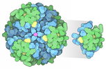

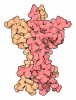





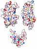
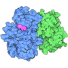
 Assembly


 Escherichia coli BL21(DE3) (bacteria) / References: UniProt: P01116, small monomeric GTPase
 Mass: 24.305 Da / Num. of mol.: 2 / Source method: obtained synthetically / Formula: Mg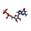
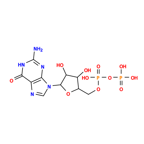 Type: RNA linking / Mass: 443.201 Da / Num. of mol.: 2 / Source method: obtained synthetically / Formula: C10H15N5O11P2 / Comment: GDP, energy-carrying molecule*YM
 Mass: 18.015 Da / Num. of mol.: 194 / Source method: isolated from a natural source / Formula: H2O
 Sample preparation
 Processing