[English] 日本語
Yorodumi
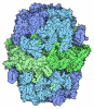
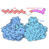
- PDB-11sz: Antibody (1B2) Bound Rifamycin Synthetase Module 2 -

+
Open data


ID or keywords:

Loading...

-
Basic information

Entry
Database: PDB / ID: 11sz
TitleAntibody (1B2) Bound Rifamycin Synthetase Module 2
Components
  • 1B2 Antibody Fragment (Fab) Heavy Chain
  • 1B2 Antibody Fragment (Fab) Light Chain
  • 6-deoxyerythronolide-B synthase, Erythronolide synthase EryA2
KeywordsBIOSYNTHETIC PROTEIN/Immune System / polyketide synthase / antibody / BIOSYNTHETIC PROTEIN-Immune System complex
Function / homology
Function and homology information


6-deoxyerythronolide-B synthase / erythronolide synthase activity / macrolide biosynthetic process / fatty acid synthase activity / phosphopantetheine binding / 3-oxoacyl-[acyl-carrier-protein] synthase activity / fatty acid biosynthetic process / oxidoreductase activity
Similarity search - Function
Erythronolide synthase, docking domain superfamily / Erythronolide synthase, docking / Erythronolide synthase, docking / Polyketide synthase dimerisation element domain / Polyketide synthase dimerisation element domain / : / Polyketide synthase, docking domain / Erythronolide synthase docking domain / Zinc-binding dehydrogenase / Polyketide synthase extender module SpnB, Rossmann fold domain ...Erythronolide synthase, docking domain superfamily / Erythronolide synthase, docking / Erythronolide synthase, docking / Polyketide synthase dimerisation element domain / Polyketide synthase dimerisation element domain / : / Polyketide synthase, docking domain / Erythronolide synthase docking domain / Zinc-binding dehydrogenase / Polyketide synthase extender module SpnB, Rossmann fold domain / : / Polyketide synthase dehydratase N-terminal domain / PKS_PP_betabranch / Polyketide synthase, dehydratase domain / PKS_DH / : / Polyketide synthase dehydratase domain / : / Polyketide and metazoan fatty acid synthase dehydratase (PKS/mFAS DH) domain profile. / Polyketide synthase, dehydratase domain superfamily / Polyketide synthase, C-terminal extension / Ketoacyl-synthetase C-terminal extension / Polyketide synthase, ketoreductase domain / KR domain / Malonyl-CoA ACP transacylase, ACP-binding / ANL, N-terminal domain / : / AMP-binding enzyme C-terminal domain / AMP-binding enzyme, C-terminal domain / Alcohol dehydrogenase, N-terminal / Alcohol dehydrogenase GroES-like domain / Acyl transferase / Acyl transferase domain / Acyl transferase domain in polyketide synthase (PKS) enzymes. / AMP-binding, conserved site / Acyl transferase domain superfamily / Putative AMP-binding domain signature. / : / Polyketide synthase, enoylreductase domain / Enoylreductase / Acyl transferase/acyl hydrolase/lysophospholipase / AMP-dependent synthetase/ligase / AMP-binding enzyme / Polyketide synthase, phosphopantetheine-binding domain / Phosphopantetheine attachment site / PKS_KR / Beta-ketoacyl synthase / Beta-ketoacyl synthase, active site / Ketosynthase family 3 (KS3) active site signature. / Ketosynthase family 3 (KS3) domain profile. / Beta-ketoacyl synthase, N-terminal / Beta-ketoacyl synthase, C-terminal / Polyketide synthase, beta-ketoacyl synthase domain / Beta-ketoacyl synthase, N-terminal domain / Beta-ketoacyl synthase, C-terminal domain / AMP-binding enzyme, C-terminal domain superfamily / GroES-like superfamily / Thiolase-like / Phosphopantetheine attachment site / Phosphopantetheine attachment site. / Phosphopantetheine attachment site / ACP-like superfamily / Carrier protein (CP) domain profile. / Phosphopantetheine binding ACP domain / NAD(P)-binding domain superfamily
Similarity search - Domain/homology
6-deoxyerythronolide-B synthase / Erythronolide synthase EryA2
Similarity search - Component
Biological speciesAmycolatopsis mediterranei (bacteria)
Homo sapiens (human)
MethodELECTRON MICROSCOPY / single particle reconstruction / cryo EM / Resolution: 2.94 Å
AuthorsCogan, D.P.
Funding support United States, 1items
OrganizationGrant numberCountry
National Institutes of Health/National Institute of General Medical Sciences (NIH/NIGMS)1R35GM160153 United States
CitationJournal: To Be Published
Title: Antibody (1B2) Bound Rifamycin Synthetase Module 2
Authors: Cogan, D.P.
History
DepositionMar 11, 2026Deposition site: RCSB / Processing site: RCSB
Revision 1.0Apr 15, 2026Provider: repository / Type: Initial release
Revision 1.0Apr 15, 2026Data content type: EM metadata / Data content type: EM metadata / Provider: repository / Type: Initial release
Revision 1.0Apr 15, 2026Data content type: Half map / Part number: 1 / Data content type: Half map / Provider: repository / Type: Initial release
Revision 1.0Apr 15, 2026Data content type: Half map / Part number: 2 / Data content type: Half map / Provider: repository / Type: Initial release
Revision 1.0Apr 15, 2026Data content type: Image / Data content type: Image / Provider: repository / Type: Initial release
Revision 1.0Apr 15, 2026Data content type: Primary map / Data content type: Primary map / Provider: repository / Type: Initial release

-
Structure visualization

Structure viewerMolecule:
MolmilJmol/JSmol

Downloads & links

-
Assembly

Deposited unit
A: 6-deoxyerythronolide-B synthase, Erythronolide synthase EryA2
B: 6-deoxyerythronolide-B synthase, Erythronolide synthase EryA2
C: 1B2 Antibody Fragment (Fab) Heavy Chain
D: 1B2 Antibody Fragment (Fab) Light Chain
H: 1B2 Antibody Fragment (Fab) Heavy Chain
L: 1B2 Antibody Fragment (Fab) Light Chain


Theoretical massNumber of molelcules
Total (without water)336,0736
Polymers336,0736
Non-polymers00
Water00
1


  • Idetical with deposited unit
  • defined by author&software
  • Evidence: electron microscopy, not applicable
TypeNameSymmetry operationNumber
identity operation1_555x,y,z1

-
Components

#1: Protein 6-deoxyerythronolide-B synthase, Erythronolide synthase EryA2 / 6-deoxyerythronolide B synthase II / 6-deoxyerythronolide-B synthase EryA2 / modules 3 and 4 / DEBS 2 / ORF B


Mass: 115872.844 Da / Num. of mol.: 2
Source method: isolated from a genetically manipulated source
Source: (gene. exp.) Amycolatopsis mediterranei (bacteria) / Gene: rifA, eryA / Plasmid: pDC71 / Production host: Escherichia coli BL21(DE3) (bacteria)
References: UniProt: O54666, UniProt: Q03132, 6-deoxyerythronolide-B synthase
#2: Antibody 1B2 Antibody Fragment (Fab) Heavy Chain


Mass: 26447.611 Da / Num. of mol.: 2
Source method: isolated from a genetically manipulated source
Source: (gene. exp.) Homo sapiens (human) / Production host: Escherichia coli BL21(DE3) (bacteria)
#3: Antibody 1B2 Antibody Fragment (Fab) Light Chain


Mass: 25715.832 Da / Num. of mol.: 2
Source method: isolated from a genetically manipulated source
Source: (gene. exp.) Homo sapiens (human) / Production host: Escherichia coli BL21(DE3) (bacteria)
Has protein modificationY

-
Experimental details

-
Experiment

ExperimentMethod: ELECTRON MICROSCOPY
EM experimentAggregation state: PARTICLE / 3D reconstruction method: single particle reconstruction

-
Sample preparation

ComponentName: rifamycin synthetase module 2 in complex with antibody fragment 1B2
Type: COMPLEX / Entity ID: all / Source: RECOMBINANT
Molecular weightValue: 335.62681 kDa/nm / Experimental value: YES
Source (natural)Organism: Amycolatopsis mediterranei (bacteria)
Source (recombinant)Organism: Escherichia coli BL21(DE3) (bacteria) / Plasmid: pDC71
Buffer solutionpH: 7.2
Buffer component
IDConc.NameBuffer-ID
10.01 MHEPES1
20.1 Mcitric acid1
SpecimenConc.: 10 mg/ml / Embedding applied: NO / Shadowing applied: NO / Staining applied: NO / Vitrification applied: YES
Specimen supportGrid material: COPPER / Grid mesh size: 300 divisions/in. / Grid type: Quantifoil R2/1
VitrificationInstrument: FEI VITROBOT MARK IV / Cryogen name: ETHANE / Humidity: 100 % / Chamber temperature: 277.15 K

-
Electron microscopy imaging

Experimental equipment
Model: Titan Krios / Image courtesy: FEI Company
MicroscopyModel: TFS KRIOS
Electron gunElectron source: FIELD EMISSION GUN / Accelerating voltage: 300 kV / Illumination mode: FLOOD BEAM
Electron lensMode: BRIGHT FIELD / Nominal defocus max: 3988 nm / Nominal defocus min: 1188 nm
Specimen holderCryogen: NITROGEN / Specimen holder model: FEI TITAN KRIOS AUTOGRID HOLDER
Image recordingElectron dose: 50 e/Å2 / Film or detector model: GATAN K3 BIOQUANTUM (6k x 4k)

-
Processing

EM software
IDNameVersionCategory
1cryoSPARC4.7.0particle selection
2PHENIX1.20.1_4487model refinement
3EPUimage acquisition
5cryoSPARC4.7.0CTF correction
10cryoSPARC4.7.0initial Euler assignment
11cryoSPARC4.7.0final Euler assignment
12cryoSPARC4.7.0classification
13cryoSPARC4.7.03D reconstruction
CTF correctionType: NONE
SymmetryPoint symmetry: C1 (asymmetric)
3D reconstructionResolution: 2.94 Å / Resolution method: FSC 0.143 CUT-OFF / Num. of particles: 852885 / Num. of class averages: 1 / Symmetry type: POINT
RefinementHighest resolution: 2.94 Å
Stereochemistry target values: REAL-SPACE (WEIGHTED MAP SUM AT ATOM CENTERS)

+
About Yorodumi

-
News

-
Feb 9, 2022. New format data for meta-information of EMDB entries

New format data for meta-information of EMDB entries

  • Version 3 of the EMDB header file is now the official format.
  • The previous official version 1.9 will be removed from the archive.

Related info.:EMDB header

External links:wwPDB to switch to version 3 of the EMDB data model

-
Aug 12, 2020. Covid-19 info

Covid-19 info

URL: https://pdbj.org/emnavi/covid19.php

New page: Covid-19 featured information page in EM Navigator.

Related info.:Covid-19 info / Mar 5, 2020. Novel coronavirus structure data

+
Mar 5, 2020. Novel coronavirus structure data

Novel coronavirus structure data

Related info.:Yorodumi Speices / Aug 12, 2020. Covid-19 info

External links:COVID-19 featured content - PDBj / Molecule of the Month (242):Coronavirus Proteases

+
Jan 31, 2019. EMDB accession codes are about to change! (news from PDBe EMDB page)

EMDB accession codes are about to change! (news from PDBe EMDB page)

  • The allocation of 4 digits for EMDB accession codes will soon come to an end. Whilst these codes will remain in use, new EMDB accession codes will include an additional digit and will expand incrementally as the available range of codes is exhausted. The current 4-digit format prefixed with “EMD-” (i.e. EMD-XXXX) will advance to a 5-digit format (i.e. EMD-XXXXX), and so on. It is currently estimated that the 4-digit codes will be depleted around Spring 2019, at which point the 5-digit format will come into force.
  • The EM Navigator/Yorodumi systems omit the EMD- prefix.

Related info.:Q: What is EMD? / ID/Accession-code notation in Yorodumi/EM Navigator

External links:EMDB Accession Codes are Changing Soon! / Contact to PDBj

+
Jul 12, 2017. Major update of PDB

Major update of PDB

  • wwPDB released updated PDB data conforming to the new PDBx/mmCIF dictionary.
  • This is a major update changing the version number from 4 to 5, and with Remediation, in which all the entries are updated.
  • In this update, many items about electron microscopy experimental information are reorganized (e.g. em_software).
  • Now, EM Navigator and Yorodumi are based on the updated data.

External links:wwPDB Remediation / Enriched Model Files Conforming to OneDep Data Standards Now Available in the PDB FTP Archive

-
Yorodumi

Thousand views of thousand structures

  • Yorodumi is a browser for structure data from EMDB, PDB, SASBDB, etc.
  • This page is also the successor to EM Navigator detail page, and also detail information page/front-end page for Omokage search.
  • The word "yorodu" (or yorozu) is an old Japanese word meaning "ten thousand". "mi" (miru) is to see.

Related info.:EMDB / PDB / SASBDB / Comparison of 3 databanks / Yorodumi Search / Aug 31, 2016. New EM Navigator & Yorodumi / Yorodumi Papers / Jmol/JSmol / Function and homology information / Changes in new EM Navigator and Yorodumi

Read more