 Movie
Movie Controller

[English] 日本語
 Yorodumi
- PDB-11ov: Cryo-EM structure of EV-D68 B3 VLP bound by neutralizing antibody 5H03 -
+ Open data

- Basic information
| Entry | Database: PDB / ID: 11ov | |||||||||||||||||||||
|---|---|---|---|---|---|---|---|---|---|---|---|---|---|---|---|---|---|---|---|---|---|---|
| Title | Cryo-EM structure of EV-D68 B3 VLP bound by neutralizing antibody 5H03 | |||||||||||||||||||||
 Components Components |
| |||||||||||||||||||||
 Keywords Keywords | VIRUS LIKE PARTICLE / EV-D68 / B3 subclade / 5H03 neutralizing antibody | |||||||||||||||||||||
| Function / homology |  Function and homology information Function and homology informationvirion component / picornain 2A / symbiont-mediated suppression of host mRNA export from nucleus / symbiont genome entry into host cell via pore formation in plasma membrane / picornain 3C / T=pseudo3 icosahedral viral capsid / host cell cytoplasmic vesicle membrane / host cell / nucleoside-triphosphate phosphatase / ribonucleoside triphosphate phosphatase activity ...virion component / picornain 2A / symbiont-mediated suppression of host mRNA export from nucleus / symbiont genome entry into host cell via pore formation in plasma membrane / picornain 3C / T=pseudo3 icosahedral viral capsid / host cell cytoplasmic vesicle membrane / host cell / nucleoside-triphosphate phosphatase / ribonucleoside triphosphate phosphatase activity / channel activity / monoatomic ion transmembrane transport / RNA helicase activity / symbiont-mediated suppression of host innate immune response / endocytosis involved in viral entry into host cell / symbiont-mediated activation of host autophagy / cysteine-type endopeptidase activity / viral RNA genome replication / RNA-directed RNA polymerase / RNA-directed RNA polymerase activity / symbiont entry into host cell / virion attachment to host cell / DNA-templated transcription / host cell nucleus / structural molecule activity / proteolysis / RNA binding / zinc ion binding / ATP binding Similarity search - Function | |||||||||||||||||||||
| Biological species |  Human enterovirus D68 Human enterovirus D68  Macaca mulatta (Rhesus monkey) Macaca mulatta (Rhesus monkey) | |||||||||||||||||||||
| Method | ELECTRON MICROSCOPY / single particle reconstruction / cryo EM / Resolution: 2.83 Å | |||||||||||||||||||||
 Authors Authors | Cheng, J. / Lei, H. / Pletnev, S. / Morano, N.C. / Zhang, B. / Du, H. / Rubin, S. / Moss, D.L. / Krug, P.W. / Kanekiyo, M. ...Cheng, J. / Lei, H. / Pletnev, S. / Morano, N.C. / Zhang, B. / Du, H. / Rubin, S. / Moss, D.L. / Krug, P.W. / Kanekiyo, M. / Pierson, T.C. / Ruckwardt, T.J. / Shapiro, L. / Kwong, P.D. / Zhou, T. | |||||||||||||||||||||
| Funding support |  United States, 1items United States, 1items
| |||||||||||||||||||||
 Citation Citation | Journal: Sci Transl Med / Year: 2026 Title: Neutralizing antibodies elicited in nonhuman primates by an enterovirus D68 virus-like particle vaccine target receptor binding sites. Authors: Daniel L Moss / Jiaxuan Cheng / Peter W Krug / Alden C Paine / Brian E Fisher / Amy R Henry / Jesmine Roberts-Torres / Timothy S Johnston / Sarah C Smith / Sergei Pletnev / Haotian Lei / ...Authors: Daniel L Moss / Jiaxuan Cheng / Peter W Krug / Alden C Paine / Brian E Fisher / Amy R Henry / Jesmine Roberts-Torres / Timothy S Johnston / Sarah C Smith / Sergei Pletnev / Haotian Lei / Nicholas C Morano / Theodore C Pierson / Lawrence Shapiro / Tongqing Zhou / Peter D Kwong / Daniel C Douek / Masaru Kanekiyo / Tracy J Ruckwardt / Abstract: Enterovirus D68 (EV-D68) is a picornavirus that causes biennial outbreaks of respiratory disease in young children that can progress to rare but severe complications, including acute flaccid myelitis ...Enterovirus D68 (EV-D68) is a picornavirus that causes biennial outbreaks of respiratory disease in young children that can progress to rare but severe complications, including acute flaccid myelitis (AFM). EV-D68 virus-like particles (VLPs) elicit potent neutralizing antibodies that are protective in animal models. Here, we report the isolation and characterization of monoclonal antibodies elicited by EV-D68 VLPs in nonhuman primates (NHPs). We identified five potently neutralizing mAbs targeting overlapping epitopes near the capsid fivefold axis of symmetry formed by pentamers of viral protein 1. Cryo-electron microscopy structures of mAbs 1E11 and 5H03 in complex with VLP revealed epitopes on the capsid that bridge the sialic acid binding site and the proteinaceous entry receptor major facilitator superfamily domain-containing protein 6 binding site. We further characterized the mechanisms by which these mAbs neutralize EV-D68 and found that mAbs can disrupt multiple steps in the EV-D68 life cycle, including promoting premature uncoating. Antibodies elicited by VLP immunization of NHP protected as well as a best-in-class human mAb in a mouse challenge model, although single-amino acid mutations in the capsid enabled viral escape. Our results demonstrate that EV-D68 VLP-elicited mAbs target major viral sites of vulnerability, provide insight into their mechanisms of neutralization, and reinforce the potential for VLP-based vaccines as a countermeasure for EV-D68. | |||||||||||||||||||||
| History |
|
- Structure visualization
| Structure viewer | Molecule: MolmilJmol/JSmol |
|---|
- Downloads & links
-Download
| PDBx/mmCIF format | 11ov.cif.gz | 188.6 KB | Display | PDBx/mmCIF format |
|---|---|---|---|---|
| PDB format | pdb11ov.ent.gz | 146.3 KB | Display | PDB format |
| PDBx/mmJSON format | 11ov.json.gz | Tree view | PDBx/mmJSON format | |
| Others |  Other downloads Other downloads |
-Validation report
| Arichive directory | https://data.pdbj.org/pub/pdb/validation_reports/1o/11ovftp://data.pdbj.org/pub/pdb/validation_reports/1o/11ov | HTTPS FTP |
|---|
-Related structure data
| Related structure data |  75894MC  11ltC M: map data used to model this data C: citing same article ( |
|---|---|
| Similar structure data |

-Links
 PDBj
PDBj


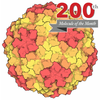
- Assembly
| Deposited unit | 
|
|---|---|
| 1 |
|
-Components
| #1: Protein | Mass: 25735.379 Da / Num. of mol.: 1 Source method: isolated from a genetically manipulated source Source: (gene. exp.) Human enterovirus D68 / Production host:  Homo sapiens (human) / References: UniProt: A0A7G9XUH9 Homo sapiens (human) / References: UniProt: A0A7G9XUH9 |
|---|---|
| #2: Protein | Mass: 23797.006 Da / Num. of mol.: 1 Source method: isolated from a genetically manipulated source Source: (gene. exp.) Human enterovirus D68 / Production host: Homo sapiens (human)References: UniProt: A0A5B9NIG2, picornain 2A, nucleoside-triphosphate phosphatase, picornain 3C, RNA-directed RNA polymerase |
| #3: Protein | Mass: 27170.895 Da / Num. of mol.: 1 Source method: isolated from a genetically manipulated source Source: (gene. exp.) Human enterovirus D68 / Production host: Homo sapiens (human)References: UniProt: A0A1L7H9D2, picornain 2A, nucleoside-triphosphate phosphatase, picornain 3C, RNA-directed RNA polymerase |
| #4: Antibody | Mass: 13768.218 Da / Num. of mol.: 1 Source method: isolated from a genetically manipulated source Source: (gene. exp.) Macaca mulatta (Rhesus monkey) / Production host: Homo sapiens (human) |
| #5: Antibody | Mass: 11764.249 Da / Num. of mol.: 1 Source method: isolated from a genetically manipulated source Source: (gene. exp.) Macaca mulatta (Rhesus monkey) / Production host: Homo sapiens (human) |
| Has protein modification | Y |
-Experimental details
-Experiment
| Experiment | Method: ELECTRON MICROSCOPY |
|---|---|
| EM experiment | Aggregation state: PARTICLE / 3D reconstruction method: single particle reconstruction |
- Sample preparation
| Component | Name: Enterovirus / Type: VIRUS / Entity ID: all / Source: RECOMBINANT |
|---|---|
| Source (natural) | Organism: Human enterovirus D68 |
| Source (recombinant) | Organism: Homo sapiens (human) |
| Details of virus | Empty: YES / Enveloped: NO / Isolate: SUBSPECIES / Type: VIRUS-LIKE PARTICLE |
| Buffer solution | pH: 7.4 |
| Specimen | Embedding applied: NO / Shadowing applied: NO / Staining applied: NO / Vitrification applied: YES |
| Vitrification | Cryogen name: ETHANE |
- Electron microscopy imaging
| Experimental equipment |  Model: Titan Krios / Image courtesy: FEI Company |
|---|---|
| Microscopy | Model: TFS KRIOS |
| Electron gun | Electron source:  FIELD EMISSION GUN / Accelerating voltage: 300 kV / Illumination mode: FLOOD BEAM FIELD EMISSION GUN / Accelerating voltage: 300 kV / Illumination mode: FLOOD BEAM |
| Electron lens | Mode: BRIGHT FIELD / Nominal defocus max: 2500 nm / Nominal defocus min: 1000 nm |
| Image recording | Electron dose: 54.5 e/Å2 / Film or detector model: GATAN K3 BIOCONTINUUM (6k x 4k) |
- Processing
| EM software |
| ||||||||||||||||||||||||
|---|---|---|---|---|---|---|---|---|---|---|---|---|---|---|---|---|---|---|---|---|---|---|---|---|---|
| CTF correction | Type: PHASE FLIPPING AND AMPLITUDE CORRECTION | ||||||||||||||||||||||||
| 3D reconstruction | Resolution: 2.83 Å / Resolution method: FSC 0.143 CUT-OFF / Num. of particles: 48000 / Symmetry type: POINT | ||||||||||||||||||||||||
| Refinement | Highest resolution: 2.83 Å Stereochemistry target values: REAL-SPACE (WEIGHTED MAP SUM AT ATOM CENTERS) | ||||||||||||||||||||||||
| Refine LS restraints |
|
