 Movie
Movie Controller


+ Open data

- Basic information
| Entry | Database: PDB / ID: 11mi | |||||||||
|---|---|---|---|---|---|---|---|---|---|---|
| Title | Crystal Structure of M. tuberculosis ClpP1P2 bound to ADEP 19 | |||||||||
 Components Components | (ATP-dependent Clp protease proteolytic subunit ...) x 2 | |||||||||
 Keywords Keywords | ANTIBIOTIC / peptidase / protease / mycobacteria / tuberculosis / acyldepsipeptide / proteostasis | |||||||||
| Function / homology |  Function and homology information Function and homology informationendopeptidase Clp / ATP-dependent peptidase activity / protein quality control for misfolded or incompletely synthesized proteins / ATPase binding / serine-type endopeptidase activity / proteolysis / plasma membrane / cytoplasm Similarity search - Function | |||||||||
| Biological species |  Mycobacterium tuberculosis H37Rv (bacteria) Mycobacterium tuberculosis H37Rv (bacteria) | |||||||||
| Method |  X-RAY DIFFRACTION / SYNCHROTRON / MOLECULAR REPLACEMENT / Resolution: 2.65 Å X-RAY DIFFRACTION / SYNCHROTRON / MOLECULAR REPLACEMENT / Resolution: 2.65 Å | |||||||||
 Authors Authors | Burnside, C.M. / Fei, F. / Schmitz, K.R. / Sello, J.K. | |||||||||
| Funding support |  United States, 2items United States, 2items
| |||||||||
 Citation Citation | Journal: J.Med.Chem. / Year: 2026 Title: Structural and Mechanistic Studies of ADEPs Yield Potent Antibacterials and a Drug Formulation Strategy for Tuberculosis. Authors: Fei, F. / Burnside, C.M. / Lun, S. / Wee, D. / Kaur, M. / Anderson, H.R. / McCarroll, M.N. / Richardson, A.E. / Neglia, S. / Liu, H.M. / Wang, X. / Gupta, S. / Rhee, K.Y. / Wright, G.D. / ...Authors: Fei, F. / Burnside, C.M. / Lun, S. / Wee, D. / Kaur, M. / Anderson, H.R. / McCarroll, M.N. / Richardson, A.E. / Neglia, S. / Liu, H.M. / Wang, X. / Gupta, S. / Rhee, K.Y. / Wright, G.D. / Bryson, B.D. / Oehlers, S.H. / Bishai, W.R. / Schmitz, K.R. / Sello, J.K. #1: Journal: Acta Crystallogr D Struct Biol / Year: 2019 Title: Macromolecular structure determination using X-rays, neutrons and electrons: recent developments in Phenix. Authors: Dorothee Liebschner / Pavel V Afonine / Matthew L Baker / Gábor Bunkóczi / Vincent B Chen / Tristan I Croll / Bradley Hintze / Li Wei Hung / Swati Jain / Airlie J McCoy / Nigel W Moriarty ...Authors: Dorothee Liebschner / Pavel V Afonine / Matthew L Baker / Gábor Bunkóczi / Vincent B Chen / Tristan I Croll / Bradley Hintze / Li Wei Hung / Swati Jain / Airlie J McCoy / Nigel W Moriarty / Robert D Oeffner / Billy K Poon / Michael G Prisant / Randy J Read / Jane S Richardson / David C Richardson / Massimo D Sammito / Oleg V Sobolev / Duncan H Stockwell / Thomas C Terwilliger / Alexandre G Urzhumtsev / Lizbeth L Videau / Christopher J Williams / Paul D Adams /   Abstract: Diffraction (X-ray, neutron and electron) and electron cryo-microscopy are powerful methods to determine three-dimensional macromolecular structures, which are required to understand biological ...Diffraction (X-ray, neutron and electron) and electron cryo-microscopy are powerful methods to determine three-dimensional macromolecular structures, which are required to understand biological processes and to develop new therapeutics against diseases. The overall structure-solution workflow is similar for these techniques, but nuances exist because the properties of the reduced experimental data are different. Software tools for structure determination should therefore be tailored for each method. Phenix is a comprehensive software package for macromolecular structure determination that handles data from any of these techniques. Tasks performed with Phenix include data-quality assessment, map improvement, model building, the validation/rebuilding/refinement cycle and deposition. Each tool caters to the type of experimental data. The design of Phenix emphasizes the automation of procedures, where possible, to minimize repetitive and time-consuming manual tasks, while default parameters are chosen to encourage best practice. A graphical user interface provides access to many command-line features of Phenix and streamlines the transition between programs, project tracking and re-running of previous tasks. | |||||||||
| History |
|
- Structure visualization
| Structure viewer | Molecule: MolmilJmol/JSmol |
|---|
- Downloads & links
-Download
| PDBx/mmCIF format | 11mi.cif.gz | 1.9 MB | Display | PDBx/mmCIF format |
|---|---|---|---|---|
| PDB format | pdb11mi.ent.gz | Display | PDB format | |
| PDBx/mmJSON format | 11mi.json.gz | Tree view | PDBx/mmJSON format | |
| Others |  Other downloads Other downloads |
-Validation report
| Arichive directory | https://data.pdbj.org/pub/pdb/validation_reports/1m/11miftp://data.pdbj.org/pub/pdb/validation_reports/1m/11mi | HTTPS FTP |
|---|
-Related structure data


-Links
 PDBj
PDBj
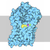
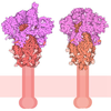
- Assembly
| Deposited unit | 
| ||||||||||||
|---|---|---|---|---|---|---|---|---|---|---|---|---|---|
| 1 | 
| ||||||||||||
| 2 | 
| ||||||||||||
| Unit cell |
|
-Components
-ATP-dependent Clp protease proteolytic subunit ... , 2 types, 28 molecules ABCDEFGOPQRSTUHIJKLMNVWXYZab
| #1: Protein | Mass: 23650.910 Da / Num. of mol.: 14 Source method: isolated from a genetically manipulated source Details: mature M. tuberculosis ClpP2 with a C-terminal 6xHis tag Source: (gene. exp.) Mycobacterium tuberculosis H37Rv (bacteria)Gene: clpP2, Rv2460c, MTV008.16c / Production host:  Escherichia coli B (bacteria) / Strain (production host): ER2566 / Variant (production host): delta-clpP / References: UniProt: P9WPC3, endopeptidase Clp Escherichia coli B (bacteria) / Strain (production host): ER2566 / Variant (production host): delta-clpP / References: UniProt: P9WPC3, endopeptidase Clp#2: Protein | Mass: 21065.934 Da / Num. of mol.: 14 Source method: isolated from a genetically manipulated source Details: mature M. tuberculosis ClpP1 Source: (gene. exp.) Mycobacterium tuberculosis H37Rv (bacteria)Gene: clpP1, clpP, Rv2461c, MTV008.17c / Production host: Escherichia coli B (bacteria) / Strain (production host): ER2566 / Variant (production host): delta-clpP / References: UniProt: P9WPC5, endopeptidase Clp |
|---|
-Non-polymers , 6 types, 562 molecules 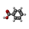

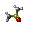
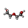

| #3: Chemical | ChemComp-A1C9P / Mass: 814.890 Da / Num. of mol.: 14 / Source method: obtained synthetically / Formula: C41H53F3N6O8 / Feature type: SUBJECT OF INVESTIGATION #4: Chemical | ChemComp-BEZ / 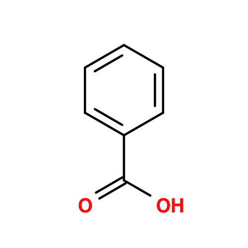 Mass: 122.121 Da / Num. of mol.: 28 / Source method: obtained synthetically / Formula: C7H6O2 Mass: 122.121 Da / Num. of mol.: 28 / Source method: obtained synthetically / Formula: C7H6O2#5: Chemical | ChemComp-LEU /  Type: L-peptide linking / Mass: 131.173 Da / Num. of mol.: 56 / Source method: obtained synthetically / Formula: C6H13NO2 Type: L-peptide linking / Mass: 131.173 Da / Num. of mol.: 56 / Source method: obtained synthetically / Formula: C6H13NO2#6: Chemical | ChemComp-DMS / 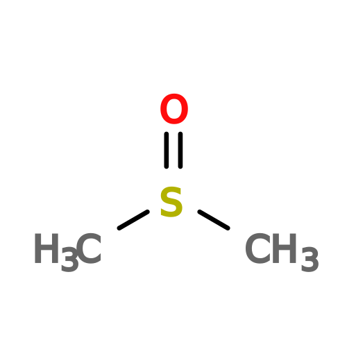 Mass: 78.133 Da / Num. of mol.: 4 / Source method: obtained synthetically / Formula: C2H6OS / Comment: DMSO, precipitant*YM Mass: 78.133 Da / Num. of mol.: 4 / Source method: obtained synthetically / Formula: C2H6OS / Comment: DMSO, precipitant*YM#7: Chemical | 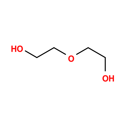 Mass: 106.120 Da / Num. of mol.: 2 / Source method: obtained synthetically / Formula: C4H10O3 Mass: 106.120 Da / Num. of mol.: 2 / Source method: obtained synthetically / Formula: C4H10O3#8: Water | ChemComp-HOH / | Mass: 18.015 Da / Num. of mol.: 458 / Source method: isolated from a natural source / Formula: H2O |
|---|
-Details
| Has ligand of interest | Y |
|---|---|
| Has protein modification | N |
-Experimental details
-Experiment
| Experiment | Method: X-RAY DIFFRACTION / Number of used crystals: 1 |
|---|
- Sample preparation
| Crystal | Density Matthews: 2.96 Å3/Da / Density % sol: 58.45 % |
|---|---|
| Crystal grow | Temperature: 293 K / Method: vapor diffusion, hanging drop / pH: 6.5 Details: 1:1 mixture of reservoir (0.1M Bis-Tris (pH 6.5), 15% PEG3350, 0.2M sodium citrate, 10% ethylene glycol) and protein solution (3.75 mg/mL ClpP1, 3.75 mg/mL ClpP2, 0.83 mM ADEP, 0.83 mM Bz- ...Details: 1:1 mixture of reservoir (0.1M Bis-Tris (pH 6.5), 15% PEG3350, 0.2M sodium citrate, 10% ethylene glycol) and protein solution (3.75 mg/mL ClpP1, 3.75 mg/mL ClpP2, 0.83 mM ADEP, 0.83 mM Bz-Leu-Leu, 10 mM HEPES (pH 7.5), 50 mM NaCl, 4.5% DMSO) |
-Data collection
| Diffraction | Mean temperature: 100 K / Serial crystal experiment: N | ||||||||||||||||||||||||||||||||||||||||||||||||||||||||||||||||||||||||||||||||||||||||||||||||||||||||||||||||||||||||||||||||||||||||||||||||||||||||||||||||||||||||||||||||||||||||||||||||||||||||||||||||||
|---|---|---|---|---|---|---|---|---|---|---|---|---|---|---|---|---|---|---|---|---|---|---|---|---|---|---|---|---|---|---|---|---|---|---|---|---|---|---|---|---|---|---|---|---|---|---|---|---|---|---|---|---|---|---|---|---|---|---|---|---|---|---|---|---|---|---|---|---|---|---|---|---|---|---|---|---|---|---|---|---|---|---|---|---|---|---|---|---|---|---|---|---|---|---|---|---|---|---|---|---|---|---|---|---|---|---|---|---|---|---|---|---|---|---|---|---|---|---|---|---|---|---|---|---|---|---|---|---|---|---|---|---|---|---|---|---|---|---|---|---|---|---|---|---|---|---|---|---|---|---|---|---|---|---|---|---|---|---|---|---|---|---|---|---|---|---|---|---|---|---|---|---|---|---|---|---|---|---|---|---|---|---|---|---|---|---|---|---|---|---|---|---|---|---|---|---|---|---|---|---|---|---|---|---|---|---|---|---|---|---|---|
| Diffraction source | Source: SYNCHROTRON / Site: NSLS-II / Beamline: 17-ID-2 / Wavelength: 0.97934 Å | ||||||||||||||||||||||||||||||||||||||||||||||||||||||||||||||||||||||||||||||||||||||||||||||||||||||||||||||||||||||||||||||||||||||||||||||||||||||||||||||||||||||||||||||||||||||||||||||||||||||||||||||||||
| Detector | Type: DECTRIS EIGER X 16M / Detector: PIXEL / Date: Oct 23, 2024 Details: horizontal bounce Si(111) double crystal monochromator | ||||||||||||||||||||||||||||||||||||||||||||||||||||||||||||||||||||||||||||||||||||||||||||||||||||||||||||||||||||||||||||||||||||||||||||||||||||||||||||||||||||||||||||||||||||||||||||||||||||||||||||||||||
| Radiation | Protocol: SINGLE WAVELENGTH / Monochromatic (M) / Laue (L): M / Scattering type: x-ray | ||||||||||||||||||||||||||||||||||||||||||||||||||||||||||||||||||||||||||||||||||||||||||||||||||||||||||||||||||||||||||||||||||||||||||||||||||||||||||||||||||||||||||||||||||||||||||||||||||||||||||||||||||
| Radiation wavelength | Wavelength: 0.97934 Å / Relative weight: 1 | ||||||||||||||||||||||||||||||||||||||||||||||||||||||||||||||||||||||||||||||||||||||||||||||||||||||||||||||||||||||||||||||||||||||||||||||||||||||||||||||||||||||||||||||||||||||||||||||||||||||||||||||||||
| Reflection | Resolution: 2.65→50 Å / Num. obs: 206665 / % possible obs: 99.1 % / Redundancy: 4.4 % / Biso Wilson estimate: 45.61 Å2 / CC1/2: 0.982 / CC star: 0.996 / Rmerge(I) obs: 0.2 / Rpim(I) all: 0.106 / Rrim(I) all: 0.227 / Χ2: 0.874 / Net I/σ(I): 4 / Num. measured all: 918826 | ||||||||||||||||||||||||||||||||||||||||||||||||||||||||||||||||||||||||||||||||||||||||||||||||||||||||||||||||||||||||||||||||||||||||||||||||||||||||||||||||||||||||||||||||||||||||||||||||||||||||||||||||||
| Reflection shell | Diffraction-ID: 1
|
- Processing
| Software |
| |||||||||||||||||||||||||||||||||||||||||||||||||||||||||||||||||||||||||||||||||||||||||||||||||||||||||||||||||||||||||||||||||||||||||||||||||||||||||||||||||||||||||||||||||||||||||||||||||||||||||||||||||||||||||
|---|---|---|---|---|---|---|---|---|---|---|---|---|---|---|---|---|---|---|---|---|---|---|---|---|---|---|---|---|---|---|---|---|---|---|---|---|---|---|---|---|---|---|---|---|---|---|---|---|---|---|---|---|---|---|---|---|---|---|---|---|---|---|---|---|---|---|---|---|---|---|---|---|---|---|---|---|---|---|---|---|---|---|---|---|---|---|---|---|---|---|---|---|---|---|---|---|---|---|---|---|---|---|---|---|---|---|---|---|---|---|---|---|---|---|---|---|---|---|---|---|---|---|---|---|---|---|---|---|---|---|---|---|---|---|---|---|---|---|---|---|---|---|---|---|---|---|---|---|---|---|---|---|---|---|---|---|---|---|---|---|---|---|---|---|---|---|---|---|---|---|---|---|---|---|---|---|---|---|---|---|---|---|---|---|---|---|---|---|---|---|---|---|---|---|---|---|---|---|---|---|---|---|---|---|---|---|---|---|---|---|---|---|---|---|---|---|---|---|
| Refinement | Method to determine structure: MOLECULAR REPLACEMENT / Resolution: 2.65→41.04 Å / SU ML: 0.3383 / Cross valid method: FREE R-VALUE / σ(F): 1.33 / Phase error: 28.9777 Stereochemistry target values: GeoStd + Monomer Library + CDL v1.2
| |||||||||||||||||||||||||||||||||||||||||||||||||||||||||||||||||||||||||||||||||||||||||||||||||||||||||||||||||||||||||||||||||||||||||||||||||||||||||||||||||||||||||||||||||||||||||||||||||||||||||||||||||||||||||
| Solvent computation | Shrinkage radii: 0.9 Å / VDW probe radii: 1.1 Å / Solvent model: FLAT BULK SOLVENT MODEL | |||||||||||||||||||||||||||||||||||||||||||||||||||||||||||||||||||||||||||||||||||||||||||||||||||||||||||||||||||||||||||||||||||||||||||||||||||||||||||||||||||||||||||||||||||||||||||||||||||||||||||||||||||||||||
| Displacement parameters | Biso mean: 46.02 Å2 | |||||||||||||||||||||||||||||||||||||||||||||||||||||||||||||||||||||||||||||||||||||||||||||||||||||||||||||||||||||||||||||||||||||||||||||||||||||||||||||||||||||||||||||||||||||||||||||||||||||||||||||||||||||||||
| Refinement step | Cycle: LAST / Resolution: 2.65→41.04 Å
| |||||||||||||||||||||||||||||||||||||||||||||||||||||||||||||||||||||||||||||||||||||||||||||||||||||||||||||||||||||||||||||||||||||||||||||||||||||||||||||||||||||||||||||||||||||||||||||||||||||||||||||||||||||||||
| Refine LS restraints |
| |||||||||||||||||||||||||||||||||||||||||||||||||||||||||||||||||||||||||||||||||||||||||||||||||||||||||||||||||||||||||||||||||||||||||||||||||||||||||||||||||||||||||||||||||||||||||||||||||||||||||||||||||||||||||
| LS refinement shell |
|
