[English] 日本語
Yorodumi
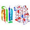
- PDB-11gv: Crystal structure of selective inhibitor 16 bound at the active s... -

+
Open data


ID or keywords:

Loading...

-
Basic information

Entry
Database: PDB / ID: 11gv
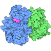
TitleCrystal structure of selective inhibitor 16 bound at the active site of CDK2
Components
  • Cyclin-A2
  • Cyclin-dependent kinase 2
KeywordsSIGNALING PROTEIN / kinase / cancer / oncology / inhibitor / selectivity
Function / homology
Function and homology information


cell cycle G1/S phase transition / cellular response to luteinizing hormone stimulus / G2/M DNA replication checkpoint / Transcription of E2F targets under negative control by p107 (RBL1) and p130 (RBL2) in complex with HDAC1 / cellular response to leptin stimulus / response to glucagon / male pronucleus / female pronucleus / cellular response to cocaine / cyclin-dependent protein serine/threonine kinase regulator activity ...cell cycle G1/S phase transition / cellular response to luteinizing hormone stimulus / G2/M DNA replication checkpoint / Transcription of E2F targets under negative control by p107 (RBL1) and p130 (RBL2) in complex with HDAC1 / cellular response to leptin stimulus / response to glucagon / male pronucleus / female pronucleus / cellular response to cocaine / cyclin-dependent protein serine/threonine kinase regulator activity / positive regulation of DNA biosynthetic process / cellular response to insulin-like growth factor stimulus / cyclin A1-CDK2 complex / cyclin E2-CDK2 complex / cyclin E1-CDK2 complex / cyclin A2-CDK2 complex / G2 Phase / Y chromosome / cyclin-dependent protein kinase activity / regulation of heterochromatin organization / Phosphorylation of proteins involved in G1/S transition by active Cyclin E:Cdk2 complexes / positive regulation of heterochromatin formation / p53-Dependent G1 DNA Damage Response / X chromosome / PTK6 Regulates Cell Cycle / regulation of anaphase-promoting complex-dependent catabolic process / Defective binding of RB1 mutants to E2F1,(E2F2, E2F3) / centriole replication / telomere maintenance in response to DNA damage / Regulation of APC/C activators between G1/S and early anaphase / animal organ regeneration / microtubule organizing center / regulation of DNA replication / G0 and Early G1 / cochlea development / Activation of the pre-replicative complex / cellular response to platelet-derived growth factor stimulus / Telomere Extension By Telomerase / cyclin-dependent protein kinase holoenzyme complex / cyclin-dependent kinase / cyclin-dependent protein serine/threonine kinase activity / TP53 Regulates Transcription of Genes Involved in G1 Cell Cycle Arrest / Regulation of MITF-M-dependent genes involved in cell cycle and proliferation / Cajal body / Cyclin E associated events during G1/S transition / Activation of ATR in response to replication stress / centrosome duplication / Cyclin A:Cdk2-associated events at S phase entry / Cyclin A/B1/B2 associated events during G2/M transition / Chk1/Chk2(Cds1) mediated inactivation of Cyclin B:Cdk1 complex / condensed chromosome / mitotic G1 DNA damage checkpoint signaling / cellular response to nitric oxide / post-translational protein modification / positive regulation of fibroblast proliferation / cyclin binding / positive regulation of DNA replication / negative regulation of protein localization to chromatin / regulation of mitotic cell cycle / cellular response to estradiol stimulus / G1/S transition of mitotic cell cycle / G2/M transition of mitotic cell cycle / Cdc20:Phospho-APC/C mediated degradation of Cyclin A / meiotic cell cycle / peptidyl-serine phosphorylation / cellular senescence / DNA Damage/Telomere Stress Induced Senescence / Meiotic recombination / CDK-mediated phosphorylation and removal of Cdc6 / SCF(Skp2)-mediated degradation of p27/p21 / Transcriptional regulation of granulopoiesis / Orc1 removal from chromatin / Cyclin D associated events in G1 / Regulation of TP53 Degradation / nuclear envelope / transcription regulator complex / Factors involved in megakaryocyte development and platelet production / ciliary basal body / Processing of DNA double-strand break ends / Senescence-Associated Secretory Phenotype (SASP) / cellular response to hypoxia / Regulation of TP53 Activity through Phosphorylation / Ras protein signal transduction / DNA replication / protein phosphorylation / chromosome, telomeric region / endosome / Ub-specific processing proteases / chromatin remodeling / cell division / protein domain specific binding / protein serine kinase activity / DNA repair / protein serine/threonine kinase activity / centrosome / positive regulation of cell population proliferation / protein kinase binding / magnesium ion binding / signal transduction / nucleoplasm
Similarity search - Function
Cyclin-A, N-terminal APC/C binding region / Cyclin-A N-terminal APC/C binding region / : / Cyclin, C-terminal domain / : / Cyclins signature. / Cyclin / Cyclin, C-terminal domain / Cyclin_C / Cyclin, N-terminal ...Cyclin-A, N-terminal APC/C binding region / Cyclin-A N-terminal APC/C binding region / : / Cyclin, C-terminal domain / : / Cyclins signature. / Cyclin / Cyclin, C-terminal domain / Cyclin_C / Cyclin, N-terminal / Cyclin, N-terminal domain / Cyclin-like / domain present in cyclins, TFIIB and Retinoblastoma / Cyclin-like superfamily / : / Serine/threonine-protein kinase, active site / Serine/Threonine protein kinases active-site signature. / Protein kinase domain / Serine/Threonine protein kinases, catalytic domain / Protein kinase, ATP binding site / Protein kinases ATP-binding region signature. / Protein kinase domain profile. / Protein kinase domain / Protein kinase-like domain superfamily
Similarity search - Domain/homology
: / ISOPROPYL ALCOHOL / Cyclin-A2 / Cyclin-dependent kinase 2
Similarity search - Component
Biological speciesHomo sapiens (human)
MethodX-RAY DIFFRACTION / SYNCHROTRON / MOLECULAR REPLACEMENT / Resolution: 1.59 Å
AuthorsMurray, J.M. / Oh, A. / Kiefer, J.R. / Verma, V.A. / Grandner, J.M. / Parr, B.T.
Funding support1items
OrganizationGrant numberCountry
Not funded
CitationJournal: J.Med.Chem. / Year: 2026
Title: Utilizing Molecular Dynamics and Mechanistic Pharmacokinetic Studies in the Design of Selective CDK2 Inhibitors.
Authors: Verma, V.A. / Grandner, J.M. / Parr, B.T. / Zeng, M. / Ashley, M. / Wang, Y. / Beroza, P. / Carione, P. / Johnson, K.M. / Oh, A.J. / Murray, J.M. / Kiefer, J.R. / Moffat, J.G. / Prangley, M. ...Authors: Verma, V.A. / Grandner, J.M. / Parr, B.T. / Zeng, M. / Ashley, M. / Wang, Y. / Beroza, P. / Carione, P. / Johnson, K.M. / Oh, A.J. / Murray, J.M. / Kiefer, J.R. / Moffat, J.G. / Prangley, M. / Merrick, K. / Vartanian, S. / Hafner, M. / Orr, C.J. / Segal, E. / Levy, E.S. / Wang, J. / Xu, Z. / Wang, S. / Liu, G. / Niu, Y. / Li, X. / Zhang, Q. / Ma, Z. / Sun, M. / Wu, Z. / Zhao, W. / Li, Y. / Zhang, L. / Magnuson, S.R. / Samy, K.E.
History
DepositionFeb 23, 2026Deposition site: RCSB / Processing site: RCSB
Revision 1.0Sep 2, 2026Provider: repository / Type: Initial release

-
Structure visualization

Structure viewerMolecule:
MolmilJmol/JSmol

Downloads & links

-
Assembly

Deposited unit
A: Cyclin-dependent kinase 2
B: Cyclin-A2
hetero molecules


Theoretical massNumber of molelcules
Total (without water)66,39834
Polymers63,9412
Non-polymers2,45732
Water11,998666
1


  • Idetical with deposited unit
  • defined by author&software
  • Evidence: gel filtration
TypeNameSymmetry operationNumber
identity operation1_555x,y,z1
Buried area8970 Å2
ΔGint-22 kcal/mol
Surface area23190 Å2
MethodPISA
Unit cell
Length a, b, c (Å)70.930, 112.230, 159.740
Angle α, β, γ (deg.)90.00, 90.00, 90.00
Int Tables number20
Space group name H-MC2221
Components on special symmetry positions
IDModelComponents
11A-614-

HOH

21A-675-

HOH

-
Components

-
Protein , 2 types, 2 molecules AB

#1: Protein Cyclin-dependent kinase 2 / Cell division protein kinase 2 / p33 protein kinase


Mass: 34056.469 Da / Num. of mol.: 1
Source method: isolated from a genetically manipulated source
Source: (gene. exp.) Homo sapiens (human) / Gene: CDK2, CDKN2 / Production host: Escherichia coli (E. coli) / References: UniProt: P24941, cyclin-dependent kinase
#2: Protein Cyclin-A2 / Cyclin-A / Cyclin A


Mass: 29884.605 Da / Num. of mol.: 1 / Fragment: residues 174-432
Source method: isolated from a genetically manipulated source
Source: (gene. exp.) Homo sapiens (human) / Gene: CCNA2, CCN1, CCNA / Production host: Escherichia coli (E. coli) / References: UniProt: P20248

-
Non-polymers , 7 types, 698 molecules

#3: Chemical ChemComp-MG / MAGNESIUM ION


Mass: 24.305 Da / Num. of mol.: 3 / Source method: obtained synthetically / Formula: Mg
#4: Chemical
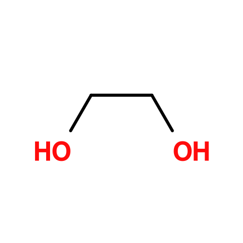
ChemComp-EDO / 1,2-ETHANEDIOL / ETHYLENE GLYCOL


Mass: 62.068 Da / Num. of mol.: 19 / Source method: obtained synthetically / Formula: C2H6O2
#5: Chemical
ChemComp-CL / CHLORIDE ION


Mass: 35.453 Da / Num. of mol.: 4 / Source method: obtained synthetically / Formula: Cl
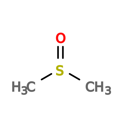
#6: Chemical ChemComp-DMS / DIMETHYL SULFOXIDE


Mass: 78.133 Da / Num. of mol.: 3 / Source method: obtained synthetically / Formula: C2H6OS / Comment: DMSO, precipitant*YM
#7: Chemical ChemComp-A1DEQ / (1R,3S)-3-{3-[(1-methyl-6-oxo-1,6-dihydropyrimidin-2-yl)amino]-1H-pyrazol-5-yl}cyclopentyl bicyclo[1.1.1]pentan-1-ylcarbamate


Mass: 384.432 Da / Num. of mol.: 2 / Source method: obtained synthetically / Formula: C19H24N6O3 / Feature type: SUBJECT OF INVESTIGATION
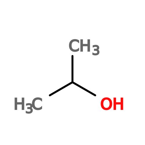
#8: Chemical ChemComp-IPA / ISOPROPYL ALCOHOL / 2-PROPANOL


Mass: 60.095 Da / Num. of mol.: 1 / Source method: obtained synthetically / Formula: C3H8O
#9: Water ChemComp-HOH / water


Mass: 18.015 Da / Num. of mol.: 666 / Source method: isolated from a natural source / Formula: H2O

-
Details

Has ligand of interestY
Has protein modificationY

-
Experimental details

-
Experiment

ExperimentMethod: X-RAY DIFFRACTION / Number of used crystals: 1

-
Sample preparation

CrystalDensity Matthews: 2.49 Å3/Da / Density % sol: 50.52 %
Crystal growTemperature: 291 K / Method: vapor diffusion
Details: 15% ethylene glycol, 10% 2-propanol, 0.2M MgCl2, 10% PEG 4000, 0.1M HEPES pH 7.5

-
Data collection

DiffractionMean temperature: 93 K / Serial crystal experiment: N
Diffraction sourceSource: SYNCHROTRON / Site: NSLS-II / Beamline: 17-ID-2 / Wavelength: 0.9793 Å
DetectorType: DECTRIS EIGER X 16M / Detector: PIXEL / Date: Nov 2, 2021
RadiationProtocol: SINGLE WAVELENGTH / Monochromatic (M) / Laue (L): M / Scattering type: x-ray
Radiation wavelengthWavelength: 0.9793 Å / Relative weight: 1
ReflectionResolution: 1.59→29.52 Å / Num. obs: 85637 / % possible obs: 99.9 % / Redundancy: 13.6 % / CC1/2: 1 / Rmerge(I) obs: 0.065 / Rpim(I) all: 0.018 / Rrim(I) all: 0.068 / Χ2: 0.97 / Net I/σ(I): 20.9 / Num. measured all: 1166101
Reflection shellResolution: 1.59→1.62 Å / % possible obs: 97.8 % / Redundancy: 13.2 % / Rmerge(I) obs: 0.746 / Num. measured all: 54598 / Num. unique obs: 4138 / CC1/2: 0.916 / Rpim(I) all: 0.208 / Rrim(I) all: 0.775 / Χ2: 0.87 / Net I/σ(I) obs: 3.2

-
Processing

Software
NameVersionClassification
PHENIX1.21.2_5419refinement
Aimlessdata scaling
XDSdata reduction
PHASERphasing
PDB_EXTRACTdata extraction
RefinementMethod to determine structure: MOLECULAR REPLACEMENT / Resolution: 1.59→29.52 Å / SU ML: 0.17 / Cross valid method: FREE R-VALUE / σ(F): 1.36 / Phase error: 18.87 / Stereochemistry target values: ML
RfactorNum. reflection% reflection
Rfree0.1942 4304 5.03 %
Rwork0.1694 --
obs0.1706 85582 99.88 %
Solvent computationShrinkage radii: 0.9 Å / VDW probe radii: 1.1 Å / Solvent model: FLAT BULK SOLVENT MODEL
Refinement stepCycle: LAST / Resolution: 1.59→29.52 Å
ProteinNucleic acidLigandSolventTotal
Num. atoms4377 0 155 666 5198
Refine LS restraints
Refine-IDTypeDev idealNumber
X-RAY DIFFRACTIONf_bond_d0.0034726
X-RAY DIFFRACTIONf_angle_d0.8136412
X-RAY DIFFRACTIONf_dihedral_angle_d14.1241725
X-RAY DIFFRACTIONf_chiral_restr0.045721
X-RAY DIFFRACTIONf_plane_restr0.005804
LS refinement shell
Resolution (Å)Rfactor RfreeNum. reflection RfreeRfactor RworkNum. reflection RworkRefine-ID% reflection obs (%)
1.59-1.610.31991290.24732608X-RAY DIFFRACTION97
1.61-1.630.25831410.23642688X-RAY DIFFRACTION100
1.63-1.650.26371570.23172679X-RAY DIFFRACTION100
1.65-1.670.23281490.21512675X-RAY DIFFRACTION100
1.67-1.690.26841430.22092682X-RAY DIFFRACTION100
1.69-1.710.23261630.21052656X-RAY DIFFRACTION100
1.71-1.740.22131580.1982677X-RAY DIFFRACTION100
1.74-1.760.21131610.19162641X-RAY DIFFRACTION100
1.76-1.790.21071270.18692747X-RAY DIFFRACTION100
1.79-1.820.22981380.18642684X-RAY DIFFRACTION100
1.82-1.850.24111530.18512679X-RAY DIFFRACTION100
1.85-1.890.18711580.17912667X-RAY DIFFRACTION100
1.89-1.920.22791380.18382699X-RAY DIFFRACTION100
1.92-1.960.24111390.18542717X-RAY DIFFRACTION100
1.96-20.20631420.18562688X-RAY DIFFRACTION100
2-2.050.20921070.18012707X-RAY DIFFRACTION100
2.05-2.10.20161250.16612737X-RAY DIFFRACTION100
2.1-2.160.20131650.16862685X-RAY DIFFRACTION100
2.16-2.220.18971360.16662731X-RAY DIFFRACTION100
2.22-2.290.20541700.16782687X-RAY DIFFRACTION100
2.29-2.370.19351600.16232690X-RAY DIFFRACTION100
2.37-2.470.20651360.16592715X-RAY DIFFRACTION100
2.47-2.580.21221300.17122712X-RAY DIFFRACTION100
2.58-2.720.20091570.17352704X-RAY DIFFRACTION100
2.72-2.890.18241300.16722763X-RAY DIFFRACTION100
2.89-3.110.18961160.16892748X-RAY DIFFRACTION100
3.11-3.420.16561260.16062763X-RAY DIFFRACTION100
3.42-3.920.1691520.14962774X-RAY DIFFRACTION100
3.92-4.930.15991550.14392763X-RAY DIFFRACTION100
4.93-29.520.1921430.17472912X-RAY DIFFRACTION100
Refinement TLS params.

Method: refined / Refine-ID: X-RAY DIFFRACTION

IDL112)L122)L132)L222)L232)L332)S11 (Å °)S12 (Å °)S13 (Å °)S21 (Å °)S22 (Å °)S23 (Å °)S31 (Å °)S32 (Å °)S33 (Å °)T112)T122)T132)T222)T232)T332)Origin x (Å)Origin y (Å)Origin z (Å)
10.1843-0.15110.10140.1814-0.02450.2646-0.0286-0.062-0.1009-0.01760.1052-0.05150.23640.03980.00110.3407-0.05650.10550.2155-0.02280.1814-14.9894-31.461230.1607
21.63450.82440.10860.5011-0.36491.04350.02680.0831-0.18220.2088-0.0559-0.12550.08040.1222-0.00360.2425-0.04860.07440.184-0.0190.1498-5.2577-23.174324.5161
34.8987-0.64011.71811.673-0.77333.46330.2906-0.0861-0.43750.09890.0050.34820.4149-0.12830.28290.2568-0.12070.17520.11770.00510.2304-18.5065-25.16723.715
40.61470.08870.12560.5867-0.15070.73390.1253-0.04340.00920.60110.00070.41310.0468-0.14240.07330.2025-0.03870.19410.17430.02630.2652-18.4123-13.546217.4392
50.51880.05740.1511.1316-0.30361.85590.08760.0898-0.0224-0.0523-0.02190.16450.11990.06880.00150.12710.00230.00970.14420.00480.1755-12.1113-16.31370.8306
60.0354-0.04230.0310.0895-0.11750.36460.14670.14790.38570.12440.00160.5770.0319-0.28930.01080.14410.00570.03670.26170.06480.4414-27.665-7.1612.2271
70.46430.19720.51420.4829-0.06370.72360.0502-0.0190.32520.18010.06790.4042-0.1836-0.1960.00220.18080.00740.09180.19670.02990.3228-19.3232-3.577.7892
80.97940.16630.09240.6950.39620.23450.1508-0.17530.2053-0.0834-0.00390.2005-0.0034-0.1940.00140.1879-0.05070.05370.2081-0.00520.2099-1.4393-0.489319.4387
91.3387-0.08830.09771.1495-0.371.0966-0.033-0.12110.10220.0989-0.0105-0.1636-0.00290.1044-0.00470.163-0.01470.0510.157-0.01120.124417.4456-16.583426.7358
101.16280.3160.04550.56910.25190.90460.018-0.0535-0.2033-0.01850.01790.00250.21510.11440.00210.2226-0.00570.06050.1776-0.00320.103212.1599-25.665723.9157
111.7521-0.0889-0.24372.2530.08451.48170.06950.03710.2852-0.2125-0.0464-0.2621-0.15570.05240.00010.1514-0.03640.08650.1195-0.0060.204418.69832.142116.4636
Refinement TLS group
IDRefine-IDRefine TLS-IDSelection details
1X-RAY DIFFRACTION1chain 'A' and (resid 1 through 28 )
2X-RAY DIFFRACTION2chain 'A' and (resid 29 through 65 )
3X-RAY DIFFRACTION3chain 'A' and (resid 66 through 100 )
4X-RAY DIFFRACTION4chain 'A' and (resid 101 through 140 )
5X-RAY DIFFRACTION5chain 'A' and (resid 141 through 247 )
6X-RAY DIFFRACTION6chain 'A' and (resid 248 through 266 )
7X-RAY DIFFRACTION7chain 'A' and (resid 267 through 290 )
8X-RAY DIFFRACTION8chain 'B' and (resid 174 through 192 )
9X-RAY DIFFRACTION9chain 'B' and (resid 193 through 268 )
10X-RAY DIFFRACTION10chain 'B' and (resid 269 through 310 )
11X-RAY DIFFRACTION11chain 'B' and (resid 311 through 432 )

+
About Yorodumi

-
News

-
Feb 9, 2022. New format data for meta-information of EMDB entries

New format data for meta-information of EMDB entries

  • Version 3 of the EMDB header file is now the official format.
  • The previous official version 1.9 will be removed from the archive.

Related info.:EMDB header

External links:wwPDB to switch to version 3 of the EMDB data model

-
Aug 12, 2020. Covid-19 info

Covid-19 info

URL: https://pdbj.org/emnavi/covid19.php

New page: Covid-19 featured information page in EM Navigator.

Related info.:Covid-19 info / Mar 5, 2020. Novel coronavirus structure data

+
Mar 5, 2020. Novel coronavirus structure data

Novel coronavirus structure data

Related info.:Yorodumi Speices / Aug 12, 2020. Covid-19 info

External links:COVID-19 featured content - PDBj / Molecule of the Month (242):Coronavirus Proteases

+
Jan 31, 2019. EMDB accession codes are about to change! (news from PDBe EMDB page)

EMDB accession codes are about to change! (news from PDBe EMDB page)

  • The allocation of 4 digits for EMDB accession codes will soon come to an end. Whilst these codes will remain in use, new EMDB accession codes will include an additional digit and will expand incrementally as the available range of codes is exhausted. The current 4-digit format prefixed with “EMD-” (i.e. EMD-XXXX) will advance to a 5-digit format (i.e. EMD-XXXXX), and so on. It is currently estimated that the 4-digit codes will be depleted around Spring 2019, at which point the 5-digit format will come into force.
  • The EM Navigator/Yorodumi systems omit the EMD- prefix.

Related info.:Q: What is EMD? / ID/Accession-code notation in Yorodumi/EM Navigator

External links:EMDB Accession Codes are Changing Soon! / Contact to PDBj

+
Jul 12, 2017. Major update of PDB

Major update of PDB

  • wwPDB released updated PDB data conforming to the new PDBx/mmCIF dictionary.
  • This is a major update changing the version number from 4 to 5, and with Remediation, in which all the entries are updated.
  • In this update, many items about electron microscopy experimental information are reorganized (e.g. em_software).
  • Now, EM Navigator and Yorodumi are based on the updated data.

External links:wwPDB Remediation / Enriched Model Files Conforming to OneDep Data Standards Now Available in the PDB FTP Archive

-
Yorodumi

Thousand views of thousand structures

  • Yorodumi is a browser for structure data from EMDB, PDB, SASBDB, etc.
  • This page is also the successor to EM Navigator detail page, and also detail information page/front-end page for Omokage search.
  • The word "yorodu" (or yorozu) is an old Japanese word meaning "ten thousand". "mi" (miru) is to see.

Related info.:EMDB / PDB / SASBDB / Comparison of 3 databanks / Yorodumi Search / Aug 31, 2016. New EM Navigator & Yorodumi / Yorodumi Papers / Jmol/JSmol / Function and homology information / Changes in new EM Navigator and Yorodumi

Read more