 Movie
Movie Controller


+ Open data

- Basic information
| Entry | Database: PDB / ID: 11ex | ||||||
|---|---|---|---|---|---|---|---|
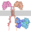
| Title | Crystal structure of hIgG1-Fc in complex with FcRL5 | ||||||
 Components Components |
| ||||||
 Keywords Keywords | IMMUNE SYSTEM / Antibody / IgG1 / Fc / Immunology / FcRL5 | ||||||
| Function / homology |  Function and homology information Function and homology informationcomplement-dependent cytotoxicity / single fertilization / antibody-dependent cellular cytotoxicity / Fc-gamma receptor I complex binding / immunoglobulin complex, circulating / Classical antibody-mediated complement activation / immunoglobulin receptor binding / complement activation, classical pathway / FCGR activation / B cell activation ...complement-dependent cytotoxicity / single fertilization / antibody-dependent cellular cytotoxicity / Fc-gamma receptor I complex binding / immunoglobulin complex, circulating / Classical antibody-mediated complement activation / immunoglobulin receptor binding / complement activation, classical pathway / FCGR activation / B cell activation / Role of phospholipids in phagocytosis / antigen binding / coreceptor activity / B cell receptor signaling pathway / FCGR3A-mediated IL10 synthesis / Regulation of Complement cascade / FCGR3A-mediated phagocytosis / Regulation of actin dynamics for phagocytic cup formation / adaptive immune response / transmembrane signaling receptor activity / Interleukin-4 and Interleukin-13 signaling / blood microparticle / antibacterial humoral response / Initial triggering of complement / cell surface receptor signaling pathway / signaling receptor complex / immune response / external side of plasma membrane / cell surface / : / extracellular exosome / extracellular region / plasma membrane Similarity search - Function | ||||||
| Biological species |  Homo sapiens (human) Homo sapiens (human) | ||||||
| Method |  X-RAY DIFFRACTION / SYNCHROTRON / MOLECULAR REPLACEMENT / Resolution: 3.42 Å X-RAY DIFFRACTION / SYNCHROTRON / MOLECULAR REPLACEMENT / Resolution: 3.42 Å | ||||||
 Authors Authors | Guo, M. / Yan, Z. | ||||||
| Funding support |  United States, 1items United States, 1items
| ||||||
 Citation Citation | Journal: To Be Published Title: Crystal structure of hIgG1-Fc in complex with FcRL5 Authors: Guo, M. / Yan, Z. | ||||||
| History |
|
- Structure visualization
| Structure viewer | Molecule: MolmilJmol/JSmol |
|---|
- Downloads & links
-Download
| PDBx/mmCIF format | 11ex.cif.gz | 186.4 KB | Display | PDBx/mmCIF format |
|---|---|---|---|---|
| PDB format | pdb11ex.ent.gz | 119 KB | Display | PDB format |
| PDBx/mmJSON format | 11ex.json.gz | Tree view | PDBx/mmJSON format | |
| Others |  Other downloads Other downloads |
-Validation report
| Arichive directory | https://data.pdbj.org/pub/pdb/validation_reports/1e/11exftp://data.pdbj.org/pub/pdb/validation_reports/1e/11ex | HTTPS FTP |
|---|
-Related structure data
| Similar structure data |
|---|
-Links
 PDBj
PDBj









- Assembly
| Deposited unit | 
| ||||||||||
|---|---|---|---|---|---|---|---|---|---|---|---|
| 1 |
| ||||||||||
| Unit cell |
|
-Components
| #1: Protein | Mass: 29625.678 Da / Num. of mol.: 1 / Mutation: F26S, K45E, L111P, E128G, H155R, W251R Source method: isolated from a genetically manipulated source Source: (gene. exp.) Homo sapiens (human) / Gene: FCRL5, FCRH5, IRTA2, UNQ503/PRO820 / Cell line (production host): HEK293 / Production host: Homo sapiens (human) / References: UniProt: Q96RD9 | ||||||||||
|---|---|---|---|---|---|---|---|---|---|---|---|
| #2: Protein | Mass: 23844.883 Da / Num. of mol.: 2 / Mutation: K320E, Q342A Source method: isolated from a genetically manipulated source Source: (gene. exp.) Homo sapiens (human) / Gene: IGHG1 / Cell line (production host): HEK293 / Production host: Homo sapiens (human) / References: UniProt: P01857#3: Polysaccharide | alpha-D-mannopyranose-(1-3)-[alpha-D-mannopyranose-(1-6)]beta-D-mannopyranose-(1-4)-2-acetamido-2- ...alpha-D-mannopyranose-(1-3)-[alpha-D-mannopyranose-(1-6)]beta-D-mannopyranose-(1-4)-2-acetamido-2-deoxy-beta-D-glucopyranose-(1-4)-[alpha-L-fucopyranose-(1-6)]2-acetamido-2-deoxy-beta-D-glucopyranose | Source method: isolated from a genetically manipulated source #4: Polysaccharide | 2-acetamido-2-deoxy-beta-D-glucopyranose-(1-2)-alpha-D-mannopyranose-(1-3)-[2-acetamido-2-deoxy- ...2-acetamido-2-deoxy-beta-D-glucopyranose-(1-2)-alpha-D-mannopyranose-(1-3)-[2-acetamido-2-deoxy-beta-D-glucopyranose-(1-2)-alpha-D-mannopyranose-(1-6)]beta-D-mannopyranose-(1-4)-2-acetamido-2-deoxy-beta-D-glucopyranose-(1-4)-[alpha-L-fucopyranose-(1-6)]2-acetamido-2-deoxy-beta-D-glucopyranose | Type: oligosaccharide / Mass: 1463.349 Da / Num. of mol.: 1 Source method: isolated from a genetically manipulated source #5: Sugar | ChemComp-NAG / |   Type: D-saccharide, beta linking / Mass: 221.208 Da / Num. of mol.: 1 / Source method: obtained synthetically / Formula: C8H15NO6 / Feature type: SUBJECT OF INVESTIGATION Type: D-saccharide, beta linking / Mass: 221.208 Da / Num. of mol.: 1 / Source method: obtained synthetically / Formula: C8H15NO6 / Feature type: SUBJECT OF INVESTIGATIONHas ligand of interest | Y | Has protein modification | Y | |
-Experimental details
-Experiment
| Experiment | Method: X-RAY DIFFRACTION / Number of used crystals: 1 |
|---|
- Sample preparation
| Crystal | Density Matthews: 4.76 Å3/Da / Density % sol: 74.14 % |
|---|---|
| Crystal grow | Temperature: 298.15 K / Method: vapor diffusion, sitting drop / pH: 6.5 / Details: 0.1MES, pH6.5, 25%(w/v) PEG 8000 |
-Data collection
| Diffraction | Mean temperature: 100 K / Serial crystal experiment: N |
|---|---|
| Diffraction source | Source: SYNCHROTRON / Site: APS / Beamline: 24-ID-E / Wavelength: 0.97905 Å |
| Detector | Type: DECTRIS EIGER X 16M / Detector: PIXEL / Date: Oct 10, 2025 |
| Radiation | Protocol: SINGLE WAVELENGTH / Monochromatic (M) / Laue (L): M / Scattering type: x-ray |
| Radiation wavelength | Wavelength: 0.97905 Å / Relative weight: 1 |
| Reflection | Resolution: 3.42→88.39 Å / Num. obs: 20062 / % possible obs: 99.9 % / Redundancy: 20.7 % / CC1/2: 0.956 / Rmerge(I) obs: 0.672 / Rpim(I) all: 0.151 / Rrim(I) all: 0.689 / Net I/σ(I): 3.6 / Num. measured all: 414740 |
| Reflection shell | Resolution: 3.42→3.48 Å / % possible obs: 96.7 % / Redundancy: 21.7 % / Rmerge(I) obs: 3.57 / Num. measured all: 20961 / Num. unique obs: 964 / CC1/2: 0.154 / Rpim(I) all: 0.785 / Rrim(I) all: 3.656 / Net I/σ(I) obs: 0.6 |
- Processing
| Software |
| ||||||||||||||||||||||||||||||||||||||||||||||||||||||||
|---|---|---|---|---|---|---|---|---|---|---|---|---|---|---|---|---|---|---|---|---|---|---|---|---|---|---|---|---|---|---|---|---|---|---|---|---|---|---|---|---|---|---|---|---|---|---|---|---|---|---|---|---|---|---|---|---|---|
| Refinement | Method to determine structure: MOLECULAR REPLACEMENT / Resolution: 3.42→88.39 Å / SU ML: 0.6801 / Cross valid method: FREE R-VALUE / σ(F): 1.33 / Phase error: 35.4466 Stereochemistry target values: GeoStd + Monomer Library + CDL v1.2
| ||||||||||||||||||||||||||||||||||||||||||||||||||||||||
| Solvent computation | Shrinkage radii: 0.9 Å / VDW probe radii: 1.1 Å / Solvent model: FLAT BULK SOLVENT MODEL | ||||||||||||||||||||||||||||||||||||||||||||||||||||||||
| Displacement parameters | Biso mean: 93.55 Å2 | ||||||||||||||||||||||||||||||||||||||||||||||||||||||||
| Refinement step | Cycle: LAST / Resolution: 3.42→88.39 Å
| ||||||||||||||||||||||||||||||||||||||||||||||||||||||||
| Refine LS restraints |
| ||||||||||||||||||||||||||||||||||||||||||||||||||||||||
| LS refinement shell |
|
