 Movie
Movie Controller


+ Open data

- Basic information
| Entry | Database: PDB / ID: 11ce | |||||||||
|---|---|---|---|---|---|---|---|---|---|---|
| Title | Crystal structure of Zn5 with Zn | |||||||||
 Components Components | The protease-Zn (II) complex of Zn5 | |||||||||
 Keywords Keywords | DE NOVO PROTEIN / de novo design / metalloproteases / enzyme design / deep learning method | |||||||||
| Biological species | synthetic construct (others) | |||||||||
| Method |  X-RAY DIFFRACTION / SYNCHROTRON / MOLECULAR REPLACEMENT / Resolution: 3.41 Å X-RAY DIFFRACTION / SYNCHROTRON / MOLECULAR REPLACEMENT / Resolution: 3.41 Å | |||||||||
 Authors Authors | Bera, A.K. / Chen, A. / Wu, K. / Kang, A. / Nguyen, H. / Baker, D. | |||||||||
| Funding support |  United States, 2items United States, 2items
| |||||||||
 Citation Citation | Journal: To Be Published Title: Computational design of metalloproteases Authors: Chen, A. / Wu, K. / Bera, A.K. / Baker, D. | |||||||||
| History |
|
- Structure visualization
| Structure viewer | Molecule:  MolmilJmol/JSmol MolmilJmol/JSmol |
|---|
- Downloads & links
-Download
| PDBx/mmCIF format | 11ce.cif.gz | 64.9 KB | Display | PDBx/mmCIF format |
|---|---|---|---|---|
| PDB format | pdb11ce.ent.gz | 37.7 KB | Display | PDB format |
| PDBx/mmJSON format | 11ce.json.gz | Tree view | PDBx/mmJSON format | |
| Others |  Other downloads Other downloads |
-Validation report
| Arichive directory | https://data.pdbj.org/pub/pdb/validation_reports/1c/11ceftp://data.pdbj.org/pub/pdb/validation_reports/1c/11ce | HTTPS FTP |
|---|
-Related structure data


-Links
 PDBj
PDBj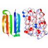
- Assembly
| Deposited unit | 
| ||||||||||||
|---|---|---|---|---|---|---|---|---|---|---|---|---|---|
| 1 |
| ||||||||||||
| Unit cell |
|
-Components
| #1: Protein | Mass: 24897.006 Da / Num. of mol.: 1 Source method: isolated from a genetically manipulated source Source: (gene. exp.) synthetic construct (others) / Production host:   Escherichia coli (E. coli) Escherichia coli (E. coli) |
|---|---|
| #2: Chemical | ChemComp-ZN /   Mass: 65.409 Da / Num. of mol.: 1 / Source method: obtained synthetically / Formula: Zn / Feature type: SUBJECT OF INVESTIGATION Mass: 65.409 Da / Num. of mol.: 1 / Source method: obtained synthetically / Formula: Zn / Feature type: SUBJECT OF INVESTIGATION |
| #3: Water | ChemComp-HOH /  Mass: 18.015 Da / Num. of mol.: 6 / Source method: isolated from a natural source / Formula: H2O Mass: 18.015 Da / Num. of mol.: 6 / Source method: isolated from a natural source / Formula: H2O |
| Has ligand of interest | Y |
| Has protein modification | N |
-Experimental details
-Experiment
| Experiment | Method: X-RAY DIFFRACTION / Number of used crystals: 1 |
|---|
- Sample preparation
| Crystal | Density Matthews: 4.08 Å3/Da / Density % sol: 69.82 % |
|---|---|
| Crystal grow | Temperature: 293 K / Method: vapor diffusion, sitting drop / pH: 7.5 Details: 21% PEG 350 MME, 0.5 M Magnesium chloride and 0.05 M Tris pH 7.5 |
-Data collection
| Diffraction | Mean temperature: 100 K / Serial crystal experiment: N |
|---|---|
| Diffraction source | Source: SYNCHROTRON / Site: APS / Beamline: 24-ID-E / Wavelength: 0.97905 Å |
| Detector | Type: DECTRIS EIGER X 16M / Detector: PIXEL / Date: Oct 17, 2025 |
| Radiation | Protocol: SINGLE WAVELENGTH / Monochromatic (M) / Laue (L): M / Scattering type: x-ray |
| Radiation wavelength | Wavelength: 0.97905 Å / Relative weight: 1 |
| Reflection | Resolution: 3.41→64.73 Å / Num. obs: 5375 / % possible obs: 99.7 % / Redundancy: 10.1 % / Biso Wilson estimate: 85.23 Å2 / CC1/2: 0.961 / Rmerge(I) obs: 0.5 / Rpim(I) all: 0.17 / Net I/σ(I): 4.2 |
| Reflection shell | Resolution: 3.41→3.68 Å / Redundancy: 9.5 % / Rmerge(I) obs: 1.04 / Mean I/σ(I) obs: 1.9 / Num. unique obs: 1113 / CC1/2: 0.479 / Rpim(I) all: 0.37 / % possible all: 99.4 |
- Processing
| Software |
| ||||||||||||||||||||||||
|---|---|---|---|---|---|---|---|---|---|---|---|---|---|---|---|---|---|---|---|---|---|---|---|---|---|
| Refinement | Method to determine structure: MOLECULAR REPLACEMENT / Resolution: 3.41→64.73 Å / SU ML: 0.1784 / Cross valid method: FREE R-VALUE / σ(F): 2 / Phase error: 18.8296 Stereochemistry target values: GeoStd + Monomer Library + CDL v1.2
| ||||||||||||||||||||||||
| Solvent computation | Shrinkage radii: 0.9 Å / VDW probe radii: 1.1 Å / Solvent model: FLAT BULK SOLVENT MODEL | ||||||||||||||||||||||||
| Displacement parameters | Biso mean: 83.46 Å2 | ||||||||||||||||||||||||
| Refinement step | Cycle: LAST / Resolution: 3.41→64.73 Å
| ||||||||||||||||||||||||
| Refine LS restraints |
| ||||||||||||||||||||||||
| LS refinement shell |
|
