National Institutes of Health/National Institute of Arthritis and Musculoskeletal and Skin Diseases (NIH/NIAMS)
United States
Citation
Journal: J Biol Chem / Year: 2026 Title: Structural basis for HIV-1 Rev recognition by the histone chaperone human Nap1. Authors: Elif Eren / Norman R Watts / Dennis C Winkler / Paul T Wingfield / Abstract: Human Nap1 (hNap1) is a histone chaperone involved in chromatin dynamics and has been shown to interact with the HIV-1 regulatory protein Rev, which is essential for nuclear export of viral RNA. ...Human Nap1 (hNap1) is a histone chaperone involved in chromatin dynamics and has been shown to interact with the HIV-1 regulatory protein Rev, which is essential for nuclear export of viral RNA. Despite the functional significance of this interaction, its structural basis has remained elusive. Here, we present the X-ray crystal structure of hNap1 and the cryo-electron microscopy structure of the core domain of hNap1-Rev complex. The structure reveals that hNap1 binds Rev dimers via its acidic concave surface, engaging the Rev arginine-rich motif and oligomerization domain, and stabilizes Rev as a dimer-of-dimers tetramer. This interaction prevents higher-order Rev aggregation and enhances Rev's cooperative binding to the Rev Response Element. Surface plasmon resonance measurements confirm the formation of a stable complex with an apparent low-micromolar affinity between hNap1 and Rev, supporting a chaperone-like, reversible association. Our findings provide molecular insight into how hNap1 modulates Rev assembly and function, suggesting a model in which hNap1 primes Rev for productive engagement with viral RNA, thereby facilitating HIV-1 replication.
A1: Protein Rev B1: Protein Rev C1: Protein Rev D1: Protein Rev E1: Protein Rev F1: Protein Rev G1: Protein Rev H1: Protein Rev I1: Protein Rev J1: Protein Rev K1: Protein Rev L1: Protein Rev M1: Protein Rev N1: Protein Rev O1: Protein Rev P1: Protein Rev R1: Protein Rev S1: Protein Rev T1: Protein Rev U1: Protein Rev V1: Protein Rev W1: Protein Rev X1: Protein Rev Y1: Protein Rev c2: Protein Rev d2: Protein Rev e2: Protein Rev f2: Protein Rev g2: Protein Rev h2: Protein Rev v2: Protein Rev w2: Protein Rev x2: Protein Rev y2: Protein Rev a3: Protein Rev b3: Protein Rev i3: Protein Rev j3: Protein Rev k3: Protein Rev l3: Protein Rev m3: Protein Rev n3: Protein Rev o3: Protein Rev p3: Protein Rev r3: Protein Rev s3: Protein Rev t3: Protein Rev u3: Protein Rev A4: Protein Rev B4: Protein Rev I4: Protein Rev J4: Protein Rev K4: Protein Rev L4: Protein Rev M4: Protein Rev N4: Protein Rev O4: Protein Rev R4: Protein Rev S4: Protein Rev T4: Protein Rev U4: Protein Rev A5: Protein Rev B5: Protein Rev C5: Protein Rev D5: Protein Rev E5: Protein Rev F5: Protein Rev G5: Protein Rev H5: Protein Rev M5: Protein Rev N5: Protein Rev O5: Protein Rev P5: Protein Rev T5: Protein Rev U5: Protein Rev V5: Protein Rev W5: Protein Rev X5: Protein Rev Y5: Protein Rev C6: Protein Rev D6: Protein Rev E6: Protein Rev F6: Protein Rev G6: Protein Rev H6: Protein Rev V6: Protein Rev W6: Protein Rev X6: Protein Rev Y6: Protein Rev a7: Protein Rev b7: Protein Rev c7: Protein Rev d7: Protein Rev e7: Protein Rev f7: Protein Rev g7: Protein Rev h7: Protein Rev i7: Protein Rev j7: Protein Rev k7: Protein Rev l7: Protein Rev m7: Protein Rev n7: Protein Rev o7: Protein Rev p7: Protein Rev r7: Protein Rev s7: Protein Rev t7: Protein Rev u7: Protein Rev v7: Protein Rev w7: Protein Rev x7: Protein Rev y7: Protein Rev a8: Protein Rev b8: Protein Rev c8: Protein Rev d8: Protein Rev e8: Protein Rev f8: Protein Rev g8: Protein Rev h8: Protein Rev m8: Protein Rev n8: Protein Rev o8: Protein Rev p8: Protein Rev t8: Protein Rev u8: Protein Rev v8: Protein Rev w8: Protein Rev x8: Protein Rev y8: Protein Rev A9: Protein Rev B9: Protein Rev C9: Protein Rev D9: Protein Rev E9: Protein Rev F9: Protein Rev G9: Protein Rev H9: Protein Rev I9: Protein Rev J9: Protein Rev K9: Protein Rev L9: Protein Rev M9: Protein Rev N9: Protein Rev O9: Protein Rev P9: Protein Rev R9: Protein Rev S9: Protein Rev T9: Protein Rev U9: Protein Rev V9: Protein Rev W9: Protein Rev X9: Protein Rev Y9: Protein Rev
In the structure databanks used in Yorodumi, some data are registered as the other names, "COVID-19 virus" and "2019-nCoV". Here are the details of the virus and the list of structure data.
Jan 31, 2019. EMDB accession codes are about to change! (news from PDBe EMDB page)
EMDB accession codes are about to change! (news from PDBe EMDB page)
The allocation of 4 digits for EMDB accession codes will soon come to an end. Whilst these codes will remain in use, new EMDB accession codes will include an additional digit and will expand incrementally as the available range of codes is exhausted. The current 4-digit format prefixed with “EMD-” (i.e. EMD-XXXX) will advance to a 5-digit format (i.e. EMD-XXXXX), and so on. It is currently estimated that the 4-digit codes will be depleted around Spring 2019, at which point the 5-digit format will come into force.
The EM Navigator/Yorodumi systems omit the EMD- prefix.
Related info.:Q: What is EMD? / ID/Accession-code notation in Yorodumi/EM Navigator
Yorodumi is a browser for structure data from EMDB, PDB, SASBDB, etc.
This page is also the successor to EM Navigator detail page, and also detail information page/front-end page for Omokage search.
The word "yorodu" (or yorozu) is an old Japanese word meaning "ten thousand". "mi" (miru) is to see.
Related info.:EMDB / PDB / SASBDB / Comparison of 3 databanks / Yorodumi Search / Aug 31, 2016. New EM Navigator & Yorodumi / Yorodumi Papers / Jmol/JSmol / Function and homology information / Changes in new EM Navigator and Yorodumi
 Movie
Movie Controller


 Open data
 Basic information
 Components
 Keywords
 Function and homology information
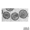 Human immunodeficiency virus 1
 Authors
 United States, 1items
  Citation
 Structure visualization
 Downloads & links
 Other downloads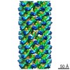


 PDBj
PDBj
 Assembly

 Escherichia coli (E. coli) / References: UniProt: Q76PP8
 Sample preparation
 Electron microscopy imaging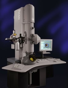
 FIELD EMISSION GUN / Accelerating voltage: 300 kV / Illumination mode: FLOOD BEAM
 Processing