 Movie
Movie Controller


+ Open data

- Basic information
| Entry | Database: PDB / ID: 10sf | ||||||||||||||||||||||||
|---|---|---|---|---|---|---|---|---|---|---|---|---|---|---|---|---|---|---|---|---|---|---|---|---|---|
| Title | The CryoEM structure of T10 type1 nanofiber | ||||||||||||||||||||||||
 Components Components | Type10 type 1 nanofiber | ||||||||||||||||||||||||
 Keywords Keywords | PROTEIN FIBRIL / nanofiber | ||||||||||||||||||||||||
| Function / homology | DECANOIC ACID Function and homology information Function and homology information | ||||||||||||||||||||||||
| Biological species | synthetic construct (others) | ||||||||||||||||||||||||
| Method | ELECTRON MICROSCOPY / helical reconstruction / cryo EM / Resolution: 3.08 Å | ||||||||||||||||||||||||
 Authors Authors | Zhang, H. / Yang, Y. | ||||||||||||||||||||||||
| Funding support | 1items
| ||||||||||||||||||||||||
 Citation Citation | Journal: ACS Appl Bio Mater / Year: 2026 Title: Tailoring Avidity through Morphology: Structure-Avidity Relationship in CD38-Binding Nanofiber Radiotracers. Authors: Jacqueline M Godbe / Hongwei Zhang / Amit K Sharma / Katelyn N Ernst / Zhenghan Jing / Michael R Dyer / Julie L Prior / Erin Teubner / Brad Manion / Rui Tang / Yang Yang / Monica Shokeen /  Abstract: The lack of targeted molecular imaging agents for multiple myeloma (MM) hinders precise disease characterization and theranostic development. We address this by engineering a tunable platform of self- ...The lack of targeted molecular imaging agents for multiple myeloma (MM) hinders precise disease characterization and theranostic development. We address this by engineering a tunable platform of self-assembled peptide nanofibers that target CD38, a key antigen in MM. Simple variation of a conjugated lipid tail length (C4-C12) dictates the supramolecular architecture, as revealed by high-resolution cryo-EM. This structural control directly modulates biological function: avidity for CD38 increases monotonically with tail length, culminating in T12 nanofibers with sub-nanomolar affinity. This optimized morphology also enables unique pH-responsive di-tyrosine cross-linking and, critically, facilitates polyvalent cell-surface engagement that outcompetes high-affinity monomers in vitro. The nanofibers are efficiently radiolabeled with Cu, exhibit exceptional serum stability, and show no toxicity at doses 20-fold above projected imaging use. By establishing lipid tail length as a simple, powerful handle for controlling nanofiber structure, avidity, and function, we present a robust, translatable platform for advancing targeted imaging and therapy in CD38-positive malignancies. | ||||||||||||||||||||||||
| History |
|
- Structure visualization
| Structure viewer | Molecule: MolmilJmol/JSmol |
|---|
- Downloads & links
-Download
| PDBx/mmCIF format | 10sf.cif.gz | 49.2 KB | Display | PDBx/mmCIF format |
|---|---|---|---|---|
| PDB format | pdb10sf.ent.gz | 37.3 KB | Display | PDB format |
| PDBx/mmJSON format | 10sf.json.gz | Tree view | PDBx/mmJSON format | |
| Others |  Other downloads Other downloads |
-Validation report
| Arichive directory | https://data.pdbj.org/pub/pdb/validation_reports/0s/10sfftp://data.pdbj.org/pub/pdb/validation_reports/0s/10sf | HTTPS FTP |
|---|
-Related structure data
| Related structure data |  75433  10sdC  10seC  10sgC  10shC M: map data used to model this data C: citing same article ( |
|---|---|
| Similar structure data |


-Links
 PDBj
PDBj- Assembly
| Deposited unit | 
| ||||||||||||||||||||||||||||||||||||
|---|---|---|---|---|---|---|---|---|---|---|---|---|---|---|---|---|---|---|---|---|---|---|---|---|---|---|---|---|---|---|---|---|---|---|---|---|---|
| 1 |
| ||||||||||||||||||||||||||||||||||||
| Noncrystallographic symmetry (NCS) | NCS oper:
|
-Components
| #1: Protein/peptide | Mass: 1070.266 Da / Num. of mol.: 18 / Source method: obtained synthetically / Details: HYPIVIGGSK(NH2) / Source: (synth.) synthetic construct (others) #2: Chemical | ChemComp-DKA / 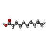  Mass: 172.265 Da / Num. of mol.: 18 / Source method: obtained synthetically / Formula: C10H20O2 / Source: (synth.) synthetic construct (others) Mass: 172.265 Da / Num. of mol.: 18 / Source method: obtained synthetically / Formula: C10H20O2 / Source: (synth.) synthetic construct (others)Has ligand of interest | N | Has protein modification | Y | |
|---|
-Experimental details
-Experiment
| Experiment | Method: ELECTRON MICROSCOPY |
|---|---|
| EM experiment | Aggregation state: FILAMENT / 3D reconstruction method: helical reconstruction |
- Sample preparation
| Component | Name: T10 type1 nanofiber / Type: COMPLEX / Entity ID: #1 / Source: SYNTHETIC |
|---|---|
| Source (natural) | Organism: synthetic construct (others) |
| Buffer solution | pH: 7.5 |
| Specimen | Embedding applied: NO / Shadowing applied: NO / Staining applied: NO / Vitrification applied: YES |
| Vitrification | Cryogen name: ETHANE |
- Electron microscopy imaging
| Experimental equipment |  Model: Titan Krios / Image courtesy: FEI Company |
|---|---|
| Microscopy | Model: TFS KRIOS |
| Electron gun | Electron source:  FIELD EMISSION GUN / Accelerating voltage: 300 kV / Illumination mode: FLOOD BEAM FIELD EMISSION GUN / Accelerating voltage: 300 kV / Illumination mode: FLOOD BEAM |
| Electron lens | Mode: BRIGHT FIELD / Nominal defocus max: 1800 nm / Nominal defocus min: 1200 nm |
| Image recording | Electron dose: 50 e/Å2 / Film or detector model: GATAN K3 (6k x 4k) |
- Processing
| EM software |
| ||||||||||||||||||||||||||||||||||||||||||||||||||||||||||||||||||||||||||||||||||||||||||||||||||||||||||
|---|---|---|---|---|---|---|---|---|---|---|---|---|---|---|---|---|---|---|---|---|---|---|---|---|---|---|---|---|---|---|---|---|---|---|---|---|---|---|---|---|---|---|---|---|---|---|---|---|---|---|---|---|---|---|---|---|---|---|---|---|---|---|---|---|---|---|---|---|---|---|---|---|---|---|---|---|---|---|---|---|---|---|---|---|---|---|---|---|---|---|---|---|---|---|---|---|---|---|---|---|---|---|---|---|---|---|---|
| CTF correction | Type: PHASE FLIPPING ONLY | ||||||||||||||||||||||||||||||||||||||||||||||||||||||||||||||||||||||||||||||||||||||||||||||||||||||||||
| Helical symmerty | Angular rotation/subunit: -0.698 ° / Axial rise/subunit: 4.79 Å / Axial symmetry: C1 | ||||||||||||||||||||||||||||||||||||||||||||||||||||||||||||||||||||||||||||||||||||||||||||||||||||||||||
| 3D reconstruction | Resolution: 3.08 Å / Resolution method: FSC 0.143 CUT-OFF / Num. of particles: 35 / Symmetry type: HELICAL | ||||||||||||||||||||||||||||||||||||||||||||||||||||||||||||||||||||||||||||||||||||||||||||||||||||||||||
| Refinement | Resolution: 3.08→3.08 Å / Cor.coef. Fo:Fc: 0.706 / SU B: 20.442 / SU ML: 0.358 / ESU R: 0.211 Stereochemistry target values: MAXIMUM LIKELIHOOD WITH PHASES Details: HYDROGENS HAVE BEEN USED IF PRESENT IN THE INPUT
| ||||||||||||||||||||||||||||||||||||||||||||||||||||||||||||||||||||||||||||||||||||||||||||||||||||||||||
| Solvent computation | Solvent model: PARAMETERS FOR MASK CACLULATION | ||||||||||||||||||||||||||||||||||||||||||||||||||||||||||||||||||||||||||||||||||||||||||||||||||||||||||
| Displacement parameters | Biso mean: 113.103 Å2 | ||||||||||||||||||||||||||||||||||||||||||||||||||||||||||||||||||||||||||||||||||||||||||||||||||||||||||
| Refinement step | Cycle: 1 / Total: 1566 | ||||||||||||||||||||||||||||||||||||||||||||||||||||||||||||||||||||||||||||||||||||||||||||||||||||||||||
| Refine LS restraints |
|