 Movie
Movie Controller

[English] 日本語
 Yorodumi
- PDB-10lw: Final Adduct of Human Ornithine Aminotransferase Inactivated by (... -
+ Open data

- Basic information
| Entry | Database: PDB / ID: 10lw | |||||||||
|---|---|---|---|---|---|---|---|---|---|---|
| Title | Final Adduct of Human Ornithine Aminotransferase Inactivated by (1R,4S)-4-Amino-3-(trifluoromethyl)cyclopent-2-ene-1-carboxylic Acid | |||||||||
 Components Components | Ornithine aminotransferase, mitochondrial | |||||||||
 Keywords Keywords | TRANSFERASE / Aminotransferase / L-ornithine / Inactivation | |||||||||
| Function / homology |  Function and homology information Function and homology information: / ornithine aminotransferase / L-ornithine transaminase activity / : / L-proline biosynthetic process / Glutamate and glutamine metabolism / visual perception / pyridoxal phosphate binding / mitochondrial matrix / mitochondrion ...: / ornithine aminotransferase / L-ornithine transaminase activity / : / L-proline biosynthetic process / Glutamate and glutamine metabolism / visual perception / pyridoxal phosphate binding / mitochondrial matrix / mitochondrion / nucleoplasm / identical protein binding / cytoplasm Similarity search - Function | |||||||||
| Biological species |  Homo sapiens (human) Homo sapiens (human) | |||||||||
| Method |  X-RAY DIFFRACTION / SYNCHROTRON / MOLECULAR REPLACEMENT / Resolution: 1.93 Å X-RAY DIFFRACTION / SYNCHROTRON / MOLECULAR REPLACEMENT / Resolution: 1.93 Å | |||||||||
 Authors Authors | Vargas, A.L. / Liu, D. | |||||||||
| Funding support |  United States, 2items United States, 2items
| |||||||||
 Citation Citation | Journal: Med.Chem.Res. / Year: 2026 Title: Inactivation of ornithine aminotransferase by (1 R ,4 S )-4-Amino-3-(trifluoromethyl)cyclopent-2-ene-1-carboxylic acid via a stable quinonoid intermediate. Authors: Kang, K.M. / Vargas, A.L. / Zhu, W. / Sokolenko, I. / Liu, D. / Silverman, R.B. | |||||||||
| History |
|
- Structure visualization
| Structure viewer | Molecule: MolmilJmol/JSmol |
|---|
- Downloads & links
-Download
| PDBx/mmCIF format | 10lw.cif.gz | 555.5 KB | Display | PDBx/mmCIF format |
|---|---|---|---|---|
| PDB format | pdb10lw.ent.gz | 407.9 KB | Display | PDB format |
| PDBx/mmJSON format | 10lw.json.gz | Tree view | PDBx/mmJSON format | |
| Others |  Other downloads Other downloads |
-Validation report
| Arichive directory | https://data.pdbj.org/pub/pdb/validation_reports/0l/10lwftp://data.pdbj.org/pub/pdb/validation_reports/0l/10lw | HTTPS FTP |
|---|
-Related structure data

-Links
 PDBj
PDBj- Assembly
| Deposited unit | 
| |||||||||||||||
|---|---|---|---|---|---|---|---|---|---|---|---|---|---|---|---|---|
| 1 | 
| |||||||||||||||
| 2 | 
| |||||||||||||||
| Unit cell |
| |||||||||||||||
| Components on special symmetry positions |
|
-Components
| #1: Protein | Mass: 48593.668 Da / Num. of mol.: 3 Source method: isolated from a genetically manipulated source Source: (gene. exp.) Homo sapiens (human) / Gene: OAT / Production host:  #2: Chemical | 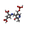  Mass: 400.277 Da / Num. of mol.: 3 / Source method: obtained synthetically / Formula: C15H17N2O9P / Feature type: SUBJECT OF INVESTIGATION Mass: 400.277 Da / Num. of mol.: 3 / Source method: obtained synthetically / Formula: C15H17N2O9P / Feature type: SUBJECT OF INVESTIGATION#3: Chemical | ChemComp-GOL /   Mass: 92.094 Da / Num. of mol.: 5 / Source method: obtained synthetically / Formula: C3H8O3 Mass: 92.094 Da / Num. of mol.: 5 / Source method: obtained synthetically / Formula: C3H8O3#4: Water | ChemComp-HOH / |  Mass: 18.015 Da / Num. of mol.: 486 / Source method: isolated from a natural source / Formula: H2O Mass: 18.015 Da / Num. of mol.: 486 / Source method: isolated from a natural source / Formula: H2OHas ligand of interest | Y | Has protein modification | N | |
|---|
-Experimental details
-Experiment
| Experiment | Method: X-RAY DIFFRACTION / Number of used crystals: 1 |
|---|
- Sample preparation
| Crystal | Density Matthews: 2.47 Å3/Da / Density % sol: 50.21 % |
|---|---|
| Crystal grow | Temperature: 298 K / Method: vapor diffusion, hanging drop / pH: 7.8 Details: 7.25%-8.50% PEG 6000, 100 mM-250 mM NaCl, 2-3.5% glycerol, and 100 mM Tricine pH 7.8. |
-Data collection
| Diffraction | Mean temperature: 100 K / Serial crystal experiment: N |
|---|---|
| Diffraction source | Source: SYNCHROTRON / Site: APS / Beamline: 21-ID-G / Wavelength: 0.97872 Å |
| Detector | Type: DECTRIS PILATUS 6M / Detector: PIXEL / Date: Sep 29, 2022 |
| Radiation | Protocol: SINGLE WAVELENGTH / Monochromatic (M) / Laue (L): M / Scattering type: x-ray |
| Radiation wavelength | Wavelength: 0.97872 Å / Relative weight: 1 |
| Reflection | Resolution: 1.93→50.01 Å / Num. obs: 102426 / % possible obs: 93.7 % / Redundancy: 6.9 % / Biso Wilson estimate: 40 Å2 / CC1/2: 1 / Net I/σ(I): 16.8 |
| Reflection shell | Resolution: 1.93→1.96 Å / Num. unique obs: 102426 / CC1/2: 0.329 |
- Processing
| Software |
| |||||||||||||||||||||||||||||||||||||||||||||||||||||||||||||||||||||||||||||||||||||||||||||||||||||||||||||||||||||||||||||||||||||||||||||||||||||||||||||||||||||||||||||||||||||||||||||||||||||||||||||||||||||||||
|---|---|---|---|---|---|---|---|---|---|---|---|---|---|---|---|---|---|---|---|---|---|---|---|---|---|---|---|---|---|---|---|---|---|---|---|---|---|---|---|---|---|---|---|---|---|---|---|---|---|---|---|---|---|---|---|---|---|---|---|---|---|---|---|---|---|---|---|---|---|---|---|---|---|---|---|---|---|---|---|---|---|---|---|---|---|---|---|---|---|---|---|---|---|---|---|---|---|---|---|---|---|---|---|---|---|---|---|---|---|---|---|---|---|---|---|---|---|---|---|---|---|---|---|---|---|---|---|---|---|---|---|---|---|---|---|---|---|---|---|---|---|---|---|---|---|---|---|---|---|---|---|---|---|---|---|---|---|---|---|---|---|---|---|---|---|---|---|---|---|---|---|---|---|---|---|---|---|---|---|---|---|---|---|---|---|---|---|---|---|---|---|---|---|---|---|---|---|---|---|---|---|---|---|---|---|---|---|---|---|---|---|---|---|---|---|---|---|---|
| Refinement | Method to determine structure: MOLECULAR REPLACEMENT / Resolution: 1.93→50.01 Å / SU ML: 0.245 / Cross valid method: FREE R-VALUE / σ(F): 1.33 / Phase error: 23.7831 Stereochemistry target values: GeoStd + Monomer Library + CDL v1.2
| |||||||||||||||||||||||||||||||||||||||||||||||||||||||||||||||||||||||||||||||||||||||||||||||||||||||||||||||||||||||||||||||||||||||||||||||||||||||||||||||||||||||||||||||||||||||||||||||||||||||||||||||||||||||||
| Solvent computation | Shrinkage radii: 0.9 Å / VDW probe radii: 1.1 Å / Solvent model: FLAT BULK SOLVENT MODEL | |||||||||||||||||||||||||||||||||||||||||||||||||||||||||||||||||||||||||||||||||||||||||||||||||||||||||||||||||||||||||||||||||||||||||||||||||||||||||||||||||||||||||||||||||||||||||||||||||||||||||||||||||||||||||
| Displacement parameters | Biso mean: 51.89 Å2 | |||||||||||||||||||||||||||||||||||||||||||||||||||||||||||||||||||||||||||||||||||||||||||||||||||||||||||||||||||||||||||||||||||||||||||||||||||||||||||||||||||||||||||||||||||||||||||||||||||||||||||||||||||||||||
| Refinement step | Cycle: LAST / Resolution: 1.93→50.01 Å
| |||||||||||||||||||||||||||||||||||||||||||||||||||||||||||||||||||||||||||||||||||||||||||||||||||||||||||||||||||||||||||||||||||||||||||||||||||||||||||||||||||||||||||||||||||||||||||||||||||||||||||||||||||||||||
| Refine LS restraints |
| |||||||||||||||||||||||||||||||||||||||||||||||||||||||||||||||||||||||||||||||||||||||||||||||||||||||||||||||||||||||||||||||||||||||||||||||||||||||||||||||||||||||||||||||||||||||||||||||||||||||||||||||||||||||||
| LS refinement shell |
|
