 Movie
Movie Controller


+ Open data

- Basic information
| Entry | Database: PDB / ID: 10es | |||||||||||||||||||||||||||
|---|---|---|---|---|---|---|---|---|---|---|---|---|---|---|---|---|---|---|---|---|---|---|---|---|---|---|---|---|
| Title | Chloroplast Glutamyl Peptidase WT in open-open conformation | |||||||||||||||||||||||||||
 Components Components | Isoform 2 of Probable glutamyl endopeptidase, chloroplastic | |||||||||||||||||||||||||||
 Keywords Keywords | PLANT PROTEIN / S9 protease / enzyme / serine protease / alpha-beta-alpha sandwich fold / beta-propeller | |||||||||||||||||||||||||||
| Function / homology |  Function and homology information Function and homology informationchloroplast stroma / Hydrolases; Acting on peptide bonds (peptidases); Serine endopeptidases / chloroplast / serine-type endopeptidase activity / proteolysis / cytosol Similarity search - Function | |||||||||||||||||||||||||||
| Biological species |   Arabidopsis thaliana (thale cress) Arabidopsis thaliana (thale cress) | |||||||||||||||||||||||||||
| Method | ELECTRON MICROSCOPY / single particle reconstruction / cryo EM / Resolution: 3 Å | |||||||||||||||||||||||||||
 Authors Authors | Ehrlich, J.J. / Routray, P. / van Wijk, K.J. / Kawate, T. | |||||||||||||||||||||||||||
| Funding support |  United States, 3items United States, 3items
| |||||||||||||||||||||||||||
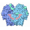
 Citation Citation | Journal: Protein Sci / Year: 2026 Title: Structural basis for dimerization, catalytic regulation, and substrate selectivity of the chloroplast S9D CGEP protease in Arabidopsis thaliana. Authors: Jacqueline J Ehrlich / Pratyush Routray / Louis Enns / Klaas J van Wijk / Toshimitsu Kawate / Abstract: S9 proteases are widely distributed across the tree-of-life and play essential roles in protein processing. However, the structural and mechanistic basis for protease activity in the S9D subfamily, ...S9 proteases are widely distributed across the tree-of-life and play essential roles in protein processing. However, the structural and mechanistic basis for protease activity in the S9D subfamily, restricted to photosynthetic eukaryotes (e.g., plants), cyanobacteria, proteobacteria and flavobacteria, is unknown. Here, we report the first high-resolution cryo-EM structures of an S9D protease, chloroplast glutamyl endopeptidase (CGEP) from the model plant Arabidopsis thaliana. CGEP adopts a dimeric architecture stabilized by two distinct interfaces: hydrophobic interactions between catalytic domains and an interdomain β-sheet linking the cap and catalytic domains. These interactions create a scaffold that supports a hinge loop, which acts as a steric gate to restrict substrate access and confine catalytic activity to the closed conformation. Unlike S9A-B-C proteases, CGEP maintains an intact catalytic triad in both open and closed states, relying on hinge-loop gating rather than catalytic disruption for regulation. Structural analysis and mutagenesis reveal that the hinge loop forms a conserved pocket favoring glutamate side chains, explaining CGEP's strong glutamate preference at cleavage sites. Together, these findings uncover a unique regulatory paradigm for S9D proteases and provide a structural framework for understanding substrate selectivity and dimerization. | |||||||||||||||||||||||||||
| History |
|
- Structure visualization
| Structure viewer | Molecule: MolmilJmol/JSmol |
|---|
- Downloads & links
-Download
| PDBx/mmCIF format | 10es.cif.gz | 321.7 KB | Display | PDBx/mmCIF format |
|---|---|---|---|---|
| PDB format | pdb10es.ent.gz | 254.3 KB | Display | PDB format |
| PDBx/mmJSON format | 10es.json.gz | Tree view | PDBx/mmJSON format | |
| Others |  Other downloads Other downloads |
-Validation report
| Arichive directory | https://data.pdbj.org/pub/pdb/validation_reports/0e/10esftp://data.pdbj.org/pub/pdb/validation_reports/0e/10es | HTTPS FTP |
|---|
-Related structure data
| Related structure data |  75117MC  10eoC  10epC  10eqC  10erC  10etC  10euC M: map data used to model this data C: citing same article ( |
|---|---|
| Similar structure data |


-Links
 PDBj
PDBj

- Assembly
| Deposited unit | 
|
|---|---|
| 1 |
|
-Components
| #1: Protein | Mass: 100358.609 Da / Num. of mol.: 2 Source method: isolated from a genetically manipulated source Details: Wild-type AtCGEP recombinantly expressed in E.coli and purified with an N-terminal 6x HIS tag followed by GG linker Source: (gene. exp.) Arabidopsis thaliana (thale cress) / Gene: GEP, At2g47390, T8I13.23 / Production host:   Escherichia coli BL21 (bacteria) Escherichia coli BL21 (bacteria)References: UniProt: Q8VZF3, Hydrolases; Acting on peptide bonds (peptidases); Serine endopeptidases Has protein modification | N | |
|---|
-Experimental details
-Experiment
| Experiment | Method: ELECTRON MICROSCOPY |
|---|---|
| EM experiment | Aggregation state: PARTICLE / 3D reconstruction method: single particle reconstruction |
- Sample preparation
| Component | Name: Chloroplast glutamyl endopeptidase dimeric protein / Type: COMPLEX / Entity ID: all / Source: RECOMBINANT | |||||||||||||||
|---|---|---|---|---|---|---|---|---|---|---|---|---|---|---|---|---|
| Molecular weight | Value: 0.104912 MDa / Experimental value: NO | |||||||||||||||
| Source (natural) | Organism: Arabidopsis thaliana (thale cress) | |||||||||||||||
| Source (recombinant) | Organism: Escherichia coli (E. coli) / Strain: BL21 | |||||||||||||||
| Buffer solution | pH: 8 | |||||||||||||||
| Buffer component |
| |||||||||||||||
| Specimen | Conc.: 1 mg/ml / Embedding applied: NO / Shadowing applied: NO / Staining applied: NO / Vitrification applied: YES Details: IMAC purified CGEP recombinantly expressed in e.coli | |||||||||||||||
| Specimen support | Grid material: COPPER / Grid mesh size: 400 divisions/in. / Grid type: Quantifoil R1.2/1.3 | |||||||||||||||
| Vitrification | Instrument: FEI VITROBOT MARK IV / Cryogen name: ETHANE / Humidity: 100 % / Chamber temperature: 277 K / Details: 7 force, 4 seconds, no wait time or drain time |
- Electron microscopy imaging
| Experimental equipment |  Model: Titan Krios / Image courtesy: FEI Company |
|---|---|
| Microscopy | Model: TFS KRIOS |
| Electron gun | Electron source:  FIELD EMISSION GUN / Accelerating voltage: 300 kV / Illumination mode: FLOOD BEAM FIELD EMISSION GUN / Accelerating voltage: 300 kV / Illumination mode: FLOOD BEAM |
| Electron lens | Mode: BRIGHT FIELD / Nominal magnification: 96000 X / Nominal defocus max: 2000 nm / Nominal defocus min: 800 nm / Cs: 2.7 mm / C2 aperture diameter: 50 µm / Alignment procedure: COMA FREE |
| Specimen holder | Cryogen: NITROGEN / Specimen holder model: FEI TITAN KRIOS AUTOGRID HOLDER |
| Image recording | Electron dose: 50 e/Å2 / Film or detector model: FEI FALCON I (4k x 4k) / Num. of grids imaged: 1 / Num. of real images: 2006 |
- Processing
| EM software |
| |||||||||||||||||||||||||||||||||||||||||||||
|---|---|---|---|---|---|---|---|---|---|---|---|---|---|---|---|---|---|---|---|---|---|---|---|---|---|---|---|---|---|---|---|---|---|---|---|---|---|---|---|---|---|---|---|---|---|---|
| CTF correction | Type: PHASE FLIPPING AND AMPLITUDE CORRECTION | |||||||||||||||||||||||||||||||||||||||||||||
| Particle selection | Num. of particles selected: 1230571 | |||||||||||||||||||||||||||||||||||||||||||||
| Symmetry | Point symmetry: C2 (2 fold cyclic) | |||||||||||||||||||||||||||||||||||||||||||||
| 3D reconstruction | Resolution: 3 Å / Resolution method: FSC 0.143 CUT-OFF / Num. of particles: 178709 / Symmetry type: POINT | |||||||||||||||||||||||||||||||||||||||||||||
| Refinement | Highest resolution: 3 Å Stereochemistry target values: REAL-SPACE (WEIGHTED MAP SUM AT ATOM CENTERS) | |||||||||||||||||||||||||||||||||||||||||||||
| Refine LS restraints |
|
