+ Open data
- Basic information
| Entry | Database: EMDB / ID: EMD-9766 | |||||||||
|---|---|---|---|---|---|---|---|---|---|---|
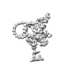
| Title | Doublet microtubule of fap45 null mutant | |||||||||
 Map data Map data | fap45_DMT | |||||||||
 Sample Sample |
| |||||||||
| Biological species |  | |||||||||
| Method | subtomogram averaging / cryo EM / Resolution: 39.6 Å | |||||||||
 Authors Authors | Owa M / Uchihashi T / Yanagisawa H / Yamano T / Iguchi H / Fukuzawa H / Wakabayashi K / Ando T / Kikkawa M | |||||||||
 Citation Citation |  Journal: Nat Commun / Year: 2019 Journal: Nat Commun / Year: 2019Title: Inner lumen proteins stabilize doublet microtubules in cilia and flagella. Authors: Mikito Owa / Takayuki Uchihashi / Haru-Aki Yanagisawa / Takashi Yamano / Hiro Iguchi / Hideya Fukuzawa / Ken-Ichi Wakabayashi / Toshio Ando / Masahide Kikkawa /  Abstract: Motile cilia are microtubule-based organelles that play important roles in most eukaryotes. Although axonemal microtubules are sufficiently stable to withstand their beating motion, it remains ...Motile cilia are microtubule-based organelles that play important roles in most eukaryotes. Although axonemal microtubules are sufficiently stable to withstand their beating motion, it remains unknown how they are stabilized while serving as tracks for axonemal dyneins. To address this question, we have identified two uncharacterized proteins, FAP45 and FAP52, as microtubule inner proteins (MIPs) in Chlamydomonas. These proteins are conserved among eukaryotes with motile cilia. Using cryo-electron tomography (cryo-ET) and high-speed atomic force microscopy (HS-AFM), we show that lack of these proteins leads to a loss of inner protrusions in B-tubules and less stable microtubules. These protrusions are located near the inner junctions of doublet microtubules and lack of both FAP52 and a known inner junction protein FAP20 results in detachment of the B-tubule from the A-tubule, as well as flagellar shortening. These results demonstrate that FAP45 and FAP52 bind to the inside of microtubules and stabilize ciliary axonemes. | |||||||||
| History |
|
- Structure visualization
| Movie |
 Movie viewer Movie viewer |
|---|---|
| Structure viewer | EM map:  SurfView SurfView Molmil Molmil Jmol/JSmol Jmol/JSmol |
| Supplemental images |
- Downloads & links
-EMDB archive
| Map data |  emd_9766.map.gz emd_9766.map.gz | 7.6 MB |  EMDB map data format EMDB map data format | |
|---|---|---|---|---|
| Header (meta data) |  emd-9766-v30.xml emd-9766-v30.xml emd-9766.xml emd-9766.xml | 8.2 KB 8.2 KB | Display Display |  EMDB header EMDB header |
| Images |  emd_9766.png emd_9766.png | 39.4 KB | ||
| Archive directory |  http://ftp.pdbj.org/pub/emdb/structures/EMD-9766 http://ftp.pdbj.org/pub/emdb/structures/EMD-9766 ftp://ftp.pdbj.org/pub/emdb/structures/EMD-9766 ftp://ftp.pdbj.org/pub/emdb/structures/EMD-9766 | HTTPS FTP |
-Validation report
| Summary document |  emd_9766_validation.pdf.gz emd_9766_validation.pdf.gz | 79 KB | Display |  EMDB validaton report EMDB validaton report |
|---|---|---|---|---|
| Full document |  emd_9766_full_validation.pdf.gz emd_9766_full_validation.pdf.gz | 78.1 KB | Display | |
| Data in XML |  emd_9766_validation.xml.gz emd_9766_validation.xml.gz | 496 B | Display | |
| Arichive directory |  https://ftp.pdbj.org/pub/emdb/validation_reports/EMD-9766 https://ftp.pdbj.org/pub/emdb/validation_reports/EMD-9766 ftp://ftp.pdbj.org/pub/emdb/validation_reports/EMD-9766 ftp://ftp.pdbj.org/pub/emdb/validation_reports/EMD-9766 | HTTPS FTP |
-Related structure data
- Links
Links
| EMDB pages |  EMDB (EBI/PDBe) / EMDB (EBI/PDBe) /  EMDataResource EMDataResource |
|---|
- Map
Map
| File |  Download / File: emd_9766.map.gz / Format: CCP4 / Size: 30.5 MB / Type: IMAGE STORED AS FLOATING POINT NUMBER (4 BYTES) Download / File: emd_9766.map.gz / Format: CCP4 / Size: 30.5 MB / Type: IMAGE STORED AS FLOATING POINT NUMBER (4 BYTES) | ||||||||||||||||||||||||||||||||||||||||||||||||||||||||||||
|---|---|---|---|---|---|---|---|---|---|---|---|---|---|---|---|---|---|---|---|---|---|---|---|---|---|---|---|---|---|---|---|---|---|---|---|---|---|---|---|---|---|---|---|---|---|---|---|---|---|---|---|---|---|---|---|---|---|---|---|---|---|
| Annotation | fap45_DMT | ||||||||||||||||||||||||||||||||||||||||||||||||||||||||||||
| Projections & slices | Image control
Images are generated by Spider. | ||||||||||||||||||||||||||||||||||||||||||||||||||||||||||||
| Voxel size | X=Y=Z: 7.1 Å | ||||||||||||||||||||||||||||||||||||||||||||||||||||||||||||
| Density |
| ||||||||||||||||||||||||||||||||||||||||||||||||||||||||||||
| Symmetry | Space group: 1 | ||||||||||||||||||||||||||||||||||||||||||||||||||||||||||||
| Details | EMDB XML:
CCP4 map header:
| ||||||||||||||||||||||||||||||||||||||||||||||||||||||||||||
-Supplemental data
- Sample components
-Entire : axoneme of Chlamydomonas reinhardtii
| Entire | Name: axoneme of Chlamydomonas reinhardtii |
|---|---|
| Components |
|
-Supramolecule #1: axoneme of Chlamydomonas reinhardtii
| Supramolecule | Name: axoneme of Chlamydomonas reinhardtii / type: organelle_or_cellular_component / ID: 1 / Parent: 0 |
|---|---|
| Source (natural) | Organism:  |
-Experimental details
-Structure determination
| Method | cryo EM |
|---|---|
 Processing Processing | subtomogram averaging |
| Aggregation state | filament |
- Sample preparation
| Concentration | 0.02 mg/mL |
|---|---|
| Buffer | pH: 7.4 |
| Vitrification | Cryogen name: ETHANE |
- Electron microscopy
| Microscope | JEOL 3100FFC |
|---|---|
| Image recording | Film or detector model: GATAN K2 SUMMIT (4k x 4k) / Average electron dose: 100.0 e/Å2 |
| Electron beam | Acceleration voltage: 300 kV / Electron source:  FIELD EMISSION GUN FIELD EMISSION GUN |
| Electron optics | Illumination mode: OTHER / Imaging mode: DIFFRACTION |
- Image processing
| Final reconstruction | Applied symmetry - Point group: C1 (asymmetric) / Resolution.type: BY AUTHOR / Resolution: 39.6 Å / Resolution method: FSC 0.5 CUT-OFF / Number subtomograms used: 2200 |
|---|---|
| Extraction | Number tomograms: 10 / Number images used: 2400 |
| Final angle assignment | Type: NOT APPLICABLE |
 Movie
Movie Controller


 Z (Sec.)
 Y (Row.)
 X (Col.)


