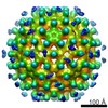
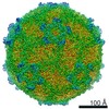
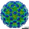
Journal: Viruses / Year: 2018 Title: T = 4 Icosahedral HIV-1 Capsid As an Immunogenic Vector for HIV-1 V3 Loop Epitope Display. Authors: Zhiqing Zhang / Maozhou He / Shimeng Bai / Feng Zhang / Jie Jiang / Qingbing Zheng / Shuangquan Gao / Xiaodong Yan / Shaowei Li / Ying Gu / Ningshao Xia / Abstract: The HIV-1 mature capsid (CA) assumes an amorphous, fullerene conical configuration due to its high flexibility. How native CA self-assembles is still unclear despite having well-defined structures of ...The HIV-1 mature capsid (CA) assumes an amorphous, fullerene conical configuration due to its high flexibility. How native CA self-assembles is still unclear despite having well-defined structures of its pentamer and hexamer building blocks. Here we explored the self-assembly of an engineered capsid protein built through artificial disulfide bonding (CA N21C/A22C) and determined the structure of one fraction of the globular particles. CA N21C/A22C was found to self-assemble into particles in relatively high ionic solutions. These particles contained disulfide-bonding hexamers as determined via non-reducing SDS-PAGE, and exhibited two major components of 57.3 S and 80.5 S in the sedimentation velocity assay. Particles had a globular morphology, approximately 40 nm in diameter, in negative-staining TEM. Through cryo-EM 3-D reconstruction, we determined a novel T = 4 icosahedral structure of CA, comprising 12 pentamers and 30 hexamers at 25 Å resolution. We engineered the HIV-1 V3 loop to the CA particles, and found the resultant particles resembled the morphology of their parental particles in TEM, had a positive reaction with V3-specific neutralizing antibodies, and conferred neutralization immunogenicity in mice. Our results shed light on HIV CA assembly and provide a particulate CA for epitope display.
History
Deposition
Nov 27, 2018
-
Header (metadata) release
Dec 19, 2018
-
Map release
Dec 19, 2018
-
Update
Dec 19, 2018
-
Current status
Dec 19, 2018
Processing site: PDBj / Status: Released
-
Structure visualization
Movie
Surface view with section colored by density value
In the structure databanks used in Yorodumi, some data are registered as the other names, "COVID-19 virus" and "2019-nCoV". Here are the details of the virus and the list of structure data.
Jan 31, 2019. EMDB accession codes are about to change! (news from PDBe EMDB page)
EMDB accession codes are about to change! (news from PDBe EMDB page)
The allocation of 4 digits for EMDB accession codes will soon come to an end. Whilst these codes will remain in use, new EMDB accession codes will include an additional digit and will expand incrementally as the available range of codes is exhausted. The current 4-digit format prefixed with “EMD-” (i.e. EMD-XXXX) will advance to a 5-digit format (i.e. EMD-XXXXX), and so on. It is currently estimated that the 4-digit codes will be depleted around Spring 2019, at which point the 5-digit format will come into force.
The EM Navigator/Yorodumi systems omit the EMD- prefix.
Related info.:Q: What is EMD? / ID/Accession-code notation in Yorodumi/EM Navigator
Yorodumi is a browser for structure data from EMDB, PDB, SASBDB, etc.
This page is also the successor to EM Navigator detail page, and also detail information page/front-end page for Omokage search.
The word "yorodu" (or yorozu) is an old Japanese word meaning "ten thousand". "mi" (miru) is to see.
Related info.:EMDB / PDB / SASBDB / Comparison of 3 databanks / Yorodumi Search / Aug 31, 2016. New EM Navigator & Yorodumi / Yorodumi Papers / Jmol/JSmol / Function and homology information / Changes in new EM Navigator and Yorodumi
 Movie
Movie Controller


 Open data
 Basic information
 Map data
 Sample
 Human immunodeficiency virus 1
 Authors
 Citation

 Structure visualization
 Movie viewer
 Downloads & links
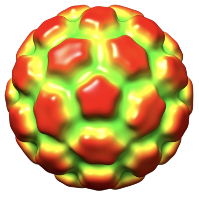 emd_9733.png
 http://ftp.pdbj.org/pub/emdb/structures/EMD-9733

 Z (Sec.)
 Y (Row.)
 X (Col.)


 Sample components
 Processing
 Electron microscopy
 FIELD EMISSION GUN
