 Movie
Movie Controller


+ Open data

- Basic information
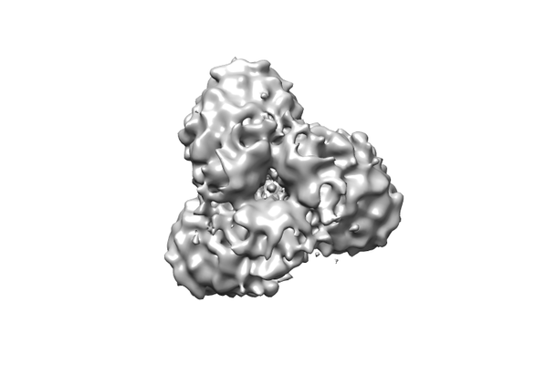
| Entry | Database: EMDB / ID: EMD-9681 | |||||||||
|---|---|---|---|---|---|---|---|---|---|---|
| Title | Arabidopsis thaliana ATG9 | |||||||||
 Map data Map data | ||||||||||
 Sample Sample |
| |||||||||
| Biological species |  | |||||||||
| Method | single particle reconstruction / cryo EM / Resolution: 9.0 Å | |||||||||
 Authors Authors | Lai LTF / Yu C / Wong JSK / Lo HS / Benlekbir S / Jiang L / Lau WCY | |||||||||
 Citation Citation | Journal: Autophagy / Year: 2020 Title: Subnanometer resolution cryo-EM structure of ATG9. Authors: Louis Tung Faat Lai / Chuanyang Yu / Jan Siu Kei Wong / Ho Sing Lo / Samir Benlekbir / Liwen Jiang / Wilson Chun Yu Lau /   Abstract: Macroautophagy/autophagy is an essential process for the maintenance of cellular homeostasis by recycling macromolecules under normal and stress conditions. ATG9 (autophagy related 9) is the only ...Macroautophagy/autophagy is an essential process for the maintenance of cellular homeostasis by recycling macromolecules under normal and stress conditions. ATG9 (autophagy related 9) is the only integral membrane protein in the autophagy core machinery and has a central role in mediating autophagosome formation. In cells, ATG9 exists on mobile vesicles that traffic to the growing phagophore, providing an essential membrane source for the formation of autophagosomes. Here we report the three-dimensional structure of ATG9 from at 7.8 Å resolution, determined by single particle cryo-electron microscopy. ATG9 organizes into a homotrimer, with each protomer contributing at least six transmembrane α-helices. At the center of the trimer, the protomers interact their membrane-embedded and C-terminal cytoplasmic regions. Combined with prediction of protein contacts using sequence co-evolutionary information, the structure provides molecular insights into the ATG9 architecture and testable hypotheses for the molecular mechanism of autophagy progression regulated by ATG9. 2D: 2-dimensional; 3D: 3-dimensional; AtATG9: ATG9; Atg: autophagy-related; ATG9: autophagy-related protein 9; cryo-EM: cryo-electron microscopy; DDM: dodecyl maltoside; GraDeR: gradient-based detergent removal; LMNG: lauryl maltose-neopentyl glycol; PAS: phagophore assembly site; PtdIns3K: phosphatidylinositol 3-kinase. | |||||||||
| History |
|
- Structure visualization
| Movie |
 Movie viewer Movie viewer |
|---|---|
| Structure viewer | EM map: SurfViewMolmilJmol/JSmol |
| Supplemental images |
- Downloads & links
-EMDB archive
| Map data | emd_9681.map.gz | 5 MB | EMDB map data format | |
|---|---|---|---|---|
| Header (meta data) | emd-9681-v30.xmlemd-9681.xml | 7.9 KB 7.9 KB | Display Display | EMDB header |
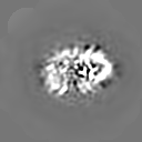
| Images |  emd_9681.png emd_9681.png | 43.7 KB | ||
| Archive directory |  http://ftp.pdbj.org/pub/emdb/structures/EMD-9681ftp://ftp.pdbj.org/pub/emdb/structures/EMD-9681 http://ftp.pdbj.org/pub/emdb/structures/EMD-9681ftp://ftp.pdbj.org/pub/emdb/structures/EMD-9681 | HTTPS FTP |
-Validation report
| Summary document | emd_9681_validation.pdf.gz | 78.7 KB | Display | EMDB validaton report |
|---|---|---|---|---|
| Full document | emd_9681_full_validation.pdf.gz | 77.8 KB | Display | |
| Data in XML | emd_9681_validation.xml.gz | 493 B | Display | |
| Arichive directory | https://ftp.pdbj.org/pub/emdb/validation_reports/EMD-9681ftp://ftp.pdbj.org/pub/emdb/validation_reports/EMD-9681 | HTTPS FTP |
-Related structure data
| Similar structure data |
|---|


-Links
| EMDB pages | EMDB (EBI/PDBe) / EMDataResource |
|---|
-Map
| File | Download / File: emd_9681.map.gz / Format: CCP4 / Size: 8 MB / Type: IMAGE STORED AS FLOATING POINT NUMBER (4 BYTES) | ||||||||||||||||||||||||||||||||||||||||||||||||||||||||||||
|---|---|---|---|---|---|---|---|---|---|---|---|---|---|---|---|---|---|---|---|---|---|---|---|---|---|---|---|---|---|---|---|---|---|---|---|---|---|---|---|---|---|---|---|---|---|---|---|---|---|---|---|---|---|---|---|---|---|---|---|---|---|
| Projections & slices | Image control
Images are generated by Spider. | ||||||||||||||||||||||||||||||||||||||||||||||||||||||||||||
| Voxel size | X=Y=Z: 2.12 Å | ||||||||||||||||||||||||||||||||||||||||||||||||||||||||||||
| Density |
| ||||||||||||||||||||||||||||||||||||||||||||||||||||||||||||
| Symmetry | Space group: 1 | ||||||||||||||||||||||||||||||||||||||||||||||||||||||||||||
| Details | EMDB XML:
CCP4 map header:
| ||||||||||||||||||||||||||||||||||||||||||||||||||||||||||||
 Z (Sec.)
 Y (Row.)
 X (Col.)


-Supplemental data
- Sample components
-Entire : ATG9
| Entire | Name: ATG9 |
|---|---|
| Components |
|
-Supramolecule #1: ATG9
| Supramolecule | Name: ATG9 / type: complex / ID: 1 / Parent: 0 |
|---|---|
| Source (natural) | Organism: |
| Recombinant expression | Organism:  Homo sapiens (human) Homo sapiens (human) |
| Molecular weight | Theoretical: 300 KDa |
-Experimental details
-Structure determination
| Method | cryo EM |
|---|---|
 Processing Processing | single particle reconstruction |
| Aggregation state | particle |
-Sample preparation
| Concentration | 0.4 mg/mL |
|---|---|
| Buffer | pH: 8.5 |
| Vitrification | Cryogen name: ETHANE |
- Electron microscopy
| Microscope | FEI TITAN KRIOS |
|---|---|
| Image recording | Film or detector model: FEI FALCON III (4k x 4k) / Detector mode: COUNTING / Average exposure time: 60.0 sec. / Average electron dose: 43.0 e/Å2 |
| Electron beam | Acceleration voltage: 300 kV / Electron source:  FIELD EMISSION GUN FIELD EMISSION GUN |
| Electron optics | Illumination mode: FLOOD BEAM / Imaging mode: BRIGHT FIELD |
| Experimental equipment |  Model: Titan Krios / Image courtesy: FEI Company |
-Image processing
| Final reconstruction | Applied symmetry - Point group: C3 (3 fold cyclic) / Resolution.type: BY AUTHOR / Resolution: 9.0 Å / Resolution method: FSC 0.143 CUT-OFF / Software - Name: RELION (ver. 2.0) / Number images used: 11335 |
|---|---|
| Initial angle assignment | Type: MAXIMUM LIKELIHOOD |
| Final angle assignment | Type: MAXIMUM LIKELIHOOD |
