Journal: Autophagy / Year: 2020 Title: Subnanometer resolution cryo-EM structure of ATG9. Authors: Louis Tung Faat Lai / Chuanyang Yu / Jan Siu Kei Wong / Ho Sing Lo / Samir Benlekbir / Liwen Jiang / Wilson Chun Yu Lau / Abstract: Macroautophagy/autophagy is an essential process for the maintenance of cellular homeostasis by recycling macromolecules under normal and stress conditions. ATG9 (autophagy related 9) is the only ...Macroautophagy/autophagy is an essential process for the maintenance of cellular homeostasis by recycling macromolecules under normal and stress conditions. ATG9 (autophagy related 9) is the only integral membrane protein in the autophagy core machinery and has a central role in mediating autophagosome formation. In cells, ATG9 exists on mobile vesicles that traffic to the growing phagophore, providing an essential membrane source for the formation of autophagosomes. Here we report the three-dimensional structure of ATG9 from at 7.8 Å resolution, determined by single particle cryo-electron microscopy. ATG9 organizes into a homotrimer, with each protomer contributing at least six transmembrane α-helices. At the center of the trimer, the protomers interact their membrane-embedded and C-terminal cytoplasmic regions. Combined with prediction of protein contacts using sequence co-evolutionary information, the structure provides molecular insights into the ATG9 architecture and testable hypotheses for the molecular mechanism of autophagy progression regulated by ATG9. 2D: 2-dimensional; 3D: 3-dimensional; AtATG9: ATG9; Atg: autophagy-related; ATG9: autophagy-related protein 9; cryo-EM: cryo-electron microscopy; DDM: dodecyl maltoside; GraDeR: gradient-based detergent removal; LMNG: lauryl maltose-neopentyl glycol; PAS: phagophore assembly site; PtdIns3K: phosphatidylinositol 3-kinase.
History
Deposition
Oct 11, 2018
-
Header (metadata) release
Oct 16, 2019
-
Map release
Oct 16, 2019
-
Update
Oct 16, 2019
-
Current status
Oct 16, 2019
Processing site: PDBj / Status: Released
-
Structure visualization
Movie
Surface view with section colored by density value
In the structure databanks used in Yorodumi, some data are registered as the other names, "COVID-19 virus" and "2019-nCoV". Here are the details of the virus and the list of structure data.
Jan 31, 2019. EMDB accession codes are about to change! (news from PDBe EMDB page)
EMDB accession codes are about to change! (news from PDBe EMDB page)
The allocation of 4 digits for EMDB accession codes will soon come to an end. Whilst these codes will remain in use, new EMDB accession codes will include an additional digit and will expand incrementally as the available range of codes is exhausted. The current 4-digit format prefixed with “EMD-” (i.e. EMD-XXXX) will advance to a 5-digit format (i.e. EMD-XXXXX), and so on. It is currently estimated that the 4-digit codes will be depleted around Spring 2019, at which point the 5-digit format will come into force.
The EM Navigator/Yorodumi systems omit the EMD- prefix.
Related info.:Q: What is EMD? / ID/Accession-code notation in Yorodumi/EM Navigator
Yorodumi is a browser for structure data from EMDB, PDB, SASBDB, etc.
This page is also the successor to EM Navigator detail page, and also detail information page/front-end page for Omokage search.
The word "yorodu" (or yorozu) is an old Japanese word meaning "ten thousand". "mi" (miru) is to see.
Related info.:EMDB / PDB / SASBDB / Comparison of 3 databanks / Yorodumi Search / Aug 31, 2016. New EM Navigator & Yorodumi / Yorodumi Papers / Jmol/JSmol / Function and homology information / Changes in new EM Navigator and Yorodumi
 Movie
Movie Controller


 Open data
 Basic information
 Map data
 Sample
 Authors
 Citation

 Structure visualization
 Movie viewer
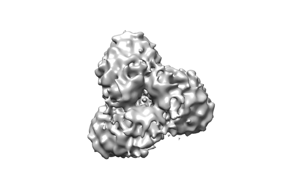
 Downloads & links
 emd_9681.png
 http://ftp.pdbj.org/pub/emdb/structures/EMD-9681


 Z (Sec.)
 Y (Row.)
 X (Col.)


 Sample components
 Homo sapiens (human)
 Processing
 Electron microscopy
 FIELD EMISSION GUN
