Journal: Proc Natl Acad Sci U S A / Year: 2019 Title: Structural bases for F plasmid conjugation and F pilus biogenesis in . Authors: Bo Hu / Pratick Khara / Peter J Christie / Abstract: Bacterial conjugation systems are members of the large type IV secretion system (T4SS) superfamily. Conjugative transfer of F plasmids residing in the was first reported in the 1940s, yet the ...Bacterial conjugation systems are members of the large type IV secretion system (T4SS) superfamily. Conjugative transfer of F plasmids residing in the was first reported in the 1940s, yet the architecture of F plasmid-encoded transfer channel and its physical relationship with the F pilus remain unknown. We visualized F-encoded structures in the native bacterial cell envelope by in situ cryoelectron tomography (CryoET). Remarkably, F plasmids encode four distinct structures, not just the translocation channel or channel-pilus complex predicted by prevailing models. The F1 structure is composed of distinct outer and inner membrane complexes and a connecting cylinder that together house the envelope-spanning translocation channel. The F2 structure is essentially the F1 complex with the F pilus attached at the outer membrane (OM). Remarkably, the F3 structure consists of the F pilus attached to a thin, cell envelope-spanning stalk, whereas the F4 structure consists of the pilus docked to the OM without an associated periplasmic density. The traffic ATPase TraC is configured as a hexamer of dimers at the cytoplasmic faces of the F1 and F2 structures, where it respectively regulates substrate transfer and F pilus biogenesis. Together, our findings present architectural renderings of the DNA conjugation or "mating" channel, the channel-pilus connection, and unprecedented pilus basal structures. These structural snapshots support a model for biogenesis of the F transfer system and allow for detailed comparisons with other structurally characterized T4SSs.
History
Deposition
Nov 14, 2018
-
Header (metadata) release
Feb 20, 2019
-
Map release
Jun 26, 2019
-
Update
Jul 24, 2019
-
Current status
Jul 24, 2019
Processing site: RCSB / Status: Released
-
Structure visualization
Movie
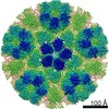
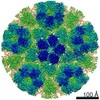
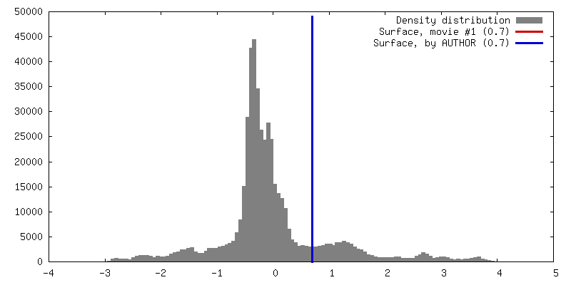
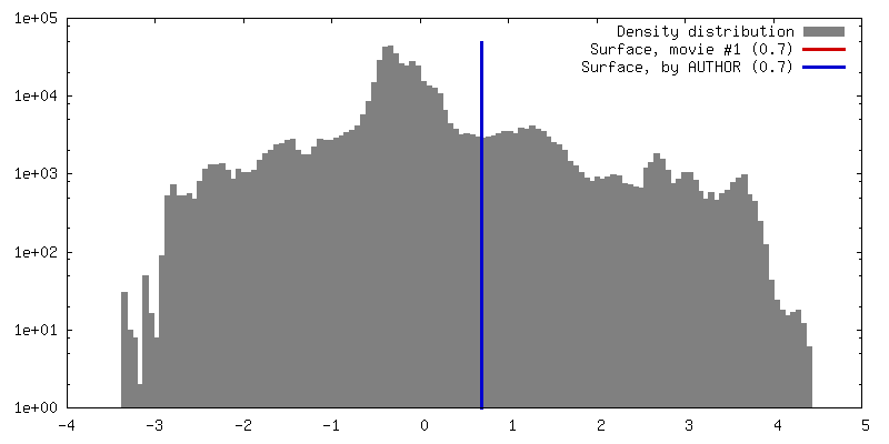
Surface view with section colored by density value
In the structure databanks used in Yorodumi, some data are registered as the other names, "COVID-19 virus" and "2019-nCoV". Here are the details of the virus and the list of structure data.
Jan 31, 2019. EMDB accession codes are about to change! (news from PDBe EMDB page)
EMDB accession codes are about to change! (news from PDBe EMDB page)
The allocation of 4 digits for EMDB accession codes will soon come to an end. Whilst these codes will remain in use, new EMDB accession codes will include an additional digit and will expand incrementally as the available range of codes is exhausted. The current 4-digit format prefixed with “EMD-” (i.e. EMD-XXXX) will advance to a 5-digit format (i.e. EMD-XXXXX), and so on. It is currently estimated that the 4-digit codes will be depleted around Spring 2019, at which point the 5-digit format will come into force.
The EM Navigator/Yorodumi systems omit the EMD- prefix.
Related info.:Q: What is EMD? / ID/Accession-code notation in Yorodumi/EM Navigator
Yorodumi is a browser for structure data from EMDB, PDB, SASBDB, etc.
This page is also the successor to EM Navigator detail page, and also detail information page/front-end page for Omokage search.
The word "yorodu" (or yorozu) is an old Japanese word meaning "ten thousand". "mi" (miru) is to see.
Related info.:EMDB / PDB / SASBDB / Comparison of 3 databanks / Yorodumi Search / Aug 31, 2016. New EM Navigator & Yorodumi / Yorodumi Papers / Jmol/JSmol / Function and homology information / Changes in new EM Navigator and Yorodumi
 Movie
Movie Controller


 Open data
 Basic information
 Map data
 Sample
 Authors
 United States, 1 items
  Citation
 Structure visualization
 Movie viewer
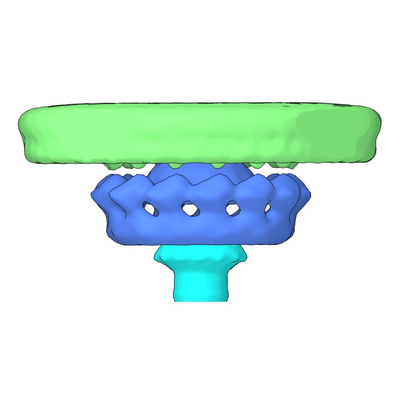
 Downloads & links
 emd_9344.png
 http://ftp.pdbj.org/pub/emdb/structures/EMD-9344

 Z (Sec.)
 Y (Row.)
 X (Col.)


 Sample components
 Processing
 Electron microscopy
 FIELD EMISSION GUN
