 ムービー
ムービー コントローラー


+ データを開く

- 基本情報
基本情報
| 登録情報 | データベース: EMDB / ID: EMD-9022 | |||||||||
|---|---|---|---|---|---|---|---|---|---|---|
| タイトル | Three-dimensional architecture of epithelial primary cilia | |||||||||
 マップデータ マップデータ | 3D map of a typical primary cilium of IMCD3 cell obtained by electron tomography of serial sections. Best to use contour level 50 with the hide-dust in UCSF Chimera for isosurface volume rending. | |||||||||
 試料 試料 |
| |||||||||
| 生物種 |  | |||||||||
| 手法 | 電子線トモグラフィー法 / ネガティブ染色法 | |||||||||
 データ登録者 データ登録者 | Sun S / Fisher RL / Bowser SS / Pentecost BT / Sui H | |||||||||
 引用 引用 | ジャーナル: Proc Natl Acad Sci U S A / 年: 2019 タイトル: Three-dimensional architecture of epithelial primary cilia. 著者: Shufeng Sun / Rebecca L Fisher / Samuel S Bowser / Brian T Pentecost / Haixin Sui /  要旨: We report a complete 3D structural model of typical epithelial primary cilia based on structural maps of full-length primary cilia obtained by serial section electron tomography. Our data demonstrate ...We report a complete 3D structural model of typical epithelial primary cilia based on structural maps of full-length primary cilia obtained by serial section electron tomography. Our data demonstrate the architecture of primary cilia differs extensively from the commonly acknowledged 9+0 paradigm. The axoneme structure is relatively stable but gradually evolves from base to tip with a decreasing number of microtubule complexes (MtCs) and a reducing diameter. The axonemal MtCs are cross-linked by previously unrecognized fibrous protein networks. Such an architecture explains why primary cilia can elastically withstand liquid flow for mechanosensing. The nine axonemal MtCs in a cilium are found to differ significantly in length indicating intraflagellar transport processes in primary cilia may be more complicated than that reported for motile cilia. The 3D maps of microtubule doublet-singlet transitions generally display longitudinal gaps at the inner junction between the A- and B-tubules, which indicates the inner junction protein is a major player in doublet-singlet transitions. In addition, vesicles releasing from kidney primary cilia were observed in the structural maps, supporting that ciliary vesicles budding may serve as ectosomes for cell-cell communication. | |||||||||
| 履歴 |
|
- 構造の表示
構造の表示
| ムービー |
 ムービービューア ムービービューア |
|---|---|
| 添付画像 |

- ダウンロードとリンク
-EMDBアーカイブ
| マップデータ | emd_9022.map.gz | 915.4 MB | EMDBマップデータ形式 | |
|---|---|---|---|---|
| ヘッダ (付随情報) | emd-9022-v30.xmlemd-9022.xml | 8.7 KB 8.7 KB | 表示 表示 | EMDBヘッダ |
| 画像 | 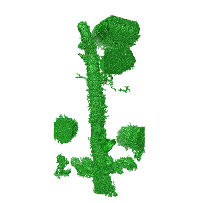 emd_9022.png emd_9022.png | 91.9 KB | ||
| アーカイブディレクトリ |  http://ftp.pdbj.org/pub/emdb/structures/EMD-9022ftp://ftp.pdbj.org/pub/emdb/structures/EMD-9022 http://ftp.pdbj.org/pub/emdb/structures/EMD-9022ftp://ftp.pdbj.org/pub/emdb/structures/EMD-9022 | HTTPS FTP |
-検証レポート
| 文書・要旨 | emd_9022_validation.pdf.gz | 77.3 KB | 表示 | EMDB検証レポート |
|---|---|---|---|---|
| 文書・詳細版 | emd_9022_full_validation.pdf.gz | 76.4 KB | 表示 | |
| XML形式データ | emd_9022_validation.xml.gz | 497 B | 表示 | |
| アーカイブディレクトリ | https://ftp.pdbj.org/pub/emdb/validation_reports/EMD-9022ftp://ftp.pdbj.org/pub/emdb/validation_reports/EMD-9022 | HTTPS FTP |
-関連構造データ
-リンク
| EMDBのページ | EMDB (EBI/PDBe) / EMDataResource |
|---|
-マップ
| ファイル | ダウンロード / ファイル: emd_9022.map.gz / 形式: CCP4 / 大きさ: 2.2 GB / タイプ: IMAGE STORED AS SIGNED BYTE | ||||||||||||||||||||||||||||||||||||||||||||||||||||||||||||||||||||
|---|---|---|---|---|---|---|---|---|---|---|---|---|---|---|---|---|---|---|---|---|---|---|---|---|---|---|---|---|---|---|---|---|---|---|---|---|---|---|---|---|---|---|---|---|---|---|---|---|---|---|---|---|---|---|---|---|---|---|---|---|---|---|---|---|---|---|---|---|---|
| 注釈 | 3D map of a typical primary cilium of IMCD3 cell obtained by electron tomography of serial sections. Best to use contour level 50 with the hide-dust in UCSF Chimera for isosurface volume rending. | ||||||||||||||||||||||||||||||||||||||||||||||||||||||||||||||||||||
| ボクセルのサイズ | X=Y=Z: 1.34128 Å | ||||||||||||||||||||||||||||||||||||||||||||||||||||||||||||||||||||
| 密度 |
| ||||||||||||||||||||||||||||||||||||||||||||||||||||||||||||||||||||
| 対称性 | 空間群: 1 | ||||||||||||||||||||||||||||||||||||||||||||||||||||||||||||||||||||
| 詳細 | EMDB XML:
CCP4マップ ヘッダ情報:
| ||||||||||||||||||||||||||||||||||||||||||||||||||||||||||||||||||||
-添付データ
- 試料の構成要素
-全体 : Primary cilium #G
| 全体 | 名称: Primary cilium #G |
|---|---|
| 要素 |
|
-超分子 #1: Primary cilium #G
| 超分子 | 名称: Primary cilium #G / タイプ: organelle_or_cellular_component / ID: 1 / 親要素: 0 詳細: IMCD3 primary cilium #G is related to Fig. S3g, Fig.S4g and movies 1-5 |
|---|---|
| 由来(天然) | 生物種: |
-実験情報
-構造解析
| 手法 | ネガティブ染色法 |
|---|---|
 解析 解析 | 電子線トモグラフィー法 |
| 試料の集合状態 | cell |
-試料調製
| 緩衝液 | pH: 7.4 / 詳細: PBS |
|---|---|
| 染色 | タイプ: POSITIVE / 材質: uranyl acetate, lead citrate 詳細: 2% uranyl acetate and Reynolds lead citrate solution |
| 糖包埋 | 材質: epoxy resin 詳細: The samples were infiltrated with a graded series of resins (SPI-PON 812, SPI-CHEM, USA) in acetone over 24 hours. After infiltration with pure resin (3 x within 24 hours), the specimens in ...詳細: The samples were infiltrated with a graded series of resins (SPI-PON 812, SPI-CHEM, USA) in acetone over 24 hours. After infiltration with pure resin (3 x within 24 hours), the specimens in resin were polymerized at 60 degrees C for 2 days. |
| グリッド | 詳細: unspecified |
| 切片作成 | ウルトラミクロトーム - 装置: UC6 microtome / ウルトラミクロトーム - 温度: 298 K / ウルトラミクロトーム - 最終 厚さ: 110 nm |
| 位置合わせマーカー | Manufacturer: Sigma-Aldrich / 直径: 10 nm |
- 電子顕微鏡法
| 顕微鏡 | FEI TECNAI 20 |
|---|---|
| 撮影 | フィルム・検出器のモデル: TVIPS TEMCAM-F415 (4k x 4k) 平均電子線量: 6.0 e/Å2 |
| 電子線 | 加速電圧: 200 kV / 電子線源:  FIELD EMISSION GUN FIELD EMISSION GUN |
| 電子光学系 | 照射モード: FLOOD BEAM / 撮影モード: BRIGHT FIELD |
-画像解析
| 最終 再構成 | アルゴリズム: SIMULTANEOUS ITERATIVE (SIRT) / ソフトウェア - 名称: TOMO3D 詳細: This SSET work used a series of dual-axis tilt data sets. 使用した粒子像数: 8316 |
|---|
