 Movie
Movie Controller


+ Open data

- Basic information
| Entry | Database: EMDB / ID: EMD-8795 | |||||||||
|---|---|---|---|---|---|---|---|---|---|---|
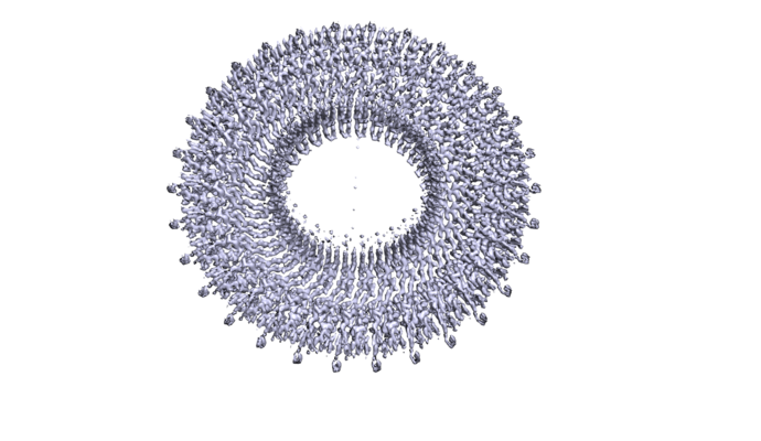
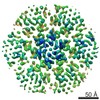
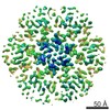
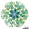
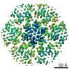
| Title | SpoIIIAG stage III sporulation engulfment assembly protein | |||||||||
 Map data Map data | ||||||||||
 Sample Sample |
| |||||||||
 Keywords Keywords | Secretion system / Sporulation channel / Ring Building Motif (RBM) / PROTEIN TRANSPORT | |||||||||
| Function / homology | Sporulation stage III, protein AG / membrane => GO:0016020 / Stage III sporulation engulfment assemblyprotein Function and homology information Function and homology information | |||||||||
| Biological species |  | |||||||||
| Method | single particle reconstruction / cryo EM / Resolution: 3.5 Å | |||||||||
 Authors Authors | Zeytuni N / Hong C | |||||||||
| Funding support |  Canada, 1 items Canada, 1 items
| |||||||||
 Citation Citation | Journal: Proc Natl Acad Sci U S A / Year: 2017 Title: Near-atomic resolution cryoelectron microscopy structure of the 30-fold homooligomeric SpoIIIAG channel essential to spore formation in . Authors: Natalie Zeytuni / Chuan Hong / Kelly A Flanagan / Liam J Worrall / Kate A Theiltges / Marija Vuckovic / Rick K Huang / Shawn C Massoni / Amy H Camp / Zhiheng Yu / Natalie C Strynadka /  Abstract: Bacterial sporulation allows starving cells to differentiate into metabolically dormant spores that can survive extreme conditions. Following asymmetric division, the mother cell engulfs the ...Bacterial sporulation allows starving cells to differentiate into metabolically dormant spores that can survive extreme conditions. Following asymmetric division, the mother cell engulfs the forespore, surrounding it with two bilayer membranes. During the engulfment process, an essential channel, the so-called feeding tube apparatus, is thought to cross both membranes to create a direct conduit between the mother cell and the forespore. At least nine proteins are required to create this channel, including SpoIIQ and SpoIIIAA-AH. Here, we present the near-atomic resolution structure of one of these proteins, SpoIIIAG, determined by single-particle cryo-EM. A 3D reconstruction revealed that SpoIIIAG assembles into a large and stable 30-fold symmetric complex with a unique mushroom-like architecture. The complex is collectively composed of three distinctive circular structures: a 60-stranded vertical β-barrel that forms a large inner channel encircled by two concentric rings, one β-mediated and the other formed by repeats of a ring-building motif (RBM) common to the architecture of various dual membrane secretion systems of distinct function. Our near-atomic resolution structure clearly shows that SpoIIIAG exhibits a unique and dramatic adaptation of the RBM fold with a unique β-triangle insertion that assembles into the prominent channel, the dimensions of which suggest the potential passage of large macromolecules between the mother cell and forespore during the feeding process. Indeed, mutation of residues located at key interfaces between monomers of this RBM resulted in severe defects both in vivo and in vitro, providing additional support for this unprecedented structure. | |||||||||
| History |
|
- Structure visualization
| Movie |
Movie viewer |
|---|---|
| Structure viewer | EM map: SurfViewMolmilJmol/JSmol |
| Supplemental images |
- Downloads & links
-EMDB archive
| Map data | emd_8795.map.gz | 9.9 MB | EMDB map data format | |
|---|---|---|---|---|
| Header (meta data) | emd-8795-v30.xmlemd-8795.xml | 14.4 KB 14.4 KB | Display Display | EMDB header |
| Images |  emd_8795.png emd_8795.png | 211.3 KB | ||
| Filedesc metadata | emd-8795.cif.gz | 5.6 KB | ||
| Archive directory |  http://ftp.pdbj.org/pub/emdb/structures/EMD-8795ftp://ftp.pdbj.org/pub/emdb/structures/EMD-8795 http://ftp.pdbj.org/pub/emdb/structures/EMD-8795ftp://ftp.pdbj.org/pub/emdb/structures/EMD-8795 | HTTPS FTP |
-Related structure data
| Related structure data |  5wc3MC 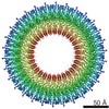 8797C  8798C  8800C M: atomic model generated by this map C: citing same article ( |
|---|---|
| Similar structure data |
-Links
| EMDB pages | EMDB (EBI/PDBe) / EMDataResource |
|---|
-Map
| File | Download / File: emd_8795.map.gz / Format: CCP4 / Size: 52.7 MB / Type: IMAGE STORED AS FLOATING POINT NUMBER (4 BYTES) | ||||||||||||||||||||||||||||||||||||||||||||||||||||||||||||
|---|---|---|---|---|---|---|---|---|---|---|---|---|---|---|---|---|---|---|---|---|---|---|---|---|---|---|---|---|---|---|---|---|---|---|---|---|---|---|---|---|---|---|---|---|---|---|---|---|---|---|---|---|---|---|---|---|---|---|---|---|---|
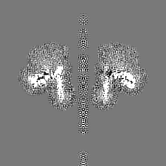
| Projections & slices | Image control
Images are generated by Spider. | ||||||||||||||||||||||||||||||||||||||||||||||||||||||||||||
| Voxel size | X=Y=Z: 1.35 Å | ||||||||||||||||||||||||||||||||||||||||||||||||||||||||||||
| Density |
| ||||||||||||||||||||||||||||||||||||||||||||||||||||||||||||
| Symmetry | Space group: 1 | ||||||||||||||||||||||||||||||||||||||||||||||||||||||||||||
| Details | EMDB XML:
CCP4 map header:
| ||||||||||||||||||||||||||||||||||||||||||||||||||||||||||||
 Z (Sec.)
 Y (Row.)
 X (Col.)


-Supplemental data
- Sample components
-Entire : SpoIIIAG
| Entire | Name: SpoIIIAG |
|---|---|
| Components |
|
-Supramolecule #1: SpoIIIAG
| Supramolecule | Name: SpoIIIAG / type: complex / ID: 1 / Parent: 0 / Macromolecule list: all / Details: residues 55-229 |
|---|---|
| Source (natural) | Organism: |
| Molecular weight | Theoretical: 580 KDa |
-Macromolecule #1: SpoIIIAG, Stage III sporulation engulfment assemblyprotein
| Macromolecule | Name: SpoIIIAG, Stage III sporulation engulfment assemblyprotein type: protein_or_peptide / ID: 1 / Number of copies: 30 / Enantiomer: LEVO |
|---|---|
| Source (natural) | Organism: |
| Molecular weight | Theoretical: 19.729086 KDa |
| Recombinant expression | Organism: |
| Sequence | String: GSHMKTENAK TITAVSSQHS ADSKEKTAEV FKASKSDKPK DSIDDYEKEY ENQLKEILET IIGVDDVSVV VNVDATSLKV YEKNKSNKN TTTEETDKEG GKRSVTDQSS EEEIVMIKNG DKETPVVVQT KKPDIRGVLV VAQGVDNVQI KQTIIEAVTR V LDVPSHRV AVAPKKIKED S UniProtKB: Stage III sporulation engulfment assemblyprotein |
-Experimental details
-Structure determination
| Method | cryo EM |
|---|---|
 Processing Processing | single particle reconstruction |
| Aggregation state | particle |
-Sample preparation
| Concentration | 13 mg/mL | |||||||||
|---|---|---|---|---|---|---|---|---|---|---|
| Buffer | pH: 8 Component:
| |||||||||
| Grid | Model: Quantifoil / Material: GOLD / Mesh: 400 / Pretreatment - Type: GLOW DISCHARGE / Pretreatment - Time: 60 sec. / Pretreatment - Atmosphere: AIR / Pretreatment - Pressure: 0.038 kPa | |||||||||
| Vitrification | Cryogen name: ETHANE / Chamber humidity: 100 % / Chamber temperature: 277 K / Instrument: FEI VITROBOT MARK IV |
- Electron microscopy
| Microscope | FEI TITAN KRIOS |
|---|---|
| Temperature | Min: 80.0 K / Max: 80.0 K |
| Specialist optics | Energy filter - Name: Gatan GIF / Energy filter - Lower energy threshold: 0 eV / Energy filter - Upper energy threshold: 20 eV |
| Details | Cs corrector |
| Image recording | Film or detector model: GATAN K2 SUMMIT (4k x 4k) / Detector mode: SUPER-RESOLUTION / Digitization - Dimensions - Width: 7676 pixel / Digitization - Dimensions - Height: 7420 pixel / Digitization - Frames/image: 1-50 / Number grids imaged: 1 / Number real images: 2876 / Average exposure time: 0.2 sec. / Average electron dose: 1.1 e/Å2 |
| Electron beam | Acceleration voltage: 300 kV / Electron source:  FIELD EMISSION GUN FIELD EMISSION GUN |
| Electron optics | C2 aperture diameter: 70.0 µm / Calibrated defocus max: 2.1 µm / Calibrated defocus min: 0.9 µm / Calibrated magnification: 37037 / Illumination mode: FLOOD BEAM / Imaging mode: BRIGHT FIELD / Cs: 0.01 mm / Nominal defocus max: 2.0 µm / Nominal defocus min: 1.0 µm / Nominal magnification: 81000 |
| Sample stage | Specimen holder model: FEI TITAN KRIOS AUTOGRID HOLDER / Cooling holder cryogen: NITROGEN |
| Experimental equipment |  Model: Titan Krios / Image courtesy: FEI Company |
