 Movie
Movie Controller

[English] 日本語
 Yorodumi
- EMDB-8753: Multi-membrane Structure of the Pandoravirus and Implications to ... -
+ Open data

- Basic information
| Entry | Database: EMDB / ID: EMD-8753 | |||||||||
|---|---|---|---|---|---|---|---|---|---|---|
| Title | Multi-membrane Structure of the Pandoravirus and Implications to Eukaryogenesis | |||||||||
 Map data Map data | Cryo-ET Tomogram of P. dulcis | |||||||||
 Sample Sample |
| |||||||||
| Biological species |  Pandoravirus salinus Pandoravirus salinus | |||||||||
| Method | electron tomography / cryo EM | |||||||||
 Authors Authors | Wang KJ / Zhou ZH | |||||||||
| Funding support |  United States, 1 items United States, 1 items
| |||||||||
 Citation Citation | Journal: To Be Published Title: In depth examination of the various structures of P. salinus through CryoET reconstructions Authors: Wang KJ / Zhou ZH | |||||||||
| History |
|
- Structure visualization
| Movie |
 Movie viewer Movie viewer |
|---|---|
| Structure viewer | EM map: SurfViewMolmilJmol/JSmol |
| Supplemental images |

- Downloads & links
-EMDB archive
| Map data | emd_8753.map.gz | 1.3 GB | EMDB map data format | |
|---|---|---|---|---|
| Header (meta data) | emd-8753-v30.xmlemd-8753.xml | 8.2 KB 8.2 KB | Display Display | EMDB header |
| Images | 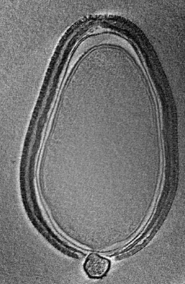 emd_8753.png emd_8753.png | 92.2 KB | ||
| Archive directory |  http://ftp.pdbj.org/pub/emdb/structures/EMD-8753ftp://ftp.pdbj.org/pub/emdb/structures/EMD-8753 http://ftp.pdbj.org/pub/emdb/structures/EMD-8753ftp://ftp.pdbj.org/pub/emdb/structures/EMD-8753 | HTTPS FTP |
-Links
| EMDB pages | EMDB (EBI/PDBe) / EMDataResource |
|---|
-Map
| File | Download / File: emd_8753.map.gz / Format: CCP4 / Size: 1.5 GB / Type: IMAGE STORED AS FLOATING POINT NUMBER (4 BYTES) | ||||||||||||||||||||||||||||||||||||||||||||||||||||||||||||||||||||
|---|---|---|---|---|---|---|---|---|---|---|---|---|---|---|---|---|---|---|---|---|---|---|---|---|---|---|---|---|---|---|---|---|---|---|---|---|---|---|---|---|---|---|---|---|---|---|---|---|---|---|---|---|---|---|---|---|---|---|---|---|---|---|---|---|---|---|---|---|---|
| Annotation | Cryo-ET Tomogram of P. dulcis | ||||||||||||||||||||||||||||||||||||||||||||||||||||||||||||||||||||
| Projections & slices | Image control
Images are generated by Spider. generated in cubic-lattice coordinate | ||||||||||||||||||||||||||||||||||||||||||||||||||||||||||||||||||||
| Voxel size | X=Y=Z: 7.066 Å | ||||||||||||||||||||||||||||||||||||||||||||||||||||||||||||||||||||
| Density |
| ||||||||||||||||||||||||||||||||||||||||||||||||||||||||||||||||||||
| Symmetry | Space group: 1 | ||||||||||||||||||||||||||||||||||||||||||||||||||||||||||||||||||||
| Details | EMDB XML:
CCP4 map header:
| ||||||||||||||||||||||||||||||||||||||||||||||||||||||||||||||||||||
 Z (Sec.)
 Y (Row.)
 X (Col.)


-Supplemental data
- Sample components
-Entire : Pandoravirus salinus
| Entire | Name: Pandoravirus salinus |
|---|---|
| Components |
|
-Supramolecule #1: Pandoravirus salinus
| Supramolecule | Name: Pandoravirus salinus / type: virus / ID: 1 / Parent: 0 / NCBI-ID: 1349410 / Sci species name: Pandoravirus salinus / Virus type: VIRION / Virus isolate: SPECIES / Virus enveloped: No / Virus empty: No |
|---|---|
| Host (natural) | Organism:  Acanthamoeba (eukaryote) Acanthamoeba (eukaryote) |
-Experimental details
-Structure determination
| Method | cryo EM |
|---|---|
 Processing Processing | electron tomography |
| Aggregation state | particle |
-Sample preparation
| Buffer | pH: 7.4 |
|---|---|
| Sugar embedding | Material: vitreous ice |
| Grid | Model: Quantifoil 200 / Material: COPPER / Mesh: 200 / Pretreatment - Type: GLOW DISCHARGE |
| Vitrification | Cryogen name: NITROGEN / Instrument: FEI VITROBOT MARK IV |
| High pressure freezing | Instrument: OTHER Details: The value given for _emd_high_pressure_freezing.instrument is Leica EM PACT2. This is not in a list of allowed values set(['LEICA EM PACT2', 'LEICA EM PACT', 'EMS-002 RAPID IMMERSION ...Details: The value given for _emd_high_pressure_freezing.instrument is Leica EM PACT2. This is not in a list of allowed values set(['LEICA EM PACT2', 'LEICA EM PACT', 'EMS-002 RAPID IMMERSION FREEZER', 'OTHER', 'LEICA EM HPM100', 'BAL-TEC HPM 010']) so OTHER is written into the XML file. |
| Sectioning | Other: NO SECTIONING |
| Fiducial marker | Manufacturer: Nanogold / Diameter: 10 nm |
- Electron microscopy
| Microscope | FEI TITAN KRIOS |
|---|---|
| Image recording | Film or detector model: GATAN ULTRASCAN 4000 (4k x 4k) / Average electron dose: 100.0 e/Å2 |
| Electron beam | Acceleration voltage: 300 kV / Electron source:  FIELD EMISSION GUN FIELD EMISSION GUN |
| Electron optics | Illumination mode: FLOOD BEAM / Imaging mode: BRIGHT FIELD |
| Experimental equipment |  Model: Titan Krios / Image courtesy: FEI Company |
-Image processing
| Final reconstruction | Algorithm: SIMULTANEOUS ITERATIVE (SIRT) / Software - Name: IMOD (ver. 4.7.15) / Number images used: 64 |
|---|
