 Movie
Movie Controller


+ Open data

- Basic information
| Entry | Database: EMDB / ID: EMD-8599 | |||||||||
|---|---|---|---|---|---|---|---|---|---|---|
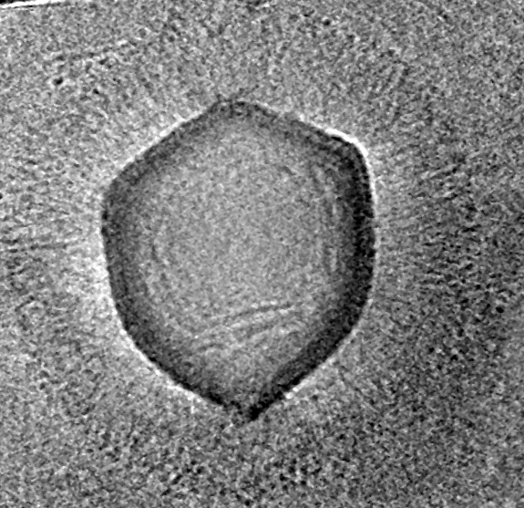
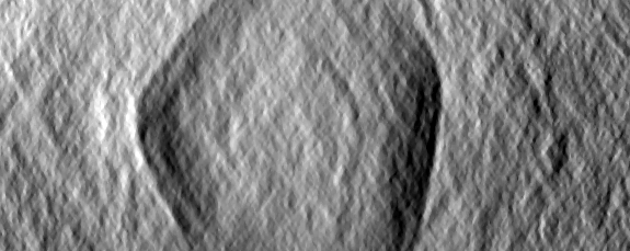
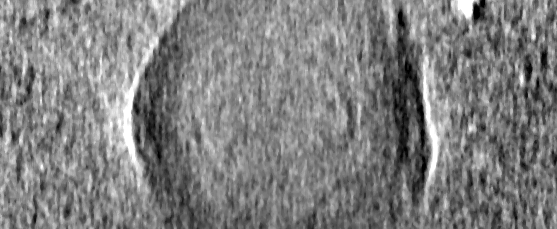
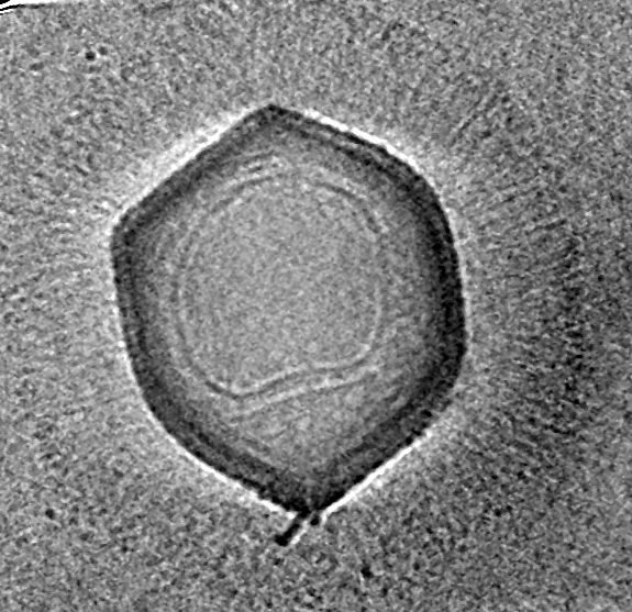
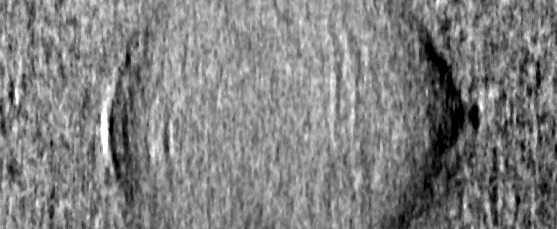
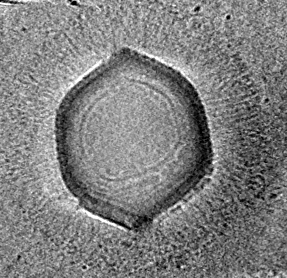
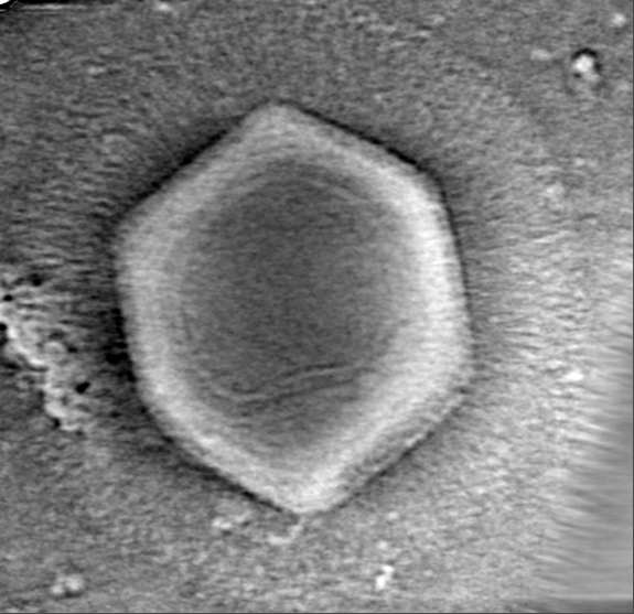
| Title | Samba Virus tomogram | |||||||||
 Map data Map data | Samba Virus particle | |||||||||
 Sample Sample |
| |||||||||
| Biological species |  Samba virus Samba virus | |||||||||
| Method | electron tomography / cryo EM | |||||||||
 Authors Authors | Schrad JR / Young EJ / Abrahao JS / Cortines JR / Parent KN | |||||||||
| Funding support |  United States, 1 items United States, 1 items
| |||||||||
 Citation Citation | Journal: Viruses / Year: 2017 Title: Microscopic Characterization of the Brazilian Giant Samba Virus. Authors: Jason R Schrad / Eric J Young / Jônatas S Abrahão / Juliana R Cortines / Kristin N Parent /   Abstract: Prior to the discovery of the mimivirus in 2003, viruses were thought to be physically small and genetically simple. Mimivirus, with its ~750-nm particle size and its ~1.2-Mbp genome, shattered these ...Prior to the discovery of the mimivirus in 2003, viruses were thought to be physically small and genetically simple. Mimivirus, with its ~750-nm particle size and its ~1.2-Mbp genome, shattered these notions and changed what it meant to be a virus. Since this discovery, the isolation and characterization of giant viruses has exploded. One of the more recently discovered giant viruses, Samba virus, is a that was isolated from the Rio Negro in the Brazilian Amazon. Initial characterization of Samba has revealed some structural information, although the preparation techniques used are prone to the generation of structural artifacts. To generate more native-like structural information for Samba, we analyzed the virus through cryo-electron microscopy, cryo-electron tomography, scanning electron microscopy, and fluorescence microscopy. These microscopy techniques demonstrated that Samba particles have a capsid diameter of ~527 nm and a fiber length of ~155 nm, making Samba the largest yet characterized. We also compared Samba to a fiberless mimivirus variant. Samba particles, unlike those of mimivirus, do not appear to be rigid, and quasi-icosahedral, although the two viruses share many common features, including a multi-layered capsid and an asymmetric nucleocapsid, which may be common amongst the . | |||||||||
| History |
|
- Structure visualization
| Movie |
 Movie viewer Movie viewer |
|---|---|
| Structure viewer | EM map: SurfViewMolmilJmol/JSmol |
| Supplemental images |

- Downloads & links
-EMDB archive
| Map data | emd_8599.map.gz | 225.8 MB | EMDB map data format | |
|---|---|---|---|---|
| Header (meta data) | emd-8599-v30.xmlemd-8599.xml | 12.5 KB 12.5 KB | Display Display | EMDB header |
| Images | 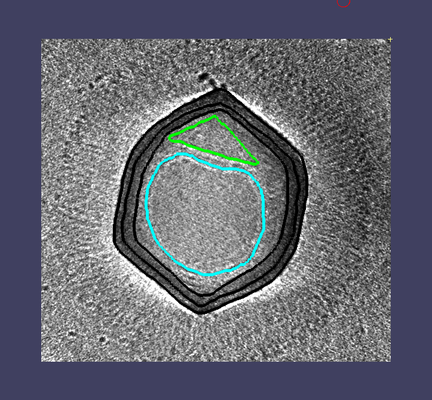 emd_8599.png emd_8599.png | 207.4 KB | ||
| Others | emd_8599_additional.map.gzemd_8599_additional_1.map.gz | 225.8 MB 225.8 MB | ||
| Archive directory |  http://ftp.pdbj.org/pub/emdb/structures/EMD-8599ftp://ftp.pdbj.org/pub/emdb/structures/EMD-8599 http://ftp.pdbj.org/pub/emdb/structures/EMD-8599ftp://ftp.pdbj.org/pub/emdb/structures/EMD-8599 | HTTPS FTP |
-Validation report
| Summary document | emd_8599_validation.pdf.gz | 257.7 KB | Display | EMDB validaton report |
|---|---|---|---|---|
| Full document | emd_8599_full_validation.pdf.gz | 257.3 KB | Display | |
| Data in XML | emd_8599_validation.xml.gz | 4.5 KB | Display | |
| Arichive directory | https://ftp.pdbj.org/pub/emdb/validation_reports/EMD-8599ftp://ftp.pdbj.org/pub/emdb/validation_reports/EMD-8599 | HTTPS FTP |
-Links
| EMDB pages | EMDB (EBI/PDBe) / EMDataResource |
|---|
-Map
| File | Download / File: emd_8599.map.gz / Format: CCP4 / Size: 279.8 MB / Type: IMAGE STORED AS FLOATING POINT NUMBER (4 BYTES) | ||||||||||||||||||||||||||||||||||||||||||||||||||||||||||||||||||||
|---|---|---|---|---|---|---|---|---|---|---|---|---|---|---|---|---|---|---|---|---|---|---|---|---|---|---|---|---|---|---|---|---|---|---|---|---|---|---|---|---|---|---|---|---|---|---|---|---|---|---|---|---|---|---|---|---|---|---|---|---|---|---|---|---|---|---|---|---|---|
| Annotation | Samba Virus particle | ||||||||||||||||||||||||||||||||||||||||||||||||||||||||||||||||||||
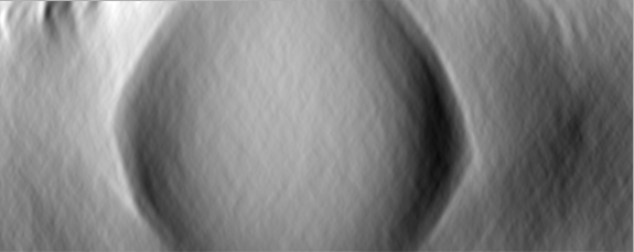
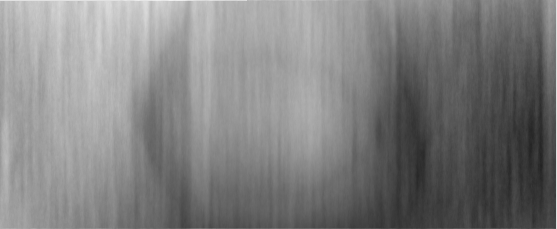
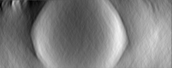
| Projections & slices | Image control
Images are generated by Spider. generated in cubic-lattice coordinate | ||||||||||||||||||||||||||||||||||||||||||||||||||||||||||||||||||||
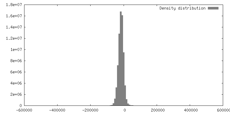
| Voxel size | X=Y=Z: 14.7 Å | ||||||||||||||||||||||||||||||||||||||||||||||||||||||||||||||||||||
| Density |
| ||||||||||||||||||||||||||||||||||||||||||||||||||||||||||||||||||||
| Symmetry | Space group: 1 | ||||||||||||||||||||||||||||||||||||||||||||||||||||||||||||||||||||
| Details | EMDB XML:
CCP4 map header:
| ||||||||||||||||||||||||||||||||||||||||||||||||||||||||||||||||||||
 Z (Sec.)
 Y (Row.)
 X (Col.)


-Supplemental data
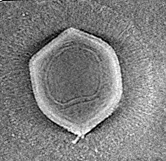
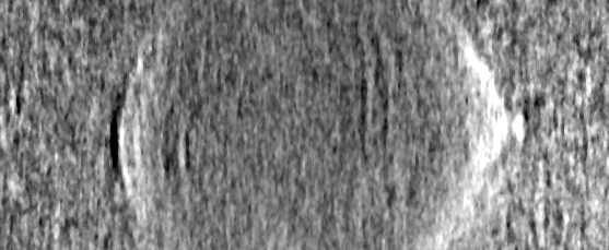
-Additional map: Samba Virus particle, inverted map
| File | emd_8599_additional.map | ||||||||||||
|---|---|---|---|---|---|---|---|---|---|---|---|---|---|
| Annotation | Samba Virus particle, inverted map | ||||||||||||
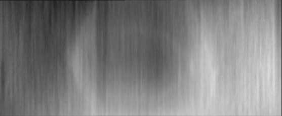
| Projections & Slices |
| ||||||||||||
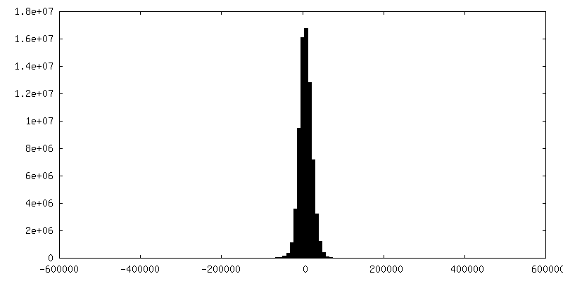
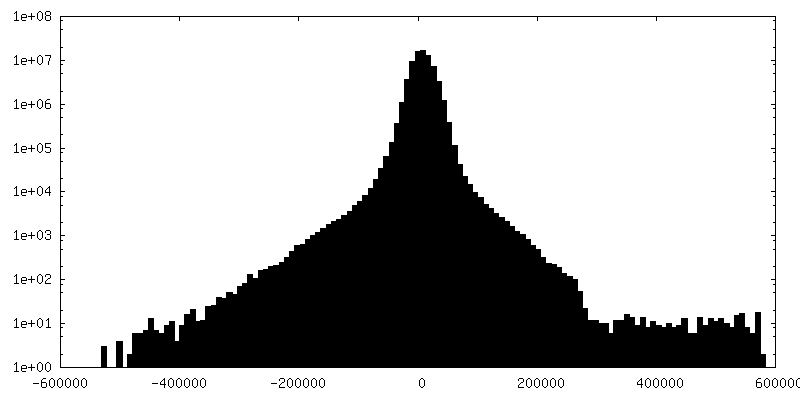
| Density Histograms |


-Additional map: Samba Virus particle, inverted map
| File | emd_8599_additional_1.map | ||||||||||||
|---|---|---|---|---|---|---|---|---|---|---|---|---|---|
| Annotation | Samba Virus particle, inverted map | ||||||||||||
| Projections & Slices |
| ||||||||||||
| Density Histograms |


- Sample components
-Entire : Samba virus
| Entire | Name: Samba virus |
|---|---|
| Components |
|
-Supramolecule #1: Samba virus
| Supramolecule | Name: Samba virus / type: virus / ID: 1 / Parent: 0 / NCBI-ID: 1461100 / Sci species name: Samba virus / Virus type: VIRION / Virus isolate: STRAIN / Virus enveloped: No / Virus empty: No |
|---|---|
| Host (natural) | Organism:  Acanthamoeba castellanii (eukaryote) Acanthamoeba castellanii (eukaryote) |
| Virus shell | Shell ID: 1 / Diameter: 8400.0 Å |
-Experimental details
-Structure determination
| Method | cryo EM |
|---|---|
 Processing Processing | electron tomography |
| Aggregation state | particle |
-Sample preparation
| Buffer | pH: 7.4 / Component - Concentration: 20.0 mM / Component - Name: sodium phosphate |
|---|---|
| Grid | Model: Quantifoil 3.5/1 / Material: COPPER / Mesh: 300 / Pretreatment - Type: PLASMA CLEANING |
| Vitrification | Cryogen name: ETHANE / Instrument: HOMEMADE PLUNGER |
| Sectioning | Other: NO SECTIONING |
| Fiducial marker | Manufacturer: Aldrich / Diameter: 10 nm |
- Electron microscopy
| Microscope | JEOL 2200FS |
|---|---|
| Temperature | Min: 83.0 K / Max: 83.0 K |
| Specialist optics | Energy filter - Name: In-column Omega Filter |
| Image recording | Film or detector model: DIRECT ELECTRON DE-20 (5k x 3k) / Detector mode: INTEGRATING / Digitization - Dimensions - Width: 5120 pixel / Digitization - Dimensions - Height: 3840 pixel / Digitization - Sampling interval: 6.4 µm / Digitization - Frames/image: 1-45 / Number grids imaged: 17 / Average exposure time: 3.0 sec. / Average electron dose: 1.1 e/Å2 Details: 63 tilt series were collected. From these, 20 complete tomograms were produced, capturing 34 particles. |
| Electron beam | Acceleration voltage: 200 kV / Electron source:  FIELD EMISSION GUN FIELD EMISSION GUN |
| Electron optics | C2 aperture diameter: 100.0 µm / Calibrated defocus max: 25.0 µm / Illumination mode: FLOOD BEAM / Imaging mode: BRIGHT FIELD / Cs: 1.4 mm / Nominal magnification: 4000 |
| Sample stage | Specimen holder model: GATAN 914 HIGH TILT LIQUID NITROGEN CRYO TRANSFER TOMOGRAPHY HOLDER Cooling holder cryogen: NITROGEN |
-Image processing
| Details | The selected images were motion-corrected and corrected for the gain and dark references prior to performance of the tomographic reconstructions. |
|---|---|
| Final reconstruction | Algorithm: SIMULTANEOUS ITERATIVE (SIRT) / Software - Name: IMOD (ver. 4.7.15) / Number images used: 67 |
