 ムービー
ムービー コントローラー


+ データを開く

- 基本情報
基本情報
| 登録情報 | データベース: EMDB / ID: EMD-8157 | |||||||||
|---|---|---|---|---|---|---|---|---|---|---|
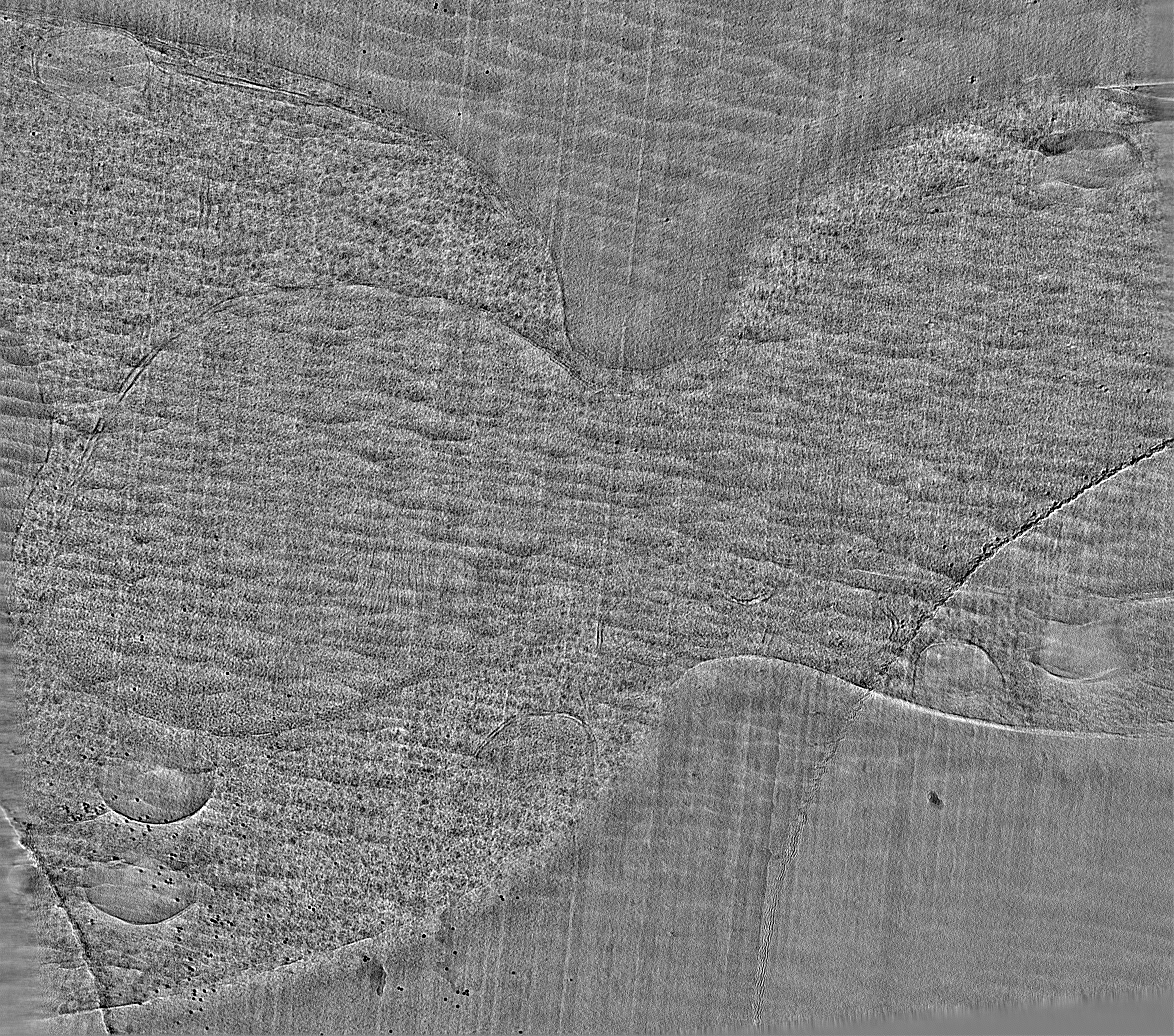
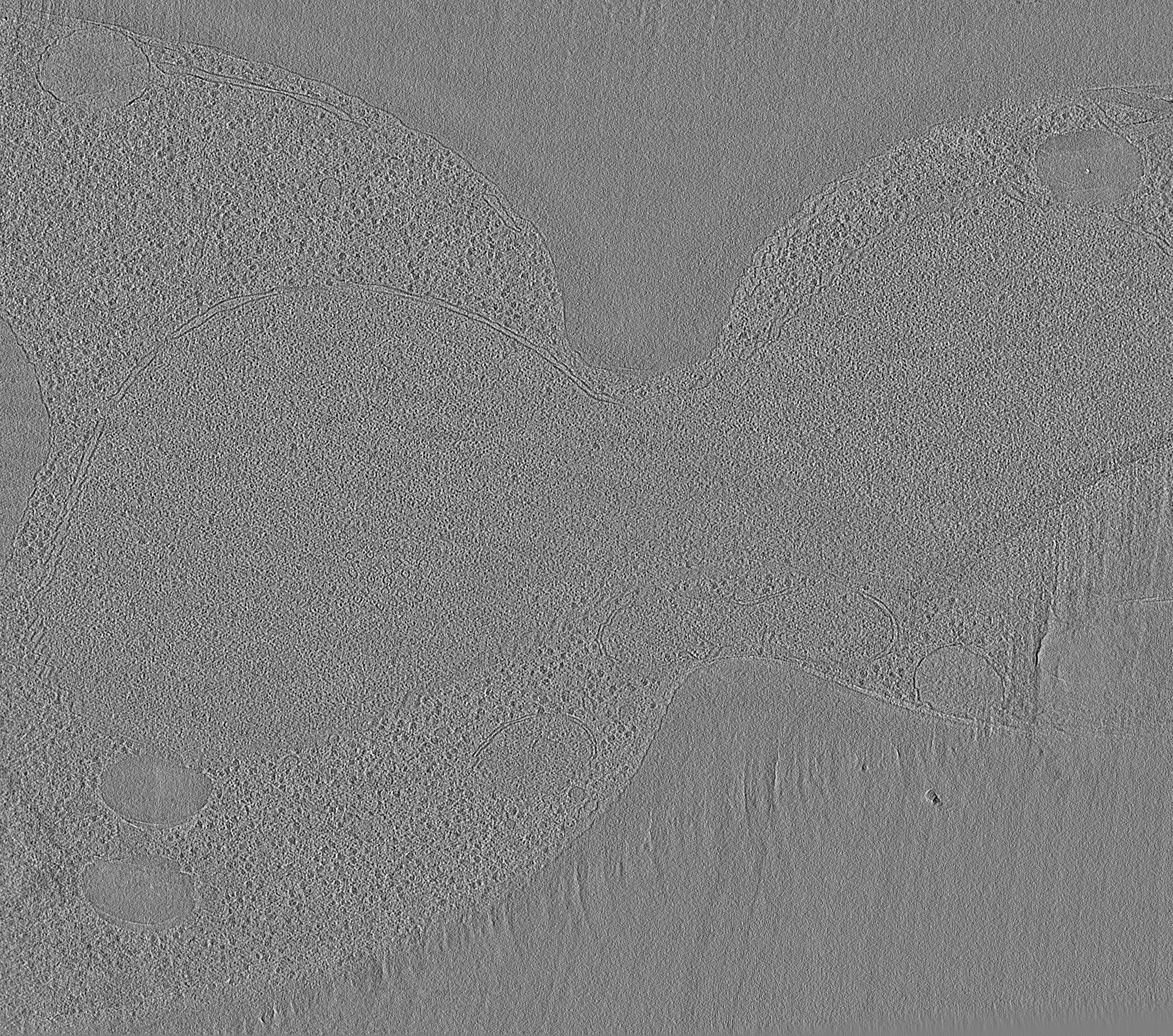
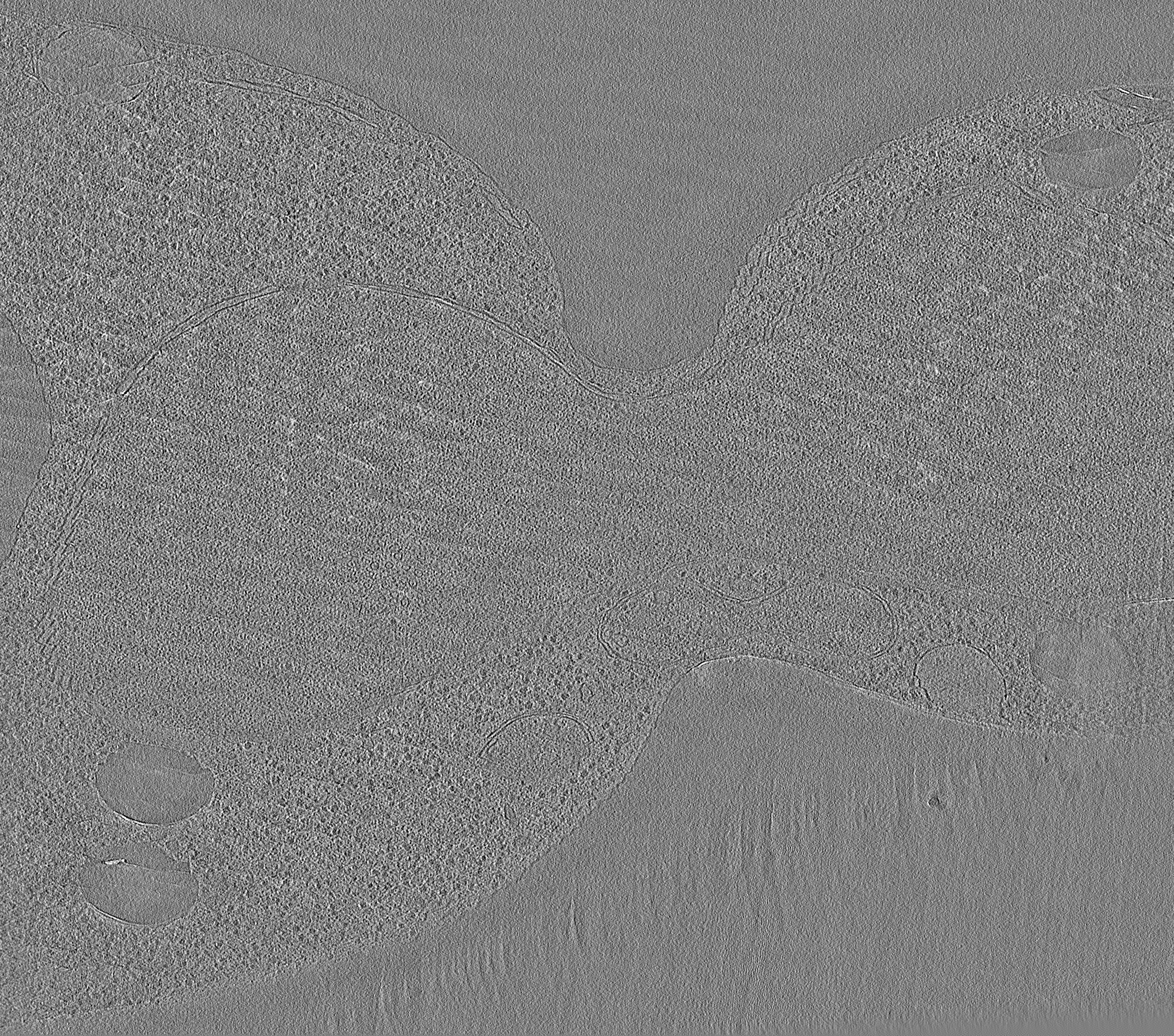
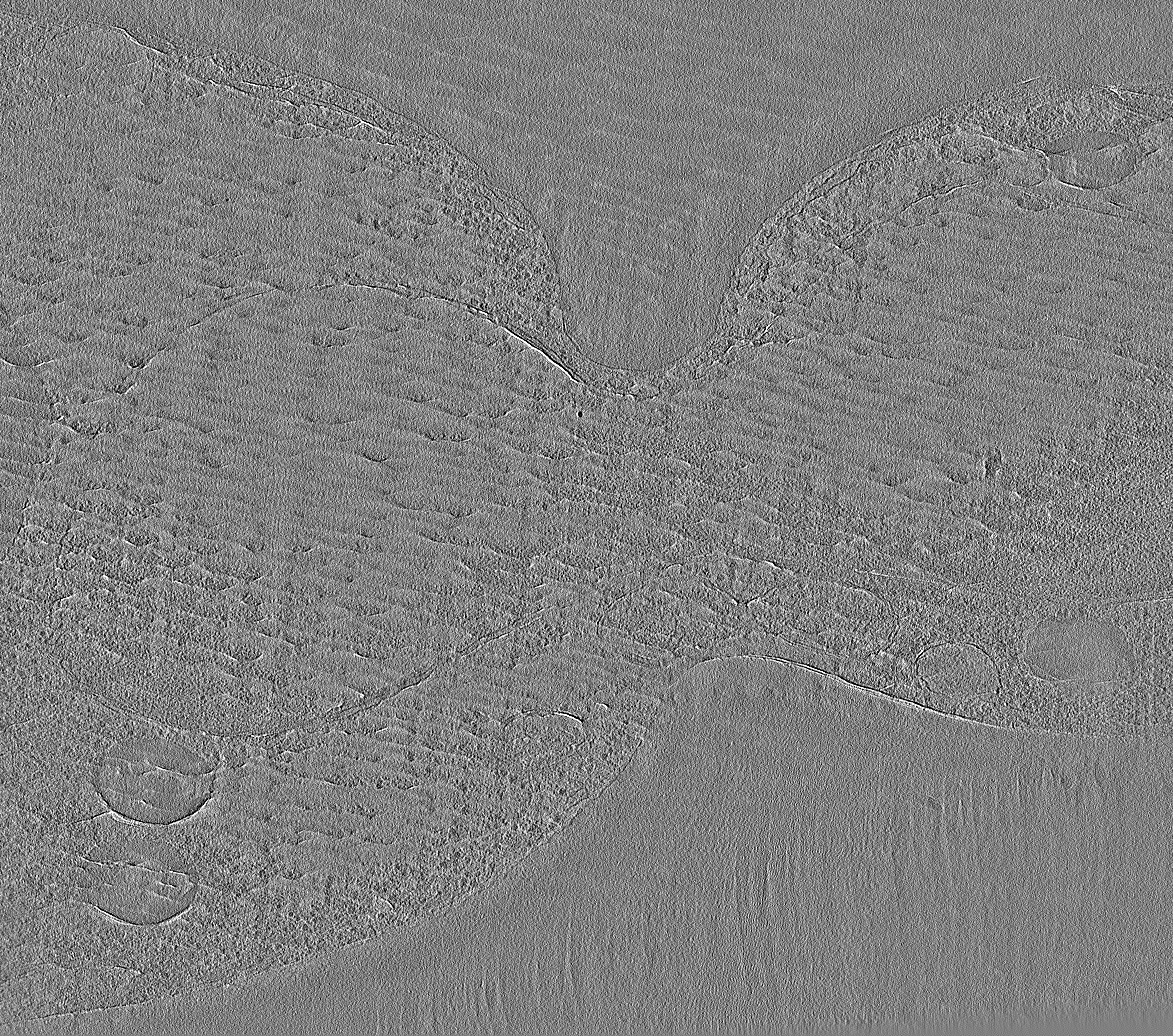
| タイトル | Electron cryotomogram of a cryosectioned budding yeast S. cerevisiae cell | |||||||||
 マップデータ マップデータ | None | |||||||||
 試料 試料 |
| |||||||||
| 生物種 |  | |||||||||
| 手法 | 電子線トモグラフィー法 / クライオ電子顕微鏡法 | |||||||||
 データ登録者 データ登録者 | Lu G | |||||||||
 引用 引用 | ジャーナル: Mol Biol Cell / 年: 2016 タイトル: Budding yeast chromatin is dispersed in a crowded nucleoplasm in vivo. 著者: Chen Chen / Hong Hwa Lim / Jian Shi / Sachiko Tamura / Kazuhiro Maeshima / Uttam Surana / Lu Gan /   要旨: Chromatin organization has an important role in the regulation of eukaryotic systems. Although recent studies have refined the three-dimensional models of chromatin organization with high resolution ...Chromatin organization has an important role in the regulation of eukaryotic systems. Although recent studies have refined the three-dimensional models of chromatin organization with high resolution at the genome sequence level, little is known about how the most fundamental units of chromatin-nucleosomes-are positioned in three dimensions in vivo. Here we use electron cryotomography to study chromatin organization in the budding yeast Saccharomyces cerevisiae Direct visualization of yeast nuclear densities shows no evidence of 30-nm fibers. Aside from preribosomes and spindle microtubules, few nuclear structures are larger than a tetranucleosome. Yeast chromatin does not form compact structures in interphase or mitosis and is consistent with being in an "open" configuration that is conducive to high levels of transcription. From our study and those of others, we propose that yeast can regulate its transcription using local nucleosome-nucleosome associations. | |||||||||
| 履歴 |
|
- 構造の表示
構造の表示
| ムービー |
 ムービービューア ムービービューア |
|---|---|
| 構造ビューア | EMマップ: SurfViewMolmilJmol/JSmol |
| 添付画像 |
- ダウンロードとリンク
-EMDBアーカイブ
| マップデータ | emd_8157.map.gz | 5 GB | EMDBマップデータ形式 | |
|---|---|---|---|---|
| ヘッダ (付随情報) | emd-8157-v30.xmlemd-8157.xml | 9.7 KB 9.7 KB | 表示 表示 | EMDBヘッダ |
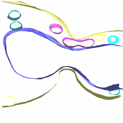
| 画像 |  emd_8157.png emd_8157.png | 57.9 KB | ||
| アーカイブディレクトリ |  http://ftp.pdbj.org/pub/emdb/structures/EMD-8157ftp://ftp.pdbj.org/pub/emdb/structures/EMD-8157 http://ftp.pdbj.org/pub/emdb/structures/EMD-8157ftp://ftp.pdbj.org/pub/emdb/structures/EMD-8157 | HTTPS FTP |
-関連構造データ
| 電子顕微鏡画像生データ | EMPIAR-10062 (タイトル: Electron cryotomography of cryosectioned budding yeast cells Data size: 125.3 Data #1: Tilt series of budding yeast cell cryosections and purified chromatin [class averages]) EMPIAR-10227 (タイトル: A collection of yeast cell cryo-ET data / Data size: 2.5 TBData #1: A collection of yeast cell cryo-ET data [tilt series]) |
|---|
-リンク
| EMDBのページ | EMDB (EBI/PDBe) / EMDataResource |
|---|
-マップ
| ファイル | ダウンロード / ファイル: emd_8157.map.gz / 形式: CCP4 / 大きさ: 1.1 GB / タイプ: IMAGE STORED AS SIGNED INTEGER (2 BYTES) | ||||||||||||||||||||||||||||||||||||||||||||||||||||||||||||||||||||
|---|---|---|---|---|---|---|---|---|---|---|---|---|---|---|---|---|---|---|---|---|---|---|---|---|---|---|---|---|---|---|---|---|---|---|---|---|---|---|---|---|---|---|---|---|---|---|---|---|---|---|---|---|---|---|---|---|---|---|---|---|---|---|---|---|---|---|---|---|---|
| 注釈 | None | ||||||||||||||||||||||||||||||||||||||||||||||||||||||||||||||||||||
| 投影像・断面図 | 画像のコントロール
画像は Spider により作成 これらの図は立方格子座標系で作成されたものです | ||||||||||||||||||||||||||||||||||||||||||||||||||||||||||||||||||||
| ボクセルのサイズ | X=Y=Z: 9.12 Å | ||||||||||||||||||||||||||||||||||||||||||||||||||||||||||||||||||||
| 密度 |
| ||||||||||||||||||||||||||||||||||||||||||||||||||||||||||||||||||||
| 対称性 | 空間群: 1 | ||||||||||||||||||||||||||||||||||||||||||||||||||||||||||||||||||||
| 詳細 | EMDB XML:
CCP4マップ ヘッダ情報:
| ||||||||||||||||||||||||||||||||||||||||||||||||||||||||||||||||||||
 Z (Sec.)
 Y (Row.)
 X (Col.)


-添付データ
- 試料の構成要素
-全体 : Saccharomyces cerevisiae cryosection
| 全体 | 名称: Saccharomyces cerevisiae cryosection |
|---|---|
| 要素 |
|
-超分子 #1: Saccharomyces cerevisiae cryosection
| 超分子 | 名称: Saccharomyces cerevisiae cryosection / タイプ: cell / ID: 1 / 親要素: 0 |
|---|---|
| 由来(天然) | 生物種: |
-実験情報
-構造解析
| 手法 | クライオ電子顕微鏡法 |
|---|---|
 解析 解析 | 電子線トモグラフィー法 |
| 試料の集合状態 | cell |
-試料調製
| 緩衝液 | pH: 6.5 |
|---|---|
| グリッド | モデル: Protochips CF200-Cu / 材質: COPPER / 支持フィルム - 材質: CARBON / 支持フィルム - トポロジー: HOLEY |
| 凍結 | 凍結剤: NITROGEN |
| 加圧凍結法 | 装置: OTHER 詳細: The value given for _emd_high_pressure_freezing.instrument is HPF Compact 01. This is not in a list of allowed values set(['LEICA EM PACT2', 'LEICA EM PACT', 'EMS-002 RAPID IMMERSION ...詳細: The value given for _emd_high_pressure_freezing.instrument is HPF Compact 01. This is not in a list of allowed values set(['LEICA EM PACT2', 'LEICA EM PACT', 'EMS-002 RAPID IMMERSION FREEZER', 'OTHER', 'LEICA EM HPM100', 'BAL-TEC HPM 010']) so OTHER is written into the XML file. |
| 切片作成 | ウルトラミクロトーム - 装置: Leica UC7/FC7 / ウルトラミクロトーム - 温度: 123 K / ウルトラミクロトーム - 最終 厚さ: 150 |
| 位置合わせマーカー | Manufacturer: Sigma / 直径: 10 nm |
- 電子顕微鏡法
| 顕微鏡 | FEI TITAN KRIOS |
|---|---|
| 撮影 | フィルム・検出器のモデル: FEI FALCON II (4k x 4k) 検出モード: INTEGRATING / デジタル化 - サイズ - 横: 4096 pixel / デジタル化 - サイズ - 縦: 4096 pixel / 撮影したグリッド数: 1 / 実像数: 62 / 平均電子線量: 1.6 e/Å2 |
| 電子線 | 加速電圧: 300 kV / 電子線源:  FIELD EMISSION GUN FIELD EMISSION GUN |
| 電子光学系 | 最小 デフォーカス(補正後): 15.0 µm / 倍率(補正後): 15678 / 照射モード: FLOOD BEAM / 撮影モード: BRIGHT FIELD / Cs: 2.7 mm / 最小 デフォーカス(公称値): 10.0 µm / 倍率(公称値): 8700 |
| 試料ステージ | 試料ホルダーモデル: FEI TITAN KRIOS AUTOGRID HOLDER ホルダー冷却材: NITROGEN |
| 実験機器 |  モデル: Titan Krios / 画像提供: FEI Company |
-画像解析
| 最終 再構成 | アルゴリズム: BACK PROJECTION / ソフトウェア - 名称: IMOD (ver. 4.8) / 使用した粒子像数: 62 |
|---|---|
| CTF補正 | ソフトウェア - 名称: IMOD (ver. 4.8) |
