 Movie
Movie Controller

[English] 日本語
 Yorodumi
- EMDB-8157: Electron cryotomogram of a cryosectioned budding yeast S. cerevis... -
+ Open data

- Basic information
| Entry | Database: EMDB / ID: EMD-8157 | |||||||||
|---|---|---|---|---|---|---|---|---|---|---|
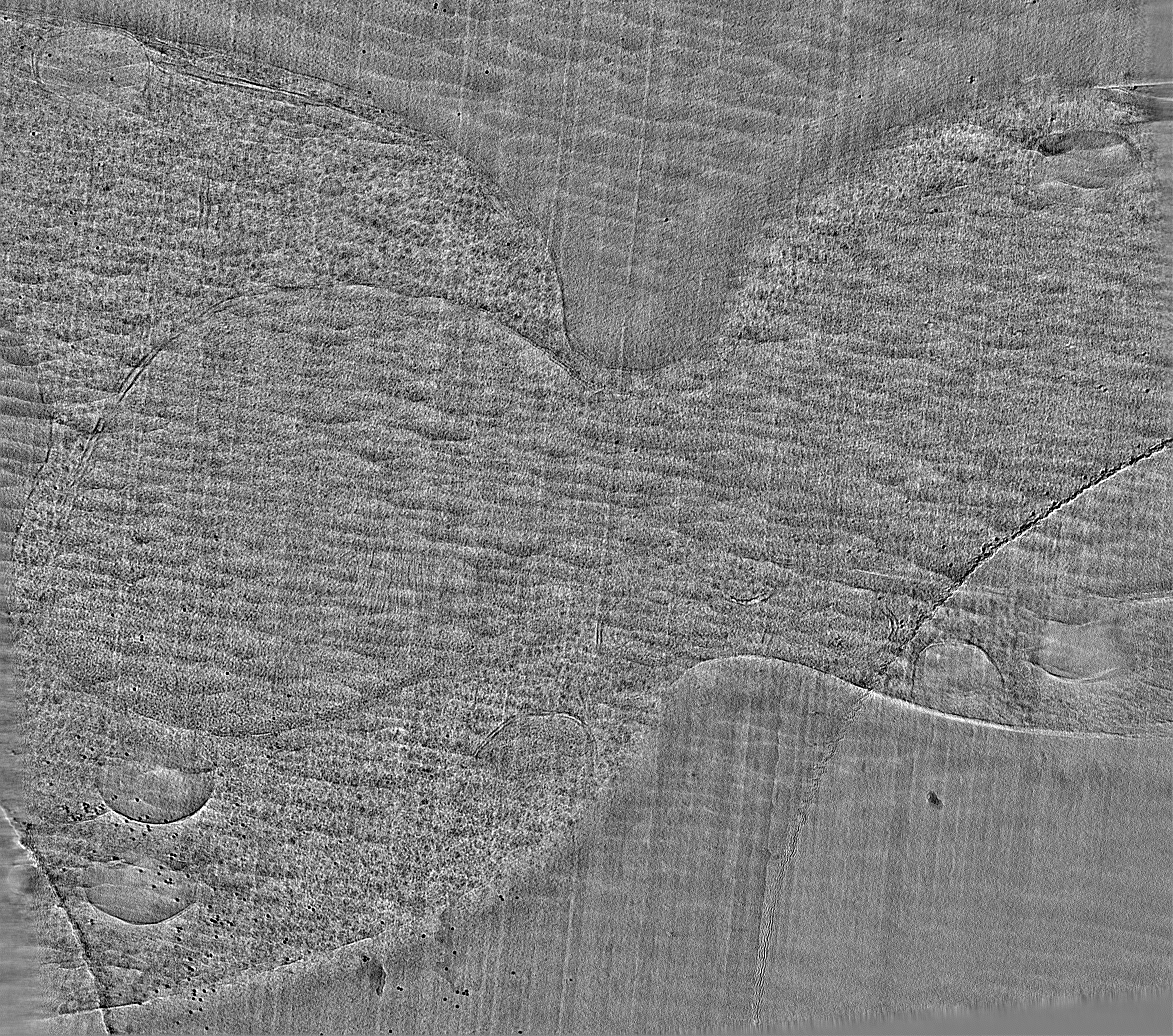
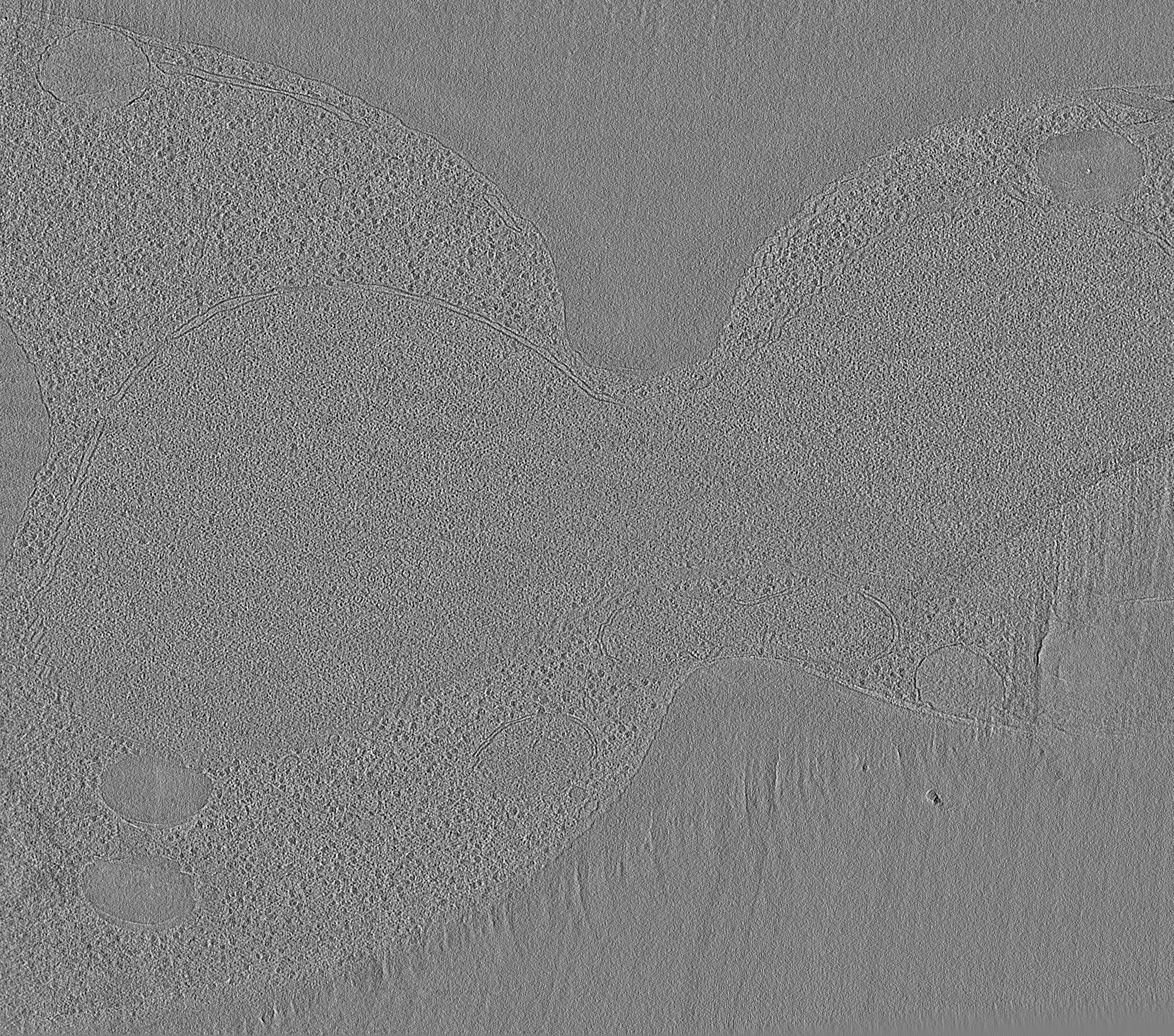
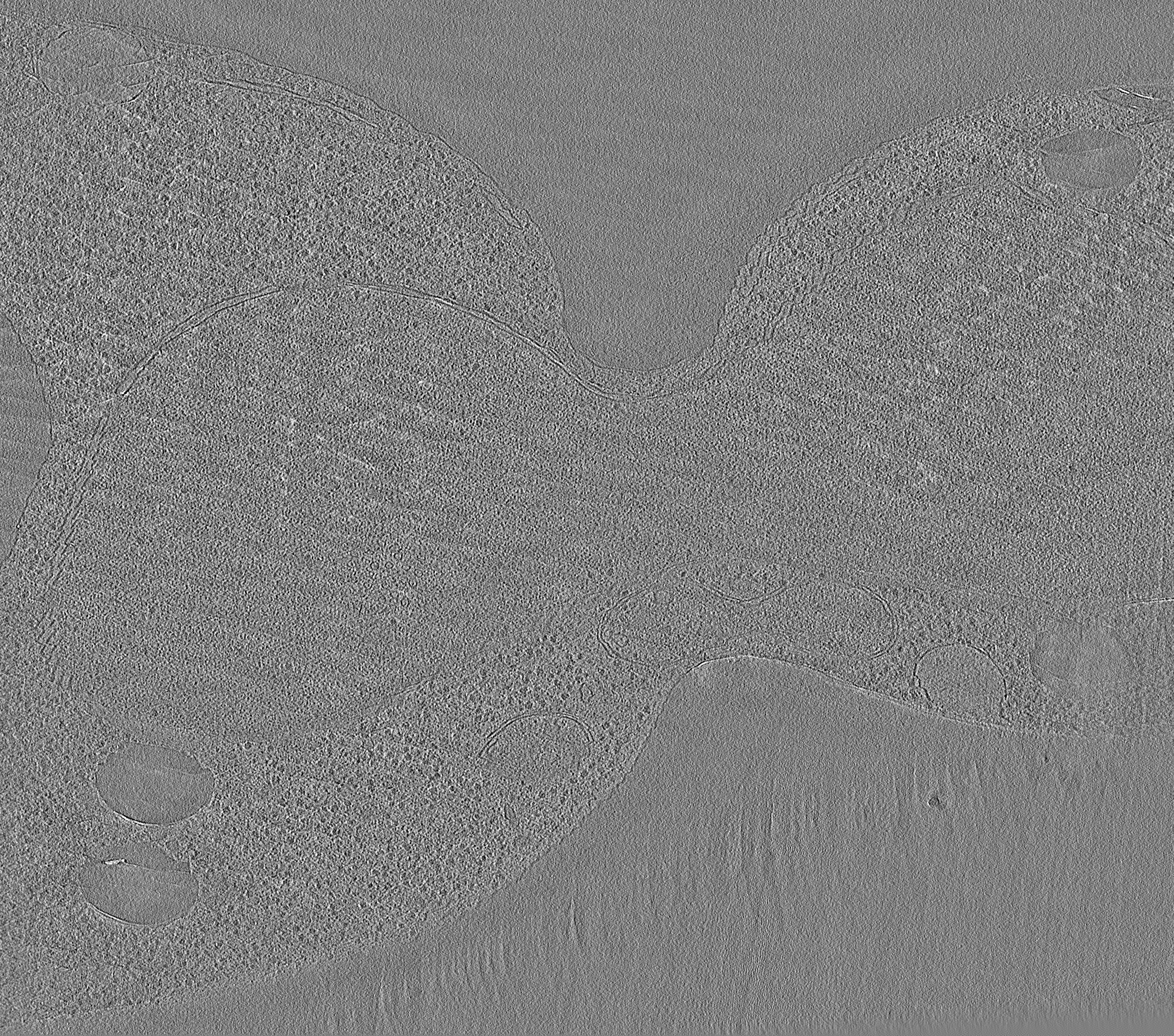
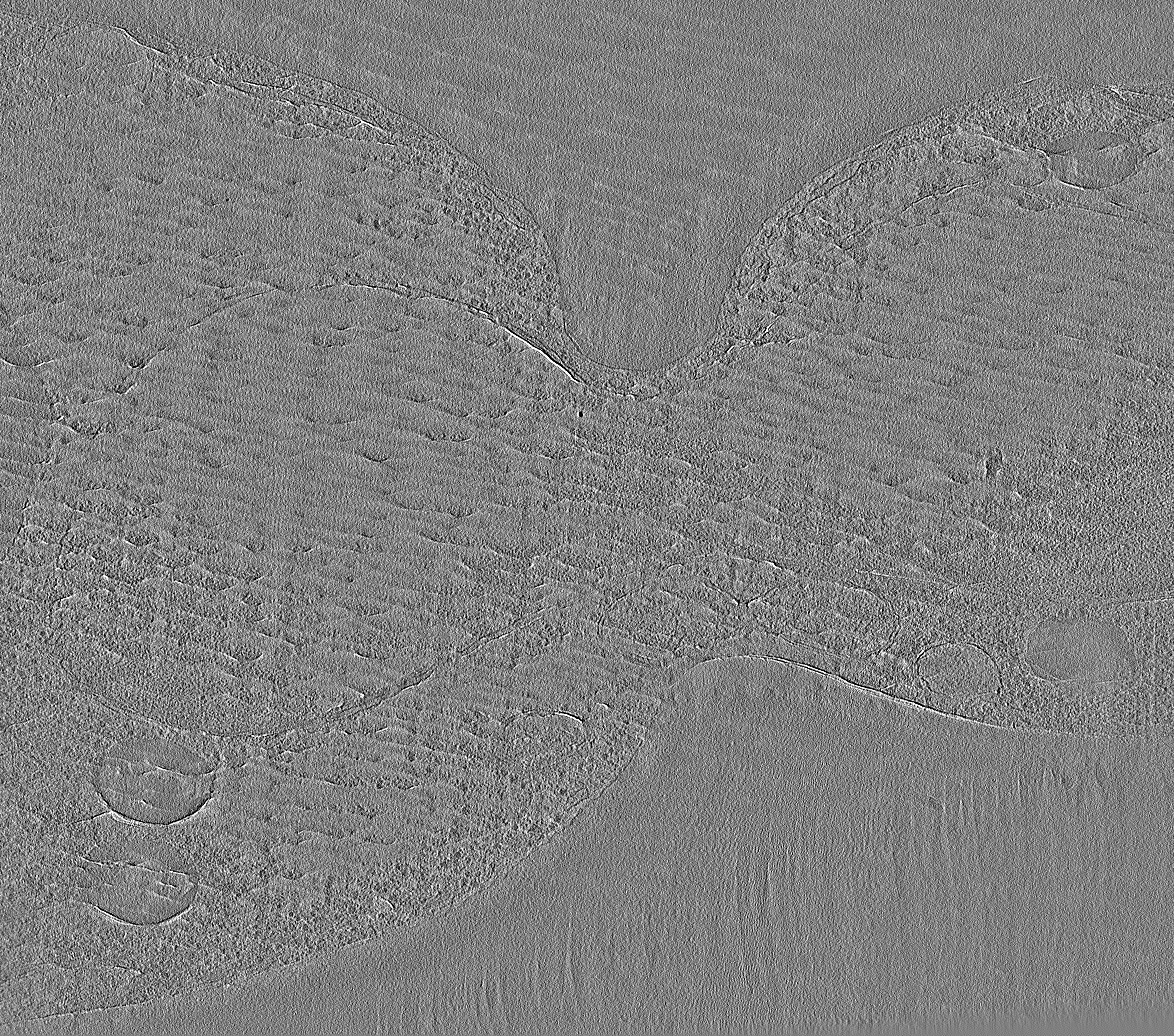
| Title | Electron cryotomogram of a cryosectioned budding yeast S. cerevisiae cell | |||||||||
 Map data Map data | None | |||||||||
 Sample Sample |
| |||||||||
| Biological species |  | |||||||||
| Method | electron tomography / cryo EM | |||||||||
 Authors Authors | Lu G | |||||||||
 Citation Citation | Journal: Mol Biol Cell / Year: 2016 Title: Budding yeast chromatin is dispersed in a crowded nucleoplasm in vivo. Authors: Chen Chen / Hong Hwa Lim / Jian Shi / Sachiko Tamura / Kazuhiro Maeshima / Uttam Surana / Lu Gan /   Abstract: Chromatin organization has an important role in the regulation of eukaryotic systems. Although recent studies have refined the three-dimensional models of chromatin organization with high resolution ...Chromatin organization has an important role in the regulation of eukaryotic systems. Although recent studies have refined the three-dimensional models of chromatin organization with high resolution at the genome sequence level, little is known about how the most fundamental units of chromatin-nucleosomes-are positioned in three dimensions in vivo. Here we use electron cryotomography to study chromatin organization in the budding yeast Saccharomyces cerevisiae Direct visualization of yeast nuclear densities shows no evidence of 30-nm fibers. Aside from preribosomes and spindle microtubules, few nuclear structures are larger than a tetranucleosome. Yeast chromatin does not form compact structures in interphase or mitosis and is consistent with being in an "open" configuration that is conducive to high levels of transcription. From our study and those of others, we propose that yeast can regulate its transcription using local nucleosome-nucleosome associations. | |||||||||
| History |
|
- Structure visualization
| Movie |
 Movie viewer Movie viewer |
|---|---|
| Structure viewer | EM map: SurfViewMolmilJmol/JSmol |
| Supplemental images |
- Downloads & links
-EMDB archive
| Map data | emd_8157.map.gz | 5 GB | EMDB map data format | |
|---|---|---|---|---|
| Header (meta data) | emd-8157-v30.xmlemd-8157.xml | 9.7 KB 9.7 KB | Display Display | EMDB header |
| Images |  emd_8157.png emd_8157.png | 57.9 KB | ||
| Archive directory |  http://ftp.pdbj.org/pub/emdb/structures/EMD-8157ftp://ftp.pdbj.org/pub/emdb/structures/EMD-8157 http://ftp.pdbj.org/pub/emdb/structures/EMD-8157ftp://ftp.pdbj.org/pub/emdb/structures/EMD-8157 | HTTPS FTP |
-Validation report
| Summary document | emd_8157_validation.pdf.gz | 77.4 KB | Display | EMDB validaton report |
|---|---|---|---|---|
| Full document | emd_8157_full_validation.pdf.gz | 76.6 KB | Display | |
| Data in XML | emd_8157_validation.xml.gz | 500 B | Display | |
| Arichive directory | https://ftp.pdbj.org/pub/emdb/validation_reports/EMD-8157ftp://ftp.pdbj.org/pub/emdb/validation_reports/EMD-8157 | HTTPS FTP |
-Related structure data
| EM raw data | EMPIAR-10062 (Title: Electron cryotomography of cryosectioned budding yeast cells Data size: 125.3 Data #1: Tilt series of budding yeast cell cryosections and purified chromatin [class averages]) EMPIAR-10227 (Title: A collection of yeast cell cryo-ET data / Data size: 2.5 TBData #1: A collection of yeast cell cryo-ET data [tilt series]) |
|---|
-Links
| EMDB pages | EMDB (EBI/PDBe) / EMDataResource |
|---|
-Map
| File | Download / File: emd_8157.map.gz / Format: CCP4 / Size: 1.1 GB / Type: IMAGE STORED AS SIGNED INTEGER (2 BYTES) | ||||||||||||||||||||||||||||||||||||||||||||||||||||||||||||||||||||
|---|---|---|---|---|---|---|---|---|---|---|---|---|---|---|---|---|---|---|---|---|---|---|---|---|---|---|---|---|---|---|---|---|---|---|---|---|---|---|---|---|---|---|---|---|---|---|---|---|---|---|---|---|---|---|---|---|---|---|---|---|---|---|---|---|---|---|---|---|---|
| Annotation | None | ||||||||||||||||||||||||||||||||||||||||||||||||||||||||||||||||||||
| Projections & slices | Image control
Images are generated by Spider. generated in cubic-lattice coordinate | ||||||||||||||||||||||||||||||||||||||||||||||||||||||||||||||||||||
| Voxel size | X=Y=Z: 9.12 Å | ||||||||||||||||||||||||||||||||||||||||||||||||||||||||||||||||||||
| Density |
| ||||||||||||||||||||||||||||||||||||||||||||||||||||||||||||||||||||
| Symmetry | Space group: 1 | ||||||||||||||||||||||||||||||||||||||||||||||||||||||||||||||||||||
| Details | EMDB XML:
CCP4 map header:
| ||||||||||||||||||||||||||||||||||||||||||||||||||||||||||||||||||||
 Z (Sec.)
 Y (Row.)
 X (Col.)


-Supplemental data
- Sample components
-Entire : Saccharomyces cerevisiae cryosection
| Entire | Name: Saccharomyces cerevisiae cryosection |
|---|---|
| Components |
|
-Supramolecule #1: Saccharomyces cerevisiae cryosection
| Supramolecule | Name: Saccharomyces cerevisiae cryosection / type: cell / ID: 1 / Parent: 0 |
|---|---|
| Source (natural) | Organism: |
-Experimental details
-Structure determination
| Method | cryo EM |
|---|---|
 Processing Processing | electron tomography |
| Aggregation state | cell |
-Sample preparation
| Buffer | pH: 6.5 |
|---|---|
| Grid | Model: Protochips CF200-Cu / Material: COPPER / Support film - Material: CARBON / Support film - topology: HOLEY |
| Vitrification | Cryogen name: NITROGEN |
| High pressure freezing | Instrument: OTHER Details: The value given for _emd_high_pressure_freezing.instrument is HPF Compact 01. This is not in a list of allowed values set(['LEICA EM PACT2', 'LEICA EM PACT', 'EMS-002 RAPID IMMERSION ...Details: The value given for _emd_high_pressure_freezing.instrument is HPF Compact 01. This is not in a list of allowed values set(['LEICA EM PACT2', 'LEICA EM PACT', 'EMS-002 RAPID IMMERSION FREEZER', 'OTHER', 'LEICA EM HPM100', 'BAL-TEC HPM 010']) so OTHER is written into the XML file. |
| Sectioning | Ultramicrotomy - Instrument: Leica UC7/FC7 / Ultramicrotomy - Temperature: 123 K / Ultramicrotomy - Final thickness: 150 |
| Fiducial marker | Manufacturer: Sigma / Diameter: 10 nm |
- Electron microscopy
| Microscope | FEI TITAN KRIOS |
|---|---|
| Image recording | Film or detector model: FEI FALCON II (4k x 4k) / Detector mode: INTEGRATING / Digitization - Dimensions - Width: 4096 pixel / Digitization - Dimensions - Height: 4096 pixel / Number grids imaged: 1 / Number real images: 62 / Average electron dose: 1.6 e/Å2 |
| Electron beam | Acceleration voltage: 300 kV / Electron source:  FIELD EMISSION GUN FIELD EMISSION GUN |
| Electron optics | Calibrated defocus min: 15.0 µm / Calibrated magnification: 15678 / Illumination mode: FLOOD BEAM / Imaging mode: BRIGHT FIELD / Cs: 2.7 mm / Nominal defocus min: 10.0 µm / Nominal magnification: 8700 |
| Sample stage | Specimen holder model: FEI TITAN KRIOS AUTOGRID HOLDER / Cooling holder cryogen: NITROGEN |
| Experimental equipment |  Model: Titan Krios / Image courtesy: FEI Company |
-Image processing
| Final reconstruction | Algorithm: BACK PROJECTION / Software - Name: IMOD (ver. 4.8) / Number images used: 62 |
|---|---|
| CTF correction | Software - Name: IMOD (ver. 4.8) |
