 Movie
Movie Controller

[English] 日本語
 Yorodumi
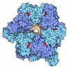
- EMDB-80335: Cryo-EM structure of the E. coli beta sliding clamp-Hda heterohexamer -
+ Open data

- Basic information
| Entry |  | |||||||||||||||
|---|---|---|---|---|---|---|---|---|---|---|---|---|---|---|---|---|
| Title | Cryo-EM structure of the E. coli beta sliding clamp-Hda heterohexamer | |||||||||||||||
 Map data Map data | Local-filtered 3DFlex refine map | |||||||||||||||
 Sample Sample |
| |||||||||||||||
 Keywords Keywords | Replication inactivator / Inhibitor / ATPase / TRANSFERASE | |||||||||||||||
| Function / homology |  Function and homology information Function and homology informationreplication inhibiting complex / Hda-beta clamp complex / DNA polymerase III complex / DNA strand elongation involved in DNA replication / DNA replication initiation / regulation of DNA-templated DNA replication initiation / 3'-5' exonuclease activity / negative regulation of DNA-templated DNA replication initiation / ADP binding / DNA replication ...replication inhibiting complex / Hda-beta clamp complex / DNA polymerase III complex / DNA strand elongation involved in DNA replication / DNA replication initiation / regulation of DNA-templated DNA replication initiation / 3'-5' exonuclease activity / negative regulation of DNA-templated DNA replication initiation / ADP binding / DNA replication / DNA-directed DNA polymerase activity / DNA binding / membrane / identical protein binding / plasma membrane / cytosol / cytoplasm Similarity search - Function | |||||||||||||||
| Biological species |   Escherichia coli (E. coli) Escherichia coli (E. coli) | |||||||||||||||
| Method | single particle reconstruction / cryo EM / Resolution: 3.43 Å | |||||||||||||||
 Authors Authors | Jiang X / Danev R / Kikkawa M | |||||||||||||||
| Funding support |  Japan, 4 items Japan, 4 items
| |||||||||||||||
 Citation Citation | Journal: Nucleic Acids Res. / Year: 2026 Title: Reversible autoinhibition of the bacterial replication-licensing Hda-beta clamp complex. Authors: Jiang X / Danev R / Luan Y / Nakakido M / Tsumoto K / Kikkawa M | |||||||||||||||
| History |
|
- Structure visualization
| Supplemental images |
|---|

- Downloads & links
-EMDB archive
| Map data | emd_80335.map.gz | 1.6 MB | EMDB map data format | |
|---|---|---|---|---|
| Header (meta data) | emd-80335-v30.xmlemd-80335.xml | 25.8 KB 25.8 KB | Display Display | EMDB header |
| FSC (resolution estimation) | emd_80335_fsc.xml | 6.4 KB | Display | FSC data file |
| Images |  emd_80335.png emd_80335.png | 73.2 KB | ||
| Masks | emd_80335_msk_1.map | 27 MB | Mask map | |
| Filedesc metadata | emd-80335.cif.gz | 6.9 KB | ||
| Others | emd_80335_additional_1.map.gzemd_80335_additional_2.map.gzemd_80335_half_map_1.map.gzemd_80335_half_map_2.map.gz | 25.4 MB 1.7 MB 2.8 MB 2.8 MB | ||
| Archive directory |  https://data.pdbj.org/pub/emdb/structures/EMD-80335ftp://data.pdbj.org/pub/emdb/structures/EMD-80335 https://data.pdbj.org/pub/emdb/structures/EMD-80335ftp://data.pdbj.org/pub/emdb/structures/EMD-80335 | HTTPS FTP |
-Related structure data
| Related structure data |  25rzMC  25ryC  25saC  25wzC M: atomic model generated by this map C: citing same article ( |
|---|---|
| Similar structure data |


-Links
| EMDB pages | EMDB (EBI/PDBe) / EMDataResource |
|---|---|
| Related items in Molecule of the Month |

-Map
| File | Download / File: emd_80335.map.gz / Format: CCP4 / Size: 27 MB / Type: IMAGE STORED AS FLOATING POINT NUMBER (4 BYTES) | ||||||||||||||||||||||||||||||||||||
|---|---|---|---|---|---|---|---|---|---|---|---|---|---|---|---|---|---|---|---|---|---|---|---|---|---|---|---|---|---|---|---|---|---|---|---|---|---|
| Annotation | Local-filtered 3DFlex refine map | ||||||||||||||||||||||||||||||||||||
| Projections & slices | Image control
Images are generated by Spider. | ||||||||||||||||||||||||||||||||||||
| Voxel size | X=Y=Z: 1.142 Å | ||||||||||||||||||||||||||||||||||||
| Density |
| ||||||||||||||||||||||||||||||||||||
| Symmetry | Space group: 1 | ||||||||||||||||||||||||||||||||||||
| Details | EMDB XML:
|
 Z (Sec.)
 Y (Row.)
 X (Col.)


-Supplemental data
-Mask #1
| File | emd_80335_msk_1.map | ||||||||||||
|---|---|---|---|---|---|---|---|---|---|---|---|---|---|
| Projections & Slices |
| ||||||||||||
| Density Histograms |


-Additional map: 3DFlex refine map
| File | emd_80335_additional_1.map | ||||||||||||
|---|---|---|---|---|---|---|---|---|---|---|---|---|---|
| Annotation | 3DFlex refine map | ||||||||||||
| Projections & Slices |
| ||||||||||||
| Density Histograms |


-Additional map: Local-filtered NU refine map
| File | emd_80335_additional_2.map | ||||||||||||
|---|---|---|---|---|---|---|---|---|---|---|---|---|---|
| Annotation | Local-filtered NU refine map | ||||||||||||
| Projections & Slices |
| ||||||||||||
| Density Histograms |


-Half map: halfmap2
| File | emd_80335_half_map_1.map | ||||||||||||
|---|---|---|---|---|---|---|---|---|---|---|---|---|---|
| Annotation | halfmap2 | ||||||||||||
| Projections & Slices |
| ||||||||||||
| Density Histograms |


-Half map: halfmap1
| File | emd_80335_half_map_2.map | ||||||||||||
|---|---|---|---|---|---|---|---|---|---|---|---|---|---|
| Annotation | halfmap1 | ||||||||||||
| Projections & Slices |
| ||||||||||||
| Density Histograms |


- Sample components
-Entire : Hetero-hexameric complex of beta sliding clamp and Hda
| Entire | Name: Hetero-hexameric complex of beta sliding clamp and Hda |
|---|---|
| Components |
|
-Supramolecule #1: Hetero-hexameric complex of beta sliding clamp and Hda
| Supramolecule | Name: Hetero-hexameric complex of beta sliding clamp and Hda type: complex / ID: 1 / Parent: 0 / Macromolecule list: #1-#2 |
|---|---|
| Source (natural) | Organism: Escherichia coli (E. coli) |
| Molecular weight | Theoretical: 215 KDa |
-Macromolecule #1: Beta sliding clamp
| Macromolecule | Name: Beta sliding clamp / type: protein_or_peptide / ID: 1 / Number of copies: 4 / Enantiomer: LEVO |
|---|---|
| Source (natural) | Organism: Escherichia coli (E. coli) |
| Molecular weight | Theoretical: 42.801863 KDa |
| Recombinant expression | Organism: Escherichia coli BL21 (bacteria) |
| Sequence | String: MGSSHHHHHH SSGLVPRGSH MKFTVEREHL LKPLQQVSGP LGGRPTLPIL GNLLLQVADG TLSLTGTDLE MEMVARVALV QPHEPGATT VPARKFFDIC RGLPEGAEIA VQLEGERMLV RSGRSRFSLS TLPAADFPNL DDWQSEVEFT LPQATMKRLI E ATQFSMAH ...String: MGSSHHHHHH SSGLVPRGSH MKFTVEREHL LKPLQQVSGP LGGRPTLPIL GNLLLQVADG TLSLTGTDLE MEMVARVALV QPHEPGATT VPARKFFDIC RGLPEGAEIA VQLEGERMLV RSGRSRFSLS TLPAADFPNL DDWQSEVEFT LPQATMKRLI E ATQFSMAH QDVRYYLNGM LFETEGEELR TVATDGHRLA VCSMPIGQSL PSHSVIVPRK GVIELMRMLD GGDNPLRVQI GS NNIRAHV GDFIFTSKLV DGRFPDYRRV LPKNPDKHLE AGCDLLKQAF ARAAILSNEK FRGVRLYVSE NQLKITANNP EQE EAEEIL DVTYSGAEME IGFNVSYVLD VLNALKCENV RMMLTDSVSS VQIEDAASQS AAYVVMPMRL UniProtKB: Beta sliding clamp |
-Macromolecule #2: DnaA regulatory inactivator Hda
| Macromolecule | Name: DnaA regulatory inactivator Hda / type: protein_or_peptide / ID: 2 / Number of copies: 2 / Enantiomer: LEVO |
|---|---|
| Source (natural) | Organism: Escherichia coli (E. coli) |
| Molecular weight | Theoretical: 26.663678 KDa |
| Recombinant expression | Organism: Escherichia coli BL21 (bacteria) |
| Sequence | String: MNTPAQLSLP LYLPDDETFA SFWPGDNSSL LAALQNVLRQ EHSGYIYLWA REGAGRSHLL HAACAELSQR GDAVGYVPLD KRTWFVPEV LDGMEHLSLV CIDNIECIAG DELWEMAIFD LYNRILESGK TRLLITGDRP PRQLNLGLPD LASRLDWGQI Y KLQPLSDE ...String: MNTPAQLSLP LYLPDDETFA SFWPGDNSSL LAALQNVLRQ EHSGYIYLWA REGAGRSHLL HAACAELSQR GDAVGYVPLD KRTWFVPEV LDGMEHLSLV CIDNIECIAG DELWEMAIFD LYNRILESGK TRLLITGDRP PRQLNLGLPD LASRLDWGQI Y KLQPLSDE DKLQALQLRA RLRGFELPED VGRFLLKRLD REMRTLFMTL DQLDRASITA QRKLTIPFVK EILKL UniProtKB: DnaA regulatory inactivator Hda |
-Macromolecule #3: ADENOSINE-5'-DIPHOSPHATE
| Macromolecule | Name: ADENOSINE-5'-DIPHOSPHATE / type: ligand / ID: 3 / Number of copies: 2 / Formula: ADP |
|---|---|
| Molecular weight | Theoretical: 427.201 Da |
| Chemical component information |  ChemComp-ADP: |
-Macromolecule #4: MAGNESIUM ION
| Macromolecule | Name: MAGNESIUM ION / type: ligand / ID: 4 / Number of copies: 2 / Formula: MG |
|---|---|
| Molecular weight | Theoretical: 24.305 Da |
-Experimental details
-Structure determination
| Method | cryo EM |
|---|---|
 Processing Processing | single particle reconstruction |
| Aggregation state | particle |
-Sample preparation
| Concentration | 1.5 mg/mL |
|---|---|
| Buffer | pH: 7.5 |
| Grid | Model: Quantifoil R1.2/1.3 / Material: COPPER / Mesh: 300 / Support film - Material: CARBON / Pretreatment - Type: GLOW DISCHARGE / Pretreatment - Time: 60 sec. |
| Vitrification | Cryogen name: ETHANE / Chamber humidity: 100 % / Chamber temperature: 279 K / Instrument: FEI VITROBOT MARK IV |
- Electron microscopy
| Microscope | JEOL CRYO ARM 200 |
|---|---|
| Specialist optics | Energy filter - Slit width: 20 eV |
| Image recording | Film or detector model: GATAN K3 (6k x 4k) / Digitization - Dimensions - Width: 5760 pixel / Digitization - Dimensions - Height: 4092 pixel / Number grids imaged: 1 / Number real images: 12396 / Average electron dose: 76.4 e/Å2 |
| Electron beam | Acceleration voltage: 200 kV / Electron source:  FIELD EMISSION GUN FIELD EMISSION GUN |
| Electron optics | C2 aperture diameter: 100.0 µm / Calibrated defocus max: 1.8 µm / Calibrated defocus min: 0.4 µm / Illumination mode: FLOOD BEAM / Imaging mode: BRIGHT FIELD / Cs: 1.55 mm / Nominal defocus max: 1.3 µm / Nominal defocus min: 1.0 µm / Nominal magnification: 80000 |
| Sample stage | Cooling holder cryogen: NITROGEN |


