 Movie
Movie Controller

[English] 日本語
 Yorodumi
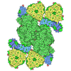
- EMDB-76459: Cryo-EM density of the [NiFe]-hydrogenase HoxEFU diaphorase subcomplex -
+ Open data

- Basic information
| Entry |  | |||||||||
|---|---|---|---|---|---|---|---|---|---|---|
| Title | Cryo-EM density of the [NiFe]-hydrogenase HoxEFU diaphorase subcomplex | |||||||||
 Map data Map data | ||||||||||
 Sample Sample |
| |||||||||
 Keywords Keywords | diaphorase module / electron transfer complex / iron-sulfur cluster protein / [NiFe]-hydrogenase subcomplex / OXIDOREDUCTASE | |||||||||
| Function / homology |  Function and homology information Function and homology informationNADH dehydrogenase (ubiquinone) activity / 2 iron, 2 sulfur cluster binding / FMN binding / 4 iron, 4 sulfur cluster binding / oxidoreductase activity / metal ion binding / membrane Similarity search - Function | |||||||||
| Biological species |   Synechocystis sp. PCC 6803 (bacteria) Synechocystis sp. PCC 6803 (bacteria) | |||||||||
| Method | single particle reconstruction / cryo EM / Resolution: 4.3 Å | |||||||||
 Authors Authors | Ziegler SJ / Gruber JN | |||||||||
| Funding support |  United States, 1 items United States, 1 items
| |||||||||
 Citation Citation | Journal: Cell Rep Phys Sci / Year: 2026 Title: Cryo-EM map and functional electron transport studies of the HoxEFU sub-complex of the HOX [NiFe]-hydrogenase from Synechocystis sp. PCC 6803 Authors: Dawson ME / Kisgeropoulos EC / Gruber JN / Ziegler SJ / Bharadwaj VS / Dahl PJ / Blahut MR / Wiley SA / Artz JH / Lubner CE / Mulder DW / King PW | |||||||||
| History |
|
- Structure visualization
| Supplemental images |
|---|

- Downloads & links
-EMDB archive
| Map data | emd_76459.map.gz | 61.8 MB | EMDB map data format | |
|---|---|---|---|---|
| Header (meta data) | emd-76459-v30.xmlemd-76459.xml | 23.9 KB 23.9 KB | Display Display | EMDB header |
| FSC (resolution estimation) | emd_76459_fsc.xml | 10.6 KB | Display | FSC data file |
| Images |  emd_76459.png emd_76459.png | 31.1 KB | ||
| Masks | emd_76459_msk_1.map | 125 MB | Mask map | |
| Filedesc metadata | emd-76459.cif.gz | 6.3 KB | ||
| Others | emd_76459_additional_1.map.gzemd_76459_half_map_1.map.gzemd_76459_half_map_2.map.gz | 117.7 MB 115.8 MB 115.8 MB | ||
| Archive directory |  https://data.pdbj.org/pub/emdb/structures/EMD-76459ftp://data.pdbj.org/pub/emdb/structures/EMD-76459 https://data.pdbj.org/pub/emdb/structures/EMD-76459ftp://data.pdbj.org/pub/emdb/structures/EMD-76459 | HTTPS FTP |
-Related structure data
| Similar structure data |
|---|
-Links
| EMDB pages | EMDB (EBI/PDBe) / EMDataResource |
|---|---|
| Related items in Molecule of the Month |


-Map
| File | Download / File: emd_76459.map.gz / Format: CCP4 / Size: 125 MB / Type: IMAGE STORED AS FLOATING POINT NUMBER (4 BYTES) | ||||||||||||||||||||||||||||||||||||
|---|---|---|---|---|---|---|---|---|---|---|---|---|---|---|---|---|---|---|---|---|---|---|---|---|---|---|---|---|---|---|---|---|---|---|---|---|---|
| Projections & slices | Image control
Images are generated by Spider. | ||||||||||||||||||||||||||||||||||||
| Voxel size | X=Y=Z: 0.8211 Å | ||||||||||||||||||||||||||||||||||||
| Density |
| ||||||||||||||||||||||||||||||||||||
| Symmetry | Space group: 1 | ||||||||||||||||||||||||||||||||||||
| Details | EMDB XML:
|
 Z (Sec.)
 Y (Row.)
 X (Col.)


-Supplemental data
-Mask #1
| File | emd_76459_msk_1.map | ||||||||||||
|---|---|---|---|---|---|---|---|---|---|---|---|---|---|
| Projections & Slices |
| ||||||||||||
| Density Histograms |


-Additional map: #1
| File | emd_76459_additional_1.map | ||||||||||||
|---|---|---|---|---|---|---|---|---|---|---|---|---|---|
| Projections & Slices |
| ||||||||||||
| Density Histograms |


-Half map: #2
| File | emd_76459_half_map_1.map | ||||||||||||
|---|---|---|---|---|---|---|---|---|---|---|---|---|---|
| Projections & Slices |
| ||||||||||||
| Density Histograms |


-Half map: #1
| File | emd_76459_half_map_2.map | ||||||||||||
|---|---|---|---|---|---|---|---|---|---|---|---|---|---|
| Projections & Slices |
| ||||||||||||
| Density Histograms |


- Sample components
-Entire : [NiFe]-hydrogenase HoxEFU diaphorase subcomplex
| Entire | Name: [NiFe]-hydrogenase HoxEFU diaphorase subcomplex |
|---|---|
| Components |
|
-Supramolecule #1: [NiFe]-hydrogenase HoxEFU diaphorase subcomplex
| Supramolecule | Name: [NiFe]-hydrogenase HoxEFU diaphorase subcomplex / type: complex / ID: 1 / Parent: 0 / Macromolecule list: all |
|---|
-Supramolecule #2: [NiFe]-hydrogenase HoxE subunit
| Supramolecule | Name: [NiFe]-hydrogenase HoxE subunit / type: complex / ID: 2 / Parent: 1 / Macromolecule list: #1 |
|---|---|
| Source (natural) | Organism: Synechocystis sp. PCC 6803 (bacteria) |
-Supramolecule #3: [NiFe]-hydrogenase HoxF subunit
| Supramolecule | Name: [NiFe]-hydrogenase HoxF subunit / type: complex / ID: 3 / Parent: 1 / Macromolecule list: #2 |
|---|---|
| Source (natural) | Organism: Synechocystis sp. PCC 6803 (bacteria) |
-Supramolecule #4: [NiFe]-hydrogenase HoxU subunit
| Supramolecule | Name: [NiFe]-hydrogenase HoxU subunit / type: complex / ID: 4 / Parent: 1 / Macromolecule list: #3 |
|---|---|
| Source (natural) | Organism: Synechocystis sp. PCC 6803 (bacteria) |
-Macromolecule #1: [NiFe]-hydrogenase HoxE subunit
| Macromolecule | Name: [NiFe]-hydrogenase HoxE subunit / type: protein_or_peptide / ID: 1 / Enantiomer: LEVO |
|---|---|
| Source (natural) | Organism: Synechocystis sp. PCC 6803 (bacteria) |
| Recombinant expression | Organism:  Escherichia coli BL21(DE3) (bacteria) Escherichia coli BL21(DE3) (bacteria) |
| Sequence | String: MTVATDRQTV PPSAAHPSGD KRFKVLDATM KRNQFNQDAL IEILHKAQEI FGYLEEDVLL YVARGLKLPL SRVFGVATFY HLFSLKPSGK HTCVVCLGTA CYVKGAGDLL KTLDQEVHLK PGETTEDGQM SLVTARCIGA CGIAPAVVYD GKVLGKQNDE AVLAAIQPWL SNS UniProtKB: Potential NAD-reducing hydrogenase subunit |
-Macromolecule #2: [NiFe]-hydrogenase HoxF subunit
| Macromolecule | Name: [NiFe]-hydrogenase HoxF subunit / type: protein_or_peptide / ID: 2 / Enantiomer: LEVO |
|---|---|
| Source (natural) | Organism: Synechocystis sp. PCC 6803 (bacteria) |
| Recombinant expression | Organism: Escherichia coli BL21(DE3) (bacteria) |
| Sequence | String: MDIKELKEIA TKSREKQTKI RIRCCSAAGC LSSEGETVKK NLTTAIAAAG LEEKVEVCGV GCMKFCGRGP LVAVDDRNQL YEFVTPDQVG DIVKKLQKPD AVAETGLISG DPHHPFYALQ RNIALENSGR IDPESIDEYI ALGGYEQLHK VVYEMTPEEV IVEMNKSGLR ...String: MDIKELKEIA TKSREKQTKI RIRCCSAAGC LSSEGETVKK NLTTAIAAAG LEEKVEVCGV GCMKFCGRGP LVAVDDRNQL YEFVTPDQVG DIVKKLQKPD AVAETGLISG DPHHPFYALQ RNIALENSGR IDPESIDEYI ALGGYEQLHK VVYEMTPEEV IVEMNKSGLR GRGGGGYPTG LKWATVAKMP GQQKYVICNA DEGDPGAFMD RSVLESDPHR ILEGMAIAAY AVGANHGYIY VRAEYPLAIQ RLQKAIQQAK RYGLMGTQIF DSPIDFKIDI RVGAGAFVCG EETALIASVE GKRGTPRPR PPYPAQSGLW QSPTLINNVE TYANVVPIIR EGGDWYGSIG TEKSKGTKVF ALTGKVENAG LIEVPMGTTV RQVVEEMGGG VPNGGQVKAV QTGGPSGGCI PADKLDTPIE YDTLLALGTM MGSGGMIVMD ESTNMVDVAQ FYMDFCKSES CGKCIPCRAG TVQLYDLLTR FLEGEATQED LIKLENLCHM VKETSLCGLG MSAPNPVIST LRYFRHEYEE LLKV UniProtKB: Hydrogenase subunit |
-Macromolecule #3: [NiFe]-hydrogenase HoxU subunit
| Macromolecule | Name: [NiFe]-hydrogenase HoxU subunit / type: protein_or_peptide / ID: 3 / Enantiomer: LEVO |
|---|---|
| Source (natural) | Organism: Synechocystis sp. PCC 6803 (bacteria) |
| Recombinant expression | Organism: Escherichia coli BL21(DE3) (bacteria) |
| Sequence | String: MSVVTLTIDD KAIAIEEGAS ILQAAKEAGV PIPTLCHLEG ISEAAACRLC MVEVEGTNKL MPACVTAVSE EMVVHTNTEK LQNYRRMTVE LLFSEGNHVC AICVANGNCE LQDMAITVGM DHSRFKYQFP KREVDLSHPM FGIDHNRCIL CTRCVRVCDE IEGAHVWDVA ...String: MSVVTLTIDD KAIAIEEGAS ILQAAKEAGV PIPTLCHLEG ISEAAACRLC MVEVEGTNKL MPACVTAVSE EMVVHTNTEK LQNYRRMTVE LLFSEGNHVC AICVANGNCE LQDMAITVGM DHSRFKYQFP KREVDLSHPM FGIDHNRCIL CTRCVRVCDE IEGAHVWDVA YRGAECKIVS GLNQPWGTVD ACTSCGKCVD ACPTGSIFHK GETTAEKIGD RRKVEFLATA RKEKEWVR UniProtKB: Hydrogenase subunit |
-Experimental details
-Structure determination
| Method | cryo EM |
|---|---|
 Processing Processing | single particle reconstruction |
| Aggregation state | particle |
-Sample preparation
| Concentration | 0.4 mg/mL | ||||||||||||
|---|---|---|---|---|---|---|---|---|---|---|---|---|---|
| Buffer | pH: 8.3 Component:
| ||||||||||||
| Grid | Model: C-flat-1.2/1.3 / Material: COPPER / Mesh: 400 / Support film - Material: CARBON / Support film - topology: HOLEY | ||||||||||||
| Vitrification | Cryogen name: ETHANE / Chamber humidity: 80 % / Chamber temperature: 298 K / Instrument: GATAN CRYOPLUNGE 3 Details: Grids were blotted for 3.0 seconds before plunging into liquid ethane.. |
- Electron microscopy
| Microscope | TFS KRIOS |
|---|---|
| Image recording | Film or detector model: GATAN K3 (6k x 4k) / Number grids imaged: 1 / Number real images: 3694 / Average electron dose: 65.0 e/Å2 |
| Electron beam | Acceleration voltage: 300 kV / Electron source:  FIELD EMISSION GUN FIELD EMISSION GUN |
| Electron optics | Illumination mode: FLOOD BEAM / Imaging mode: BRIGHT FIELD / Nominal defocus max: 2.5 µm / Nominal defocus min: 1.5 µm / Nominal magnification: 29000 |
| Sample stage | Specimen holder model: FEI TITAN KRIOS AUTOGRID HOLDER / Cooling holder cryogen: NITROGEN |
| Experimental equipment |  Model: Titan Krios / Image courtesy: FEI Company |

