[English] 日本語
Yorodumi
- EMDB-76151: Cryo-EM of Xcp endopilus -

+
Open data


ID or keywords:

Loading...

-
Basic information

Entry
Database: EMDB / ID: EMD-76151
TitleCryo-EM of Xcp endopilus
Map data
Sample
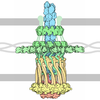
  • Complex: Hxc endo pilus filament
    • Protein or peptide: Type II secretion system core protein G
  • Ligand: CALCIUM ION
Keywordsendopilus / cryo-EM / Xcp / T2SS / PROTEIN FIBRIL
Function / homology
Function and homology information


protein secretion by the type II secretion system / type II protein secretion system complex / plasma membrane
Similarity search - Function
Type II secretion system protein GspG / Type II secretion system protein GspG, C-terminal / Type II secretion system (T2SS), protein G / Bacterial general secretion pathway protein G-type pilin / Prokaryotic N-terminal methylation site. / Prokaryotic N-terminal methylation motif / Prokaryotic N-terminal methylation site / Pilin-like
Similarity search - Domain/homology
Type II secretion system core protein G
Similarity search - Component
Biological speciesPseudomonas aeruginosa PAO1 (bacteria)
Methodhelical reconstruction / cryo EM / Resolution: 4.6 Å
AuthorsSonani RR / Ball G / Chouikha I / Durand E / Voulhoux R / Egelman EH
Funding support United States, 1 items
OrganizationGrant numberCountry
National Institutes of Health/National Institute of General Medical Sciences (NIH/NIGMS)GM122510 United States
CitationJournal: Structure / Year: 2026
Title: Structures of the Xcp and Hxc T2SS endopili provide new insights into type IV pili subfamilies.
Authors: Ravi R Sonani / Iman Chouikha / Geneviève Ball / Mathilde Tribout / Edward H Egelman / Romé Voulhoux /
Abstract: Type IV filaments (T4F) are widespread bacterial appendages involved in adhesion, motility, DNA uptake, and protein secretion. In the type II secretion system (T2SS), a short pilus-like structure ...Type IV filaments (T4F) are widespread bacterial appendages involved in adhesion, motility, DNA uptake, and protein secretion. In the type II secretion system (T2SS), a short pilus-like structure termed the endopilus drives protein export across the outer membrane. Despite its important function, the specific molecular determinants of endopili remain poorly understood. Here, we used cryo-electron microscopy to determine the structures of two endopili assembled by the independent Hxc and Xcp T2SS systems of Pseudomonas aeruginosa. Comparative analyses reveal defining structural and chemical features of T2SS endopili, including a unique surface property of the XcpG endopilus that explains its distinctive bundling behavior. Mapping inter-subunit interactions further identifies residues governing selective assembly within coexisting secretion systems. Comparisons with related type IVa pili define a conserved architectural framework and constrained helical parameters characteristic of the T2SS endopilus family, providing insight into how subtle chemical variations encode functional specificity and efficient protein export.
History
DepositionMar 17, 2026-
Header (metadata) releaseSep 23, 2026-
Map releaseSep 23, 2026-
UpdateSep 23, 2026-
Current statusSep 23, 2026Processing site: RCSB / Status: Released

-
Structure visualization

Supplemental images

Downloads & links

-
Map

FileDownload / File: emd_76151.map.gz / Format: CCP4 / Size: 64 MB / Type: IMAGE STORED AS FLOATING POINT NUMBER (4 BYTES)
Projections & slices

Image control

Size
Brightness
Contrast
Others
AxesZ (Sec.)Y (Row.)X (Col.)
1.07 Å/pix.
x 256 pix.
= 272.64 Å
1.07 Å/pix.
x 256 pix.
= 272.64 Å
1.07 Å/pix.
x 256 pix.
= 272.64 Å

Surface

Projections

Slices (1/3)

Slices (1/2)

Slices (2/3)

Images are generated by Spider.

Voxel sizeX=Y=Z: 1.065 Å
Density
Contour LevelBy AUTHOR: 0.15
Minimum - Maximum-0.7545917 - 0.8142611
Average (Standard dev.)0.0017153055 (±0.03322058)
SymmetrySpace group: 1
Details

EMDB XML:

Map geometry
Axis orderXYZ
Origin000
Dimensions256256256
Spacing256256256
CellA=B=C: 272.64 Å
α=β=γ: 90.0 °

-
Supplemental data

-
Additional map: #1

Fileemd_76151_additional_1.map
Projections & Slices
AxesZYX

Projections

Slices (1/2)
Density Histograms

-
Half map: #1

Fileemd_76151_half_map_1.map
Projections & Slices
AxesZYX

Projections

Slices (1/2)
Density Histograms

-
Half map: #2

Fileemd_76151_half_map_2.map
Projections & Slices
AxesZYX

Projections

Slices (1/2)
Density Histograms

-
Sample components

-
Entire : Hxc endo pilus filament

EntireName: Hxc endo pilus filament
Components
  • Complex: Hxc endo pilus filament
    • Protein or peptide: Type II secretion system core protein G
  • Ligand: CALCIUM ION

-
Supramolecule #1: Hxc endo pilus filament

SupramoleculeName: Hxc endo pilus filament / type: complex / ID: 1 / Parent: 0 / Macromolecule list: #1
Source (natural)Organism: Pseudomonas aeruginosa PAO1 (bacteria)

-
Macromolecule #1: Type II secretion system core protein G

MacromoleculeName: Type II secretion system core protein G / type: protein_or_peptide / ID: 1 / Number of copies: 4 / Enantiomer: LEVO
Source (natural)Organism: Pseudomonas aeruginosa PAO1 (bacteria)
Molecular weightTheoretical: 14.546561 KDa
Recombinant expressionOrganism: Pseudomonas aeruginosa PAK (bacteria)
SequenceString:
GFTLIEIMVV VVILGILAAL VVPQVMSRPD QAKVTVAKGD IKAIAAALDM YKLDNFAYPS TQQGLEALVK KPTGNPQPKN WNKDGYLKK LPVDPWGNPY QYLAPGTKGP FDLYSLGADG KEGGSDNDAD IGNWDN

UniProtKB: Type II secretion system core protein G

-
Macromolecule #2: CALCIUM ION

MacromoleculeName: CALCIUM ION / type: ligand / ID: 2 / Number of copies: 4 / Formula: CA
Molecular weightTheoretical: 40.078 Da

-
Experimental details

-
Structure determination

Methodcryo EM
Processinghelical reconstruction
Aggregation statehelical array

-
Sample preparation

BufferpH: 7
VitrificationCryogen name: ETHANE

-
Electron microscopy

MicroscopeTFS KRIOS
Image recordingFilm or detector model: GATAN K3 BIOQUANTUM (6k x 4k) / Average electron dose: 50.0 e/Å2
Electron beamAcceleration voltage: 300 kV / Electron source: FIELD EMISSION GUN
Electron opticsIllumination mode: FLOOD BEAM / Imaging mode: BRIGHT FIELD / Nominal defocus max: 2.4 µm / Nominal defocus min: 0.8 µm
Experimental equipment
Model: Titan Krios / Image courtesy: FEI Company

+
Image processing

Final reconstructionApplied symmetry - Helical parameters - Δz: 83.2 Å
Applied symmetry - Helical parameters - Δ&Phi: 10.42 °
Applied symmetry - Helical parameters - Axial symmetry: C1 (asymmetric)
Resolution.type: BY AUTHOR / Resolution: 4.6 Å / Resolution method: FSC 0.143 CUT-OFF / Software - Name: cryoSPARC / Number images used: 31076
CTF correctionType: PHASE FLIPPING AND AMPLITUDE CORRECTION
Startup modelType of model: OTHER
Final angle assignmentType: NOT APPLICABLE
FSC plot (resolution estimation)

+
About Yorodumi

-
News

-
Feb 9, 2022. New format data for meta-information of EMDB entries

New format data for meta-information of EMDB entries

  • Version 3 of the EMDB header file is now the official format.
  • The previous official version 1.9 will be removed from the archive.

Related info.:EMDB header

External links:wwPDB to switch to version 3 of the EMDB data model

-
Aug 12, 2020. Covid-19 info

Covid-19 info

URL: https://pdbj.org/emnavi/covid19.php

New page: Covid-19 featured information page in EM Navigator.

Related info.:Covid-19 info / Mar 5, 2020. Novel coronavirus structure data

+
Mar 5, 2020. Novel coronavirus structure data

Novel coronavirus structure data

Related info.:Yorodumi Speices / Aug 12, 2020. Covid-19 info

External links:COVID-19 featured content - PDBj / Molecule of the Month (242):Coronavirus Proteases

+
Jan 31, 2019. EMDB accession codes are about to change! (news from PDBe EMDB page)

EMDB accession codes are about to change! (news from PDBe EMDB page)

  • The allocation of 4 digits for EMDB accession codes will soon come to an end. Whilst these codes will remain in use, new EMDB accession codes will include an additional digit and will expand incrementally as the available range of codes is exhausted. The current 4-digit format prefixed with “EMD-” (i.e. EMD-XXXX) will advance to a 5-digit format (i.e. EMD-XXXXX), and so on. It is currently estimated that the 4-digit codes will be depleted around Spring 2019, at which point the 5-digit format will come into force.
  • The EM Navigator/Yorodumi systems omit the EMD- prefix.

Related info.:Q: What is EMD? / ID/Accession-code notation in Yorodumi/EM Navigator

External links:EMDB Accession Codes are Changing Soon! / Contact to PDBj

+
Jul 12, 2017. Major update of PDB

Major update of PDB

  • wwPDB released updated PDB data conforming to the new PDBx/mmCIF dictionary.
  • This is a major update changing the version number from 4 to 5, and with Remediation, in which all the entries are updated.
  • In this update, many items about electron microscopy experimental information are reorganized (e.g. em_software).
  • Now, EM Navigator and Yorodumi are based on the updated data.

External links:wwPDB Remediation / Enriched Model Files Conforming to OneDep Data Standards Now Available in the PDB FTP Archive

-
Yorodumi

Thousand views of thousand structures

  • Yorodumi is a browser for structure data from EMDB, PDB, SASBDB, etc.
  • This page is also the successor to EM Navigator detail page, and also detail information page/front-end page for Omokage search.
  • The word "yorodu" (or yorozu) is an old Japanese word meaning "ten thousand". "mi" (miru) is to see.

Related info.:EMDB / PDB / SASBDB / Comparison of 3 databanks / Yorodumi Search / Aug 31, 2016. New EM Navigator & Yorodumi / Yorodumi Papers / Jmol/JSmol / Function and homology information / Changes in new EM Navigator and Yorodumi

Read more