guanosine tetraphosphate binding / negative regulation of cytoplasmic translational initiation / transcriptional attenuation / endoribonuclease inhibitor activity / positive regulation of ribosome biogenesis / RNA-binding transcription regulator activity / negative regulation of cytoplasmic translation / translation repressor activity / translation initiation factor activity / negative regulation of translational initiation ...guanosine tetraphosphate binding / negative regulation of cytoplasmic translational initiation / transcriptional attenuation / endoribonuclease inhibitor activity / positive regulation of ribosome biogenesis / RNA-binding transcription regulator activity / negative regulation of cytoplasmic translation / translation repressor activity / translation initiation factor activity / negative regulation of translational initiation / negative regulation of DNA-templated DNA replication initiation / mRNA regulatory element binding translation repressor activity / cytosolic ribosome assembly / ribosome assembly / assembly of large subunit precursor of preribosome / transcription antitermination / regulation of cell growth / translational initiation / DNA-templated transcription termination / response to radiation / maintenance of translational fidelity / mRNA 5'-UTR binding / transferase activity / large ribosomal subunit / ribosomal small subunit assembly / ribosome binding / ribosomal small subunit biogenesis / small ribosomal subunit rRNA binding / 5S rRNA binding / ribosomal large subunit assembly / small ribosomal subunit / cytosolic small ribosomal subunit / large ribosomal subunit rRNA binding / tRNA binding / cytosolic large ribosomal subunit / cytoplasmic translation / rRNA binding / negative regulation of translation / ribonucleoprotein complex / ribosome / translation / structural constituent of ribosome / response to antibiotic / negative regulation of DNA-templated transcription / mRNA binding / GTPase activity / GTP binding / DNA binding / RNA binding / metal ion binding / zinc ion binding / membrane / cytosol / cytoplasm Similarity search - Function
Initiation factor 2 associated domain, bacterial / Bacterial translation initiation factor IF-2 associated region / Translation initiation factor IF-2, N-terminal / Translation initiation factor IF-2, N-terminal region / Translation initiation factor IF-1 / Translation initiation factor IF-2, domain II / Initiation factor 2 signature. / Translation initiation factor IF-2, bacterial-like / : / Elongation factor G domain 2 ...Initiation factor 2 associated domain, bacterial / Bacterial translation initiation factor IF-2 associated region / Translation initiation factor IF-2, N-terminal / Translation initiation factor IF-2, N-terminal region / Translation initiation factor IF-1 / Translation initiation factor IF-2, domain II / Initiation factor 2 signature. / Translation initiation factor IF-2, bacterial-like / : / Elongation factor G domain 2 / Translation initiation factor IF- 2, domain 3 / Translation-initiation factor 2 / Translation initiation factor IF- 2 / Translation initiation factor IF-2, domain 3 superfamily / RNA-binding domain, S1, IF1 type / Translation initiation factor 1A / IF-1 / S1 domain IF1 type profile. / Putative DNA-binding domain superfamily / : / Ribosomal protein uL15, N-terminal region / Translation elongation factor EFTu-like, domain 2 / Ribosomal protein S21, conserved site / Elongation factor Tu domain 2 / Ribosomal protein S21 signature. / Ribosomal protein L25, short-form / Ribosomal protein S14, bacterial/plastid / Ribosomal protein L31 type A / Ribosomal protein S1-like RNA-binding domain / Translational (tr)-type GTP-binding domain / Elongation factor Tu GTP binding domain / Translational (tr)-type guanine nucleotide-binding (G) domain profile. / Ribosomal protein S16, conserved site / Ribosomal protein S16 signature. / Ribosomal protein S21 superfamily / Ribosomal protein S21 / S1 domain / Ribosomal protein L31 signature. / Ribosomal protein L31 / Ribosomal protein L31 superfamily / Ribosomal protein L31 / Ribosomal protein S21 / Ribosomal protein L9 signature. / Ribosomal protein L16 signature 1. / Ribosomal protein L21, conserved site / Ribosomal protein L21 signature. / Ribosomal protein L9, bacteria/chloroplast / Ribosomal protein L6, conserved site / Ribosomal protein L9, C-terminal / Ribosomal protein L9, C-terminal domain / Ribosomal protein L6 signature 1. / Ribosomal protein L9, C-terminal domain superfamily / : / Ribosomal protein L16 signature 2. / Ribosomal protein L16, conserved site / Ribosomal protein L17 signature. / Ribosomal L25p family / Ribosomal protein L25 / Ribosomal protein L25/Gln-tRNA synthetase, N-terminal / Ribosomal protein L36 signature. / Ribosomal protein L25/Gln-tRNA synthetase, anti-codon-binding domain superfamily / : / Ribosomal protein L33, conserved site / Ribosomal protein L33 signature. / Ribosomal protein L28/L24 superfamily / Ribosomal protein L32p, bacterial type / Ribosomal protein L35, conserved site / Ribosomal protein L35 signature. / Ribosomal protein L9 / Ribosomal protein L9, N-terminal domain superfamily / Ribosomal protein L9, N-terminal / Ribosomal protein L9, N-terminal domain / Ribosomal protein L28 / Ribosomal protein L35, non-mitochondrial / Ribosomal protein L18, bacterial-type / Ribosomal protein S6, conserved site / Ribosomal protein S6 signature. / Ribosomal protein S3, bacterial-type / : / Ribosomal protein S13, bacterial-type / Ribosomal protein S19, bacterial-type / Ribosomal protein L6, bacterial-type / Ribosomal protein S7, bacterial/organellar-type / Ribosomal protein S11, bacterial-type / Ribosomal protein L5, bacterial-type / Ribosomal protein L9/RNase H1, N-terminal / Ribosomal protein S20 / Ribosomal protein S20 superfamily / Ribosomal protein S20 / Ribosomal protein L19, conserved site / Ribosomal protein L19 signature. / Ribosomal protein S4, bacterial-type / Ribosomal protein S5, bacterial-type / 30S ribosomal protein S17 / : / Ribosomal protein S6, plastid/chloroplast / Ribosomal protein L20 signature. / Ribosomal protein L36 / Ribosomal protein L36 superfamily / Ribosomal protein L36 / Ribosomal protein L34, conserved site Similarity search - Domain/homology
Small ribosomal subunit protein bS18 / Large ribosomal subunit protein bL36 / Small ribosomal subunit protein bS21 / Small ribosomal subunit protein uS12 / Large ribosomal subunit protein bL28 / Small ribosomal subunit protein uS14 / Small ribosomal subunit protein uS13 / Translation initiation factor IF-1 / Translation initiation factor IF-2 / Small ribosomal subunit protein uS11 ...Small ribosomal subunit protein bS18 / Large ribosomal subunit protein bL36 / Small ribosomal subunit protein bS21 / Small ribosomal subunit protein uS12 / Large ribosomal subunit protein bL28 / Small ribosomal subunit protein uS14 / Small ribosomal subunit protein uS13 / Translation initiation factor IF-1 / Translation initiation factor IF-2 / Small ribosomal subunit protein uS11 / Large ribosomal subunit protein uL16 / Large ribosomal subunit protein bL20 / Large ribosomal subunit protein bL34 / Small ribosomal subunit protein bS6 / Large ribosomal subunit protein bL33 / Small ribosomal subunit protein uS10 / Large ribosomal subunit protein uL3 / Large ribosomal subunit protein uL23 / Small ribosomal subunit protein uS19 / Large ribosomal subunit protein uL29 / Small ribosomal subunit protein uS17 / Large ribosomal subunit protein uL24 / Small ribosomal subunit protein uS8 / Large ribosomal subunit protein uL30 / Small ribosomal subunit protein uS9 / Large ribosomal subunit protein bL21 / Small ribosomal subunit protein uS15 / Small ribosomal subunit protein bS16 / Large ribosomal subunit protein bL32 / Small ribosomal subunit protein bS20 / Small ribosomal subunit protein uS2 / Small ribosomal subunit protein uS7 / Large ribosomal subunit protein bL19 / Large ribosomal subunit protein bL27 / Large ribosomal subunit protein bL31 / Large ribosomal subunit protein bL35 / Large ribosomal subunit protein bL9 / Small ribosomal subunit protein uS3 / Small ribosomal subunit protein uS4 / Small ribosomal subunit protein uS5 / Large ribosomal subunit protein uL13 / Large ribosomal subunit protein uL14 / Large ribosomal subunit protein bL17 / Large ribosomal subunit protein uL6 / Large ribosomal subunit protein uL18 / Large ribosomal subunit protein uL2 / Large ribosomal subunit protein uL4 / Large ribosomal subunit protein uL22 / Large ribosomal subunit protein uL5 / Large ribosomal subunit protein bL25 / Large ribosomal subunit protein uL15 Similarity search - Component
Biological species
Escherichia coli (E. coli) / Escherichia phage T4 (virus)
Method
single particle reconstruction / cryo EM / Resolution: 3.0 Å
Model: Quantifoil R2/1 / Material: GOLD / Mesh: 200 / Support film - Material: CARBON / Support film - topology: HOLEY / Pretreatment - Type: GLOW DISCHARGE / Pretreatment - Time: 50 sec. / Pretreatment - Atmosphere: OTHER
Vitrification
Cryogen name: ETHANE / Chamber humidity: 85 % / Chamber temperature: 295 K / Instrument: LEICA EM GP
-
Electron microscopy
Microscope
TFS KRIOS
Specialist optics
Energy filter - Name: GIF Bioquantum / Energy filter - Slit width: 20 eV
Image recording
Film or detector model: GATAN K3 BIOQUANTUM (6k x 4k) / Detector mode: COUNTING / Number grids imaged: 1 / Number real images: 12290 / Average exposure time: 1.68 sec. / Average electron dose: 40.299 e/Å2
Electron beam
Acceleration voltage: 300 kV / Electron source: FIELD EMISSION GUN
In the structure databanks used in Yorodumi, some data are registered as the other names, "COVID-19 virus" and "2019-nCoV". Here are the details of the virus and the list of structure data.
Jan 31, 2019. EMDB accession codes are about to change! (news from PDBe EMDB page)
EMDB accession codes are about to change! (news from PDBe EMDB page)
The allocation of 4 digits for EMDB accession codes will soon come to an end. Whilst these codes will remain in use, new EMDB accession codes will include an additional digit and will expand incrementally as the available range of codes is exhausted. The current 4-digit format prefixed with “EMD-” (i.e. EMD-XXXX) will advance to a 5-digit format (i.e. EMD-XXXXX), and so on. It is currently estimated that the 4-digit codes will be depleted around Spring 2019, at which point the 5-digit format will come into force.
The EM Navigator/Yorodumi systems omit the EMD- prefix.
Related info.:Q: What is EMD? / ID/Accession-code notation in Yorodumi/EM Navigator
Yorodumi is a browser for structure data from EMDB, PDB, SASBDB, etc.
This page is also the successor to EM Navigator detail page, and also detail information page/front-end page for Omokage search.
The word "yorodu" (or yorozu) is an old Japanese word meaning "ten thousand". "mi" (miru) is to see.
Related info.:EMDB / PDB / SASBDB / Comparison of 3 databanks / Yorodumi Search / Aug 31, 2016. New EM Navigator & Yorodumi / Yorodumi Papers / Jmol/JSmol / Function and homology information / Changes in new EM Navigator and Yorodumi
 Movie
Movie Controller

 Yorodumi
 Open data
 Basic information
 Map data
 Sample
 Keywords
 Function and homology information
 Escherichia coli (E. coli) /
  Escherichia phage T4 (virus)
 Authors
 United States,
  Czech Republic, 4 items
  Citation
 Structure visualization
 Downloads & links
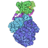
 emd_74431.png
 https://data.pdbj.org/pub/emdb/structures/EMD-74431





 Z (Sec.)
 Y (Row.)
 X (Col.)


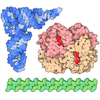
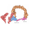
 Sample components

 Processing
 Electron microscopy
 FIELD EMISSION GUN

