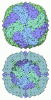
- EMDB-73901: Human Ferritin Heavy Chain in the presence of Mg-ATP -
+
Open data
ID or keywords:
Loading...
-
Basic information
Entry
Database: EMDB / ID: EMD-73901
Title
Human Ferritin Heavy Chain in the presence of Mg-ATP
Map data
Human Ferritin Heavy Chain in the presence of Mg-ATP - Sharpened map
Sample
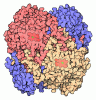
Complex: Human Heavy Chain Ferritin
Protein or peptide: Ferritin heavy chain
Ligand: water
Keywords
iron storage / METAL BINDING PROTEIN
Function / homology
Function and homology information
iron ion sequestering activity / ferritin complex / Scavenging by Class A Receptors / Golgi Associated Vesicle Biogenesis / ferroxidase / negative regulation of ferroptosis / ferroxidase activity / autolysosome / negative regulation of fibroblast proliferation / ferric iron binding ...iron ion sequestering activity / ferritin complex / Scavenging by Class A Receptors / Golgi Associated Vesicle Biogenesis / ferroxidase / negative regulation of ferroptosis / ferroxidase activity / autolysosome / negative regulation of fibroblast proliferation / ferric iron binding / autophagosome / iron ion transport / ferrous iron binding / Iron uptake and transport / tertiary granule lumen / ficolin-1-rich granule lumen / intracellular iron ion homeostasis / immune response / iron ion binding / negative regulation of cell population proliferation / Neutrophil degranulation / extracellular exosome / extracellular region / identical protein binding / nucleus / cytosol / cytoplasm Similarity search - Function
Journal: Int J Biol Macromol / Year: 2026 Title: Ferritin iron uptake and oxidation are dynamically modulated by nucleotide phosphate architecture via electrostatic gating. Authors: Anitha Rajendran / Sean Henley / Brent L Nannenga / Genki Terashi / Ayush Srivastava / Daisuke Kihara / Fadi Bou-Abdallah / Abstract: Ferritin safeguards cells from iron-induced oxidative stress by oxidizing and storing Fe within its nanocage, yet how its macromolecular architecture enables responsiveness to the cellular chemical ...Ferritin safeguards cells from iron-induced oxidative stress by oxidizing and storing Fe within its nanocage, yet how its macromolecular architecture enables responsiveness to the cellular chemical environment remains unclear. Here, we show that ferritin's iron-oxidation activity is modulated by an electrostatic gating mechanism centered at its 3-fold channels and sensitive to solution charge conditions representative of intracellular metabolites. At physiologically relevant nucleotide concentrations, ferritin-catalyzed Fe oxidation is strongly attenuated in the presence of triphosphate nucleotides, while diphosphates and monophosphates exert progressively weaker effects, indicating that ferritin responds selectively to the charge density and geometry of the phosphate chain, rather than nucleotide identity. High-resolution cryo-electron microscopy identifies condition-dependent differences in non-protein density within and near the ferritin 3-fold channels, consistent with changes in the local solvent and/or ion environment, rather than discrete ligand binding. Fluorescence and calorimetric measurements reveal weak, reversible nucleotide association (K ∼ 1 mM), supporting a low-affinity, dynamic electrostatic interaction mode. The inhibitory trend persists under reduced oxygen conditions and across ferritin assemblies with varying H/L composition, supporting physiological relevance across cellular oxygen tensions and native ferritin heteropolymers. Ferritin activity is similarly modulated in bacterial, yeast, and human cell lysates under near-physiological conditions, demonstrating the robustness of this behavior in complex environments. Together, these findings establish ferritin as a biological macromolecule whose intrinsic channel electrostatics enable reversible modulation of iron uptake and oxidation in response to its chemical environment.
In the structure databanks used in Yorodumi, some data are registered as the other names, "COVID-19 virus" and "2019-nCoV". Here are the details of the virus and the list of structure data.
Jan 31, 2019. EMDB accession codes are about to change! (news from PDBe EMDB page)
EMDB accession codes are about to change! (news from PDBe EMDB page)
The allocation of 4 digits for EMDB accession codes will soon come to an end. Whilst these codes will remain in use, new EMDB accession codes will include an additional digit and will expand incrementally as the available range of codes is exhausted. The current 4-digit format prefixed with “EMD-” (i.e. EMD-XXXX) will advance to a 5-digit format (i.e. EMD-XXXXX), and so on. It is currently estimated that the 4-digit codes will be depleted around Spring 2019, at which point the 5-digit format will come into force.
The EM Navigator/Yorodumi systems omit the EMD- prefix.
Related info.:Q: What is EMD? / ID/Accession-code notation in Yorodumi/EM Navigator
Yorodumi is a browser for structure data from EMDB, PDB, SASBDB, etc.
This page is also the successor to EM Navigator detail page, and also detail information page/front-end page for Omokage search.
The word "yorodu" (or yorozu) is an old Japanese word meaning "ten thousand". "mi" (miru) is to see.
Related info.:EMDB / PDB / SASBDB / Comparison of 3 databanks / Yorodumi Search / Aug 31, 2016. New EM Navigator & Yorodumi / Yorodumi Papers / Jmol/JSmol / Function and homology information / Changes in new EM Navigator and Yorodumi
 Movie
Movie Controller


 Open data
 Basic information
 Map data
 Sample
 Keywords
 Function and homology information
 Homo sapiens (human)
 Authors
 United States, 2 items
  Citation
 Structure visualization
 Downloads & links
 emd_73901.png
 https://data.pdbj.org/pub/emdb/structures/EMD-73901
 Z (Sec.)
 Y (Row.)
 X (Col.)


 Sample components
 Escherichia coli B (bacteria)
 Processing
 Electron microscopy
 FIELD EMISSION GUN

