 Movie
Movie Controller

[English] 日本語
 Yorodumi
- EMDB-73706: Trypanosoma brucei mitochondrial RNA-editing catalytic complex 2,... -
+ Open data

- Basic information
| Entry |  | ||||||||||||||||||||||||
|---|---|---|---|---|---|---|---|---|---|---|---|---|---|---|---|---|---|---|---|---|---|---|---|---|---|
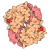
| Title | Trypanosoma brucei mitochondrial RNA-editing catalytic complex 2, U-insertion (RECC2), composite map | ||||||||||||||||||||||||
 Map data Map data | RNA-editing catalytic complex 2 (RECC2), composite map | ||||||||||||||||||||||||
 Sample Sample |
| ||||||||||||||||||||||||
 Keywords Keywords | RNA editing / tRNA / OB-fold / mitochondria / RNA BINDING PROTEIN | ||||||||||||||||||||||||
| Function / homology |  Function and homology information Function and homology informationmRNA editing complex / RNA nucleotide insertion / RNA nucleotide deletion / RNA modification / mitochondrial mRNA editing complex / RNA endonuclease activity producing 5'-phosphomonoesters, hydrolytic mechanism / mitochondrial RNA modification / kinetoplast / alpha-catenin binding / ribonuclease III activity ...mRNA editing complex / RNA nucleotide insertion / RNA nucleotide deletion / RNA modification / mitochondrial mRNA editing complex / RNA endonuclease activity producing 5'-phosphomonoesters, hydrolytic mechanism / mitochondrial RNA modification / kinetoplast / alpha-catenin binding / ribonuclease III activity / response to metal ion / mRNA modification / RNA processing / single-stranded DNA binding / 3'-5'-RNA exonuclease activity / mitochondrion / RNA binding / zinc ion binding / cytoplasm Similarity search - Function | ||||||||||||||||||||||||
| Biological species |   Trypanosoma brucei (eukaryote) Trypanosoma brucei (eukaryote) | ||||||||||||||||||||||||
| Method | single particle reconstruction / cryo EM / Resolution: 2.79 Å | ||||||||||||||||||||||||
 Authors Authors | Liu YT / Jih J / Zhou ZH / Aphasizhev R | ||||||||||||||||||||||||
| Funding support |  United States, United States,  China, 7 items China, 7 items
| ||||||||||||||||||||||||
 Citation Citation | Journal: Nature / Year: 2026 Title: Structural basis of the RNA-editing cascade in trypanosome mitochondria. Authors: Yun-Tao Liu / Andres F Vacas / Jonathan Jih / Xiaojing Zhao / Clinton Yu / Jane K J Lee / Takuma Suematsu / Md Solayman / Hong Wang / Xiaorong Wang / Lan Huang / Liye Zhang / Inna ...Authors: Yun-Tao Liu / Andres F Vacas / Jonathan Jih / Xiaojing Zhao / Clinton Yu / Jane K J Lee / Takuma Suematsu / Md Solayman / Hong Wang / Xiaorong Wang / Lan Huang / Liye Zhang / Inna Aphasizheva / Z Hong Zhou / Ruslan Aphasizhev / Abstract: The molecular mechanism of uridine insertion-and-deletion mRNA editing in trypanosome mitochondria has remained unclear because of the highly dynamic nature of the underlying multi-enzyme machinery. ...The molecular mechanism of uridine insertion-and-deletion mRNA editing in trypanosome mitochondria has remained unclear because of the highly dynamic nature of the underlying multi-enzyme machinery. Here, we define editosomes as supramolecular assemblies formed by the RNA-editing substrate-binding complex (RESC) and either RNA-editing catalytic complex 1 or 2 (RECC1 or RECC2), and present cryo-electron microscopy structures of the approximately 1-MDa RECC1 and RECC2. Resembling dragonflies, with a head, thorax-like core, tail and wings, these ribonucleoproteins mediate the uridine deletion and uridine insertion cascades, respectively. In each RECC, a tetrameric core containing one active and three inactive RNase III domains captures the guide RNA (gRNA)-mRNA duplex, while auxiliary zinc fingers distinguish deletion sites from insertion sites and position the substrate for mRNA cleavage (step I). Three peripheral oligonucleotide-binding-fold heterotetramers are flexibly attached to the core, forming a spatially adaptable reaction chamber. The tail recruits the exonuclease and uridylyltransferase that remove or add uridines (step II), whereas the wings, coordinated by an architectural tRNA, position RNA ligases to seal the edited mRNA (step III). Together, these structures reveal how gRNA-directed substrate recognition, mRNA cleavage, uridine deletion and insertion and ligation are integrated in a single macromolecular machine. This architecture defines the mechanism of information transfer in RNA editing. | ||||||||||||||||||||||||
| History |
|
- Structure visualization
| Supplemental images |
|---|

- Downloads & links
-EMDB archive
| Map data | emd_73706.map.gz | 195.2 MB | EMDB map data format | |
|---|---|---|---|---|
| Header (meta data) | emd-73706-v30.xmlemd-73706.xml | 40.3 KB 40.3 KB | Display Display | EMDB header |
| Images |  emd_73706.png emd_73706.png | 91.2 KB | ||
| Filedesc metadata | emd-73706.cif.gz | 9.9 KB | ||
| Archive directory |  https://data.pdbj.org/pub/emdb/structures/EMD-73706ftp://data.pdbj.org/pub/emdb/structures/EMD-73706 https://data.pdbj.org/pub/emdb/structures/EMD-73706ftp://data.pdbj.org/pub/emdb/structures/EMD-73706 | HTTPS FTP |
-Related structure data
| Related structure data |  9z0hMC  9deiC C: citing same article ( M: atomic model generated by this map |
|---|---|
| Similar structure data |


-Links
| EMDB pages | EMDB (EBI/PDBe) / EMDataResource |
|---|---|
| Related items in Molecule of the Month |


-Map
| File | Download / File: emd_73706.map.gz / Format: CCP4 / Size: 216 MB / Type: IMAGE STORED AS FLOATING POINT NUMBER (4 BYTES) | ||||||||||||||||||||||||||||||||||||
|---|---|---|---|---|---|---|---|---|---|---|---|---|---|---|---|---|---|---|---|---|---|---|---|---|---|---|---|---|---|---|---|---|---|---|---|---|---|
| Annotation | RNA-editing catalytic complex 2 (RECC2), composite map | ||||||||||||||||||||||||||||||||||||
| Projections & slices | Image control
Images are generated by Spider. | ||||||||||||||||||||||||||||||||||||
| Voxel size | X=Y=Z: 1.1 Å | ||||||||||||||||||||||||||||||||||||
| Density |
| ||||||||||||||||||||||||||||||||||||
| Symmetry | Space group: 1 | ||||||||||||||||||||||||||||||||||||
| Details | EMDB XML:
|
 X (Sec.)
 Y (Row.)
 Z (Col.)


-Supplemental data
- Sample components
+Entire : RECC2
+Supramolecule #1: RECC2
+Supramolecule #2: RECC2 core
+Supramolecule #3: RECC2 left wing
+Supramolecule #4: RECC2 right wing
+Supramolecule #5: tRNA
+Macromolecule #1: RNA editing complex protein MP44
+Macromolecule #2: KREPB7
+Macromolecule #3: RNA editing complex protein MP61
+Macromolecule #4: RNA editing complex protein MP46
+Macromolecule #5: MP18 RNA editing complex protein, putative
+Macromolecule #6: RNA-editing complex protein MP42
+Macromolecule #7: KREPA2
+Macromolecule #8: RNA editing complex protein
+Macromolecule #9: KREPA5
+Macromolecule #10: RNA-editing complex protein MP81
+Macromolecule #11: tRNA-valine (anticodon AAC)
+Macromolecule #12: ZINC ION
+Macromolecule #13: MAGNESIUM ION
+Macromolecule #14: water

-Experimental details
-Structure determination
| Method | cryo EM |
|---|---|
 Processing Processing | single particle reconstruction |
| Aggregation state | particle |
-Sample preparation
| Buffer | pH: 7.6 Component:
| ||||||||||||||||||
|---|---|---|---|---|---|---|---|---|---|---|---|---|---|---|---|---|---|---|---|
| Grid | Model: Quantifoil R1.2/1.3 / Material: GOLD / Mesh: 300 / Support film - Material: GRAPHENE / Support film - topology: CONTINUOUS / Pretreatment - Type: PLASMA CLEANING / Pretreatment - Time: 300 sec. | ||||||||||||||||||
| Vitrification | Cryogen name: ETHANE / Chamber humidity: 100 % / Chamber temperature: 277 K / Instrument: FEI VITROBOT MARK IV | ||||||||||||||||||
| Details | From T. brucei mitochondrial T2 isolate |
- Electron microscopy #1
| Microscopy ID | 1 |
|---|---|
| Microscope | TFS KRIOS |
| Specialist optics | Energy filter - Name: GIF Bioquantum / Energy filter - Slit width: 20 eV |
| Image recording | Image recording ID: 1 / Film or detector model: GATAN K3 BIOQUANTUM (6k x 4k) / Number real images: 23092 / Average exposure time: 2.0 sec. / Average electron dose: 45.0 e/Å2 |
| Electron beam | Acceleration voltage: 300 kV / Electron source:  FIELD EMISSION GUN FIELD EMISSION GUN |
| Electron optics | C2 aperture diameter: 50.0 µm / Illumination mode: FLOOD BEAM / Imaging mode: BRIGHT FIELD / Cs: 2.7 mm / Nominal defocus max: 3.0 µm / Nominal defocus min: 1.0 µm / Nominal magnification: 81000 |
| Sample stage | Specimen holder model: FEI TITAN KRIOS AUTOGRID HOLDER / Cooling holder cryogen: NITROGEN |
| Experimental equipment |  Model: Titan Krios / Image courtesy: FEI Company |
-Electron microscopy #1~
| Microscopy ID | 1 |
|---|---|
| Microscope | TFS KRIOS |
| Specialist optics | Energy filter - Name: TFS Selectris X / Energy filter - Slit width: 10 eV |
| Image recording | Image recording ID: 2 / Film or detector model: FEI FALCON IV (4k x 4k) / Number real images: 40988 / Average exposure time: 3.5 sec. / Average electron dose: 45.0 e/Å2 |
| Electron beam | Acceleration voltage: 300 kV / Electron source: FIELD EMISSION GUN |
| Electron optics | C2 aperture diameter: 50.0 µm / Illumination mode: FLOOD BEAM / Imaging mode: BRIGHT FIELD / Cs: 2.7 mm / Nominal defocus max: 2.8000000000000003 µm / Nominal defocus min: 0.6 µm / Nominal magnification: 130000 |
| Sample stage | Specimen holder model: FEI TITAN KRIOS AUTOGRID HOLDER / Cooling holder cryogen: NITROGEN |
| Experimental equipment | Model: Titan Krios / Image courtesy: FEI Company |
+Image processing
-Atomic model buiding 1
| Details | Consensus map and focused refinement maps were combined to generate a composite map for final model refinement. |
|---|---|
| Refinement | Space: REAL / Protocol: AB INITIO MODEL |
| Output model | PDB-9z0h: |
