Movie
Movie Controller

[English] 日本語
 Yorodumi
- EMDB-73699: Structure of disulfide-crosslinked S. cerevisiae Hrd1 dimer bound... -
+ Open data

- Basic information
| Entry |  | |||||||||||||||
|---|---|---|---|---|---|---|---|---|---|---|---|---|---|---|---|---|
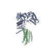
| Title | Structure of disulfide-crosslinked S. cerevisiae Hrd1 dimer bound to one copy of Hrd3 in MSP1D1 nanodisc | |||||||||||||||
 Map data Map data | Structure of a disulfide-crosslinked S. cerevisiae Hrd1 dimer bound to one copy of Hrd3 | |||||||||||||||
 Sample Sample |
| |||||||||||||||
 Keywords Keywords | MEMBRANE PROTEIN / Hrd1 ubiquitin ligase / Hrd3 / MSP1D1 nanodisc | |||||||||||||||
| Function / homology |  Function and homology information Function and homology informationHrd1p ubiquitin ligase ERAD-M complex / detection of unfolded protein / luminal surveillance complex / Hrd1p ubiquitin ligase complex / Hrd1p ubiquitin ligase ERAD-L complex / fungal-type cell wall organization / negative regulation of protein autoubiquitination / retrograde protein transport, ER to cytosol / protein autoubiquitination / endoplasmic reticulum unfolded protein response ...Hrd1p ubiquitin ligase ERAD-M complex / detection of unfolded protein / luminal surveillance complex / Hrd1p ubiquitin ligase complex / Hrd1p ubiquitin ligase ERAD-L complex / fungal-type cell wall organization / negative regulation of protein autoubiquitination / retrograde protein transport, ER to cytosol / protein autoubiquitination / endoplasmic reticulum unfolded protein response / ERAD pathway / protein K48-linked ubiquitination / RING-type E3 ubiquitin transferase / ubiquitin-protein transferase activity / ubiquitin protein ligase activity / ubiquitin-dependent protein catabolic process / endoplasmic reticulum membrane / endoplasmic reticulum / zinc ion binding / identical protein binding Similarity search - Function | |||||||||||||||
| Biological species |  | |||||||||||||||
| Method | single particle reconstruction / cryo EM / Resolution: 3.7 Å | |||||||||||||||
 Authors Authors | Pisa R / Rapoport TA | |||||||||||||||
| Funding support |  United States, 4 items United States, 4 items
| |||||||||||||||
 Citation Citation | Journal: bioarchive / Year: 2025 Title: Lipid bilayer thinning near a ubiquitin ligase selects ER membrane proteins for degradation Authors: Pisa R / Rapoport TA | |||||||||||||||
| History |
|
- Structure visualization
| Supplemental images |
|---|

- Downloads & links
-EMDB archive
| Map data | emd_73699.map.gz | 71.6 MB | EMDB map data format | |
|---|---|---|---|---|
| Header (meta data) | emd-73699-v30.xmlemd-73699.xml | 19.2 KB 19.2 KB | Display Display | EMDB header |
| FSC (resolution estimation) | emd_73699_fsc.xml | 11.1 KB | Display | FSC data file |
| Images |  emd_73699.png emd_73699.png | 73.6 KB | ||
| Filedesc metadata | emd-73699.cif.gz | 6.7 KB | ||
| Others | emd_73699_half_map_1.map.gzemd_73699_half_map_2.map.gz | 134.3 MB 134.3 MB | ||
| Archive directory |  http://ftp.pdbj.org/pub/emdb/structures/EMD-73699ftp://ftp.pdbj.org/pub/emdb/structures/EMD-73699 http://ftp.pdbj.org/pub/emdb/structures/EMD-73699ftp://ftp.pdbj.org/pub/emdb/structures/EMD-73699 | HTTPS FTP |
-Related structure data
| Related structure data |  9z0eMC M: atomic model generated by this map C: citing same article ( |
|---|---|
| Similar structure data |
-Links
| EMDB pages | EMDB (EBI/PDBe) / EMDataResource |
|---|---|
| Related items in Molecule of the Month |


-Map
| File | Download / File: emd_73699.map.gz / Format: CCP4 / Size: 144.7 MB / Type: IMAGE STORED AS FLOATING POINT NUMBER (4 BYTES) | ||||||||||||||||||||
|---|---|---|---|---|---|---|---|---|---|---|---|---|---|---|---|---|---|---|---|---|---|
| Annotation | Structure of a disulfide-crosslinked S. cerevisiae Hrd1 dimer bound to one copy of Hrd3 | ||||||||||||||||||||
| Voxel size | X=Y=Z: 0.857 Å | ||||||||||||||||||||
| Density |
| ||||||||||||||||||||
| Symmetry | Space group: 1 | ||||||||||||||||||||
| Details | EMDB XML:
|
-Supplemental data
-Half map: half map B
| File | emd_73699_half_map_1.map | ||||||||||||
|---|---|---|---|---|---|---|---|---|---|---|---|---|---|
| Annotation | half map B | ||||||||||||
| Projections & Slices |
| ||||||||||||
| Density Histograms |
 Z
Z Y
Y X
X


-Half map: half map A
| File | emd_73699_half_map_2.map | ||||||||||||
|---|---|---|---|---|---|---|---|---|---|---|---|---|---|
| Annotation | half map A | ||||||||||||
| Projections & Slices |
| ||||||||||||
| Density Histograms |


- Sample components
-Entire : Structure of disulfide-crosslinked S. cerevisiae Hrd1 dimer bound...
| Entire | Name: Structure of disulfide-crosslinked S. cerevisiae Hrd1 dimer bound to one copy of Hrd3 in MSP1D1 nanodisc |
|---|---|
| Components |
|
-Supramolecule #1: Structure of disulfide-crosslinked S. cerevisiae Hrd1 dimer bound...
| Supramolecule | Name: Structure of disulfide-crosslinked S. cerevisiae Hrd1 dimer bound to one copy of Hrd3 in MSP1D1 nanodisc type: complex / ID: 1 / Parent: 0 / Macromolecule list: all |
|---|---|
| Source (natural) | Organism: |
-Macromolecule #1: ERAD-associated E3 ubiquitin-protein ligase HRD1
| Macromolecule | Name: ERAD-associated E3 ubiquitin-protein ligase HRD1 / type: protein_or_peptide / ID: 1 / Number of copies: 2 / Enantiomer: LEVO / EC number: RING-type E3 ubiquitin transferase |
|---|---|
| Source (natural) | Organism: |
| Molecular weight | Theoretical: 40.129148 KDa |
| Recombinant expression | Organism: |
| Sequence | String: MVPENRRKQL AIFVVVTYLL TFYCVYSATK TSVSFLQVTL KLNEGFNLMV LSIFILLNST LLWQLLTKLL FGELRLIEHE HIFERLPFT ICNTLFMSSL FHERYFFTVA FFGLLLLYLK VFHWILKDRL EALLQSINDS TTMKTLIFSR FSFNLVLLAV V DYQIITRC ...String: MVPENRRKQL AIFVVVTYLL TFYCVYSATK TSVSFLQVTL KLNEGFNLMV LSIFILLNST LLWQLLTKLL FGELRLIEHE HIFERLPFT ICNTLFMSSL FHERYFFTVA FFGLLLLYLK VFHWILKDRL EALLQSINDS TTMKTLIFSR FSFNLVLLAV V DYQIITRC ISSIYTNQKS DIESTSLYLI QVMEFTMLLI DLLNLFLQTC LNFWEFYRSQ QSLSNENNHI VHGDPTDENT VE SDQSQPV LNDDDDDDDD DRQFTGLEGK FMYEKAIDVF TRFLKTALHL SMLIPFRMPM MLLKDVVWDI LALYQSGTSL WKI WRNNKQ GSGGASGGSG DYKDDDDK UniProtKB: ERAD-associated E3 ubiquitin-protein ligase HRD1 |
-Macromolecule #2: ERAD-associated E3 ubiquitin-protein ligase component HRD3
| Macromolecule | Name: ERAD-associated E3 ubiquitin-protein ligase component HRD3 type: protein_or_peptide / ID: 2 / Number of copies: 1 / Enantiomer: LEVO |
|---|---|
| Source (natural) | Organism: |
| Molecular weight | Theoretical: 93.787172 KDa |
| Recombinant expression | Organism: |
| Sequence | String: MITLLLYLCV ICNAIVLIRA DSIADPWPEA RHLLNTIAKS RDPMKEAAME PNADEFVGFY VPMDYSPRNE EKNYQSIWQN EITDSQRHI YELLVQSSEQ FNNSEATYTL SQIHLWSQYN FPHNMTLAHK YLEKFNDLTH FTNHSAIFDL AVMYATGGCA S GNDQTVIP ...String: MITLLLYLCV ICNAIVLIRA DSIADPWPEA RHLLNTIAKS RDPMKEAAME PNADEFVGFY VPMDYSPRNE EKNYQSIWQN EITDSQRHI YELLVQSSEQ FNNSEATYTL SQIHLWSQYN FPHNMTLAHK YLEKFNDLTH FTNHSAIFDL AVMYATGGCA S GNDQTVIP QDSAKALLYY QRAAQLGNLK AKQVLAYKYY SGFNVPRNFH KSLVLYRDIA EQLRKSYSRD EWDIVFPYWE SY NVRISDF ESGLLGKGLN SVPSSTVRKR TTRPDIGSPF IAQVNGVQMT LQIEPMGRFA FNGNDGNING DEDDEDASER RII RIYYAA LNDYKGTYSQ SRNCERAKNL LELTYKEFQP HVDNLDPLQV FYYVRCLQLL GHMYFTGEGS SKPNIHMAEE ILTT SLEIS RRAQGPIGRA CIDLGLINQY ITNNISQAIS YYMKAMKTQA NNGIVEFQLS KLATSFPEEK IGDPFNLMET AYLNG FIPA IYEFAVMIES GMNSKSSVEN TAYLFKTFVD KNEAIMAPKL RTAFAALIND RSEVALWAYS QLAEQGYETA QVSAAY LMY QLPYEFEDPP RTTDQRKTLA ISYYTRAFKQ GNIDAGVVAG DIYFQMQNYS KAMALYQGAA LKYSIQAIWN LGYMHEH GL GVNRDFHLAK RYYDQVSEHD HRFYLASKLS VLKLHLKSWL TWITREKVNY WKPSSPLNPN EDTQHSKTSW YKQLTKIL Q RMRHKEDSDK AAEDSHKHRT VVQNGANHRG DDQEEASEIL GFQMEDGGGE NLYFQSGGGM DERTTGWRGG HVVEGLAGE LEQLRARLEH HPQGQREP UniProtKB: ERAD-associated E3 ubiquitin-protein ligase component HRD3 |
-Experimental details
-Structure determination
| Method | cryo EM |
|---|---|
 Processing Processing | single particle reconstruction |
| Aggregation state | particle |
-Sample preparation
| Buffer | pH: 7.5 Component:
| |||||||||
|---|---|---|---|---|---|---|---|---|---|---|
| Vitrification | Cryogen name: ETHANE |
- Electron microscopy
| Microscope | TFS KRIOS |
|---|---|
| Image recording | Film or detector model: GATAN K3 BIOCONTINUUM (6k x 4k) / Number grids imaged: 1 / Number real images: 4101 / Average electron dose: 55.0 e/Å2 Details: ---------Data Collection Parameters------------ Voltage 300kv Cs 0.01 mm ac 0.07 Magnification 81kx Physical pixel size 0.857 A Total dose 55 e/A2 50 frames per movie Dose per frame 1.1 e/A2 ...Details: ---------Data Collection Parameters------------ Voltage 300kv Cs 0.01 mm ac 0.07 Magnification 81kx Physical pixel size 0.857 A Total dose 55 e/A2 50 frames per movie Dose per frame 1.1 e/A2 Data collection strategy 3 by 3 + 3 shots per hole Defocus range -0.8um ~ -2.0um Energy filter slit width 10eV |
| Electron beam | Acceleration voltage: 300 kV / Electron source:  FIELD EMISSION GUN FIELD EMISSION GUN |
| Electron optics | Illumination mode: OTHER / Imaging mode: OTHER / Nominal defocus max: 2.0 µm / Nominal defocus min: 0.8 µm |
| Experimental equipment |  Model: Titan Krios / Image courtesy: FEI Company |