[English] 日本語
Yorodumi
- EMDB-72667: Cryo-EM map of Corynebacterium glutamicum pyruvate dehydrogenase ... -

+
Open data


ID or keywords:

Loading...

-
Basic information

Entry
Database: EMDB / ID: EMD-72667
TitleCryo-EM map of Corynebacterium glutamicum pyruvate dehydrogenase complex E2p core in a trimer state
Map data
Sample
  • Complex: trimer of Dihydrolipoyllysine-residue acetyltransferase of Corynebacterium glutamicum
    • Protein or peptide: Dihydrolipoyllysine-residue acetyltransferase component of pyruvate dehydrogenase complex
Keywordstrimer dihydrolipoamide acetyltransferase Corynebacterium glutamicum / TRANSFERASE
Function / homology
Function and homology information


lipoic acid binding / dihydrolipoyllysine-residue acetyltransferase / dihydrolipoyllysine-residue acetyltransferase activity / cytoplasm
Similarity search - Function
2-oxoglutarate dehydrogenase, E2 component / : / Peripheral subunit-binding domain / e3 binding domain / Peripheral subunit-binding (PSBD) domain profile. / E3-binding domain superfamily / 2-oxo acid dehydrogenase, lipoyl-binding site / 2-oxo acid dehydrogenases acyltransferase component lipoyl binding site. / 2-oxoacid dehydrogenase acyltransferase, catalytic domain / 2-oxoacid dehydrogenases acyltransferase (catalytic domain) ...2-oxoglutarate dehydrogenase, E2 component / : / Peripheral subunit-binding domain / e3 binding domain / Peripheral subunit-binding (PSBD) domain profile. / E3-binding domain superfamily / 2-oxo acid dehydrogenase, lipoyl-binding site / 2-oxo acid dehydrogenases acyltransferase component lipoyl binding site. / 2-oxoacid dehydrogenase acyltransferase, catalytic domain / 2-oxoacid dehydrogenases acyltransferase (catalytic domain) / Biotin-requiring enzyme / Biotinyl/lipoyl domain profile. / Biotin/lipoyl attachment / Single hybrid motif / Chloramphenicol acetyltransferase-like domain superfamily
Similarity search - Domain/homology
Dihydrolipoyllysine-residue acetyltransferase component of pyruvate dehydrogenase complex
Similarity search - Component
Biological speciesMycobacterium tuberculosis (bacteria) / Corynebacterium glutamicum (bacteria)
Methodsingle particle reconstruction / cryo EM / Resolution: 4.51 Å
AuthorsHsu HC / Li H
Funding support United States, 1 items
OrganizationGrant numberCountry
National Institutes of Health/National Institute Of Allergy and Infectious Diseases (NIH/NIAID)AI175342 United States
Citation
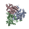
Journal: J Biol Chem / Year: 2026
Title: Mycobacterium tuberculosis assembles a unique hexameric E2p core of the pyruvate dehydrogenase complex.
Authors: Hao-Chi Hsu / Isabelle Bonnet / Ruslana Bryk / Huilin Li /
Abstract: The pyruvate dehydrogenase complex (PDHc) is a universally conserved multienzyme system that converts pyruvate into acetyl-CoA for entry into the TCA cycle and for NADH production. Its central ...The pyruvate dehydrogenase complex (PDHc) is a universally conserved multienzyme system that converts pyruvate into acetyl-CoA for entry into the TCA cycle and for NADH production. Its central scaffold, the dihydrolipoyl transacetylase (E2p), forms an oligomeric inner core that recruits pyruvate dehydrogenase (E1p) and dihydrolipoyl dehydrogenase (E3). All previously characterized PDHc assemblies adopt either an octahedral 24-mer or an icosahedral 60-mer E2p core, each constructed from trimeric building blocks. We recently showed that the Mycobacterium tuberculosis (Mtb) E2p protein DlaT also functions as the core of the pathogen's peroxynitrite reductase/peroxidase (PNR/P) complex. Here, using cryo-EM, we demonstrate that DlaT assembles into discrete hexamers and dodecamers at micromolar concentrations, which approximate intracellular DlaT concentrations in Mtb. Structure-guided mutagenesis combined with in vitro activity assays indicate that the hexamer represents the functional E2p core of the Mtb PDHc. This noncanonical architecture arises from unique interfaces between DlaT trimers that preclude formation of the classic spherical 24- or 60-mer structures. We propose that this specialized E2p organization enables Mtb to regulate metabolic activities and to remodel the E2p core for engagement in the PNR/P antioxidant pathway under stress. Our findings reveal an unexpected diversity in PDHc architecture and uncover a distinct organization principle for the core metabolic complex in mycobacteria.
History
DepositionSep 11, 2025-
Header (metadata) releaseFeb 25, 2026-
Map releaseFeb 25, 2026-
UpdateFeb 25, 2026-
Current statusFeb 25, 2026Processing site: RCSB / Status: Released

-
Structure visualization

Supplemental images

Downloads & links

-
Map

FileDownload / File: emd_72667.map.gz / Format: CCP4 / Size: 27 MB / Type: IMAGE STORED AS FLOATING POINT NUMBER (4 BYTES)
Projections & slices

Image control

Size
Brightness
Contrast
Others
AxesZ (Sec.)Y (Row.)X (Col.)
0.92 Å/pix.
x 192 pix.
= 176.64 Å
0.92 Å/pix.
x 192 pix.
= 176.64 Å
0.92 Å/pix.
x 192 pix.
= 176.64 Å

Surface

Projections

Slices (1/3)

Slices (1/2)

Slices (2/3)

Images are generated by Spider.

Voxel sizeX=Y=Z: 0.92 Å
Density
Contour LevelBy AUTHOR: 0.203
Minimum - Maximum-1.2909732 - 1.9193522
Average (Standard dev.)0.00080139324 (±0.05840263)
SymmetrySpace group: 1
Details

EMDB XML:

Map geometry
Axis orderXYZ
Origin000
Dimensions192192192
Spacing192192192
CellA=B=C: 176.64 Å
α=β=γ: 90.0 °

-
Supplemental data

-
Mask #1

Fileemd_72667_msk_1.map
Projections & Slices
AxesZYX

Projections

Slices (1/2)
Density Histograms

-
Half map: #1

Fileemd_72667_half_map_1.map
Projections & Slices
AxesZYX

Projections

Slices (1/2)
Density Histograms

-
Half map: #2

Fileemd_72667_half_map_2.map
Projections & Slices
AxesZYX

Projections

Slices (1/2)
Density Histograms

-
Sample components

-
Entire : trimer of Dihydrolipoyllysine-residue acetyltransferase of Coryne...

EntireName: trimer of Dihydrolipoyllysine-residue acetyltransferase of Corynebacterium glutamicum
Components
  • Complex: trimer of Dihydrolipoyllysine-residue acetyltransferase of Corynebacterium glutamicum
    • Protein or peptide: Dihydrolipoyllysine-residue acetyltransferase component of pyruvate dehydrogenase complex

-
Supramolecule #1: trimer of Dihydrolipoyllysine-residue acetyltransferase of Coryne...

SupramoleculeName: trimer of Dihydrolipoyllysine-residue acetyltransferase of Corynebacterium glutamicum
type: complex / ID: 1 / Parent: 0 / Macromolecule list: all
Source (natural)Organism: Mycobacterium tuberculosis (bacteria)
Molecular weightTheoretical: 210 KDa

-
Macromolecule #1: Dihydrolipoyllysine-residue acetyltransferase component of pyruva...

MacromoleculeName: Dihydrolipoyllysine-residue acetyltransferase component of pyruvate dehydrogenase complex
type: protein_or_peptide / ID: 1 / Details: Trimer / Enantiomer: LEVO
Source (natural)Organism: Corynebacterium glutamicum (bacteria)
Recombinant expressionOrganism: Escherichia coli BL21 (bacteria)
SequenceString: MAFSVEMPEL GESVTEGTIT QWLKSVGDTV EVDEPLLEVS TDKVDTEIPS PVAGVILEIK AEEDDTVDVG GVIAIIGDAD ETPANEAPAD EAPAPAEEEE PVKEEPKKEA APEAPAATGA ATDVEMPELG ESVTEGTITQ WLKAVGDTVE VDEPLLEVST DKVDTEIPSP ...String:
MAFSVEMPEL GESVTEGTIT QWLKSVGDTV EVDEPLLEVS TDKVDTEIPS PVAGVILEIK AEEDDTVDVG GVIAIIGDAD ETPANEAPAD EAPAPAEEEE PVKEEPKKEA APEAPAATGA ATDVEMPELG ESVTEGTITQ WLKAVGDTVE VDEPLLEVST DKVDTEIPSP VAGTIVEILA DEDDTVDVGA VIARIGDANA AAAPAEEEAA PAEEEEPVKE EPKKEAAPEA PAATGAATDV EMPELGESVT EGTITQWLKA VGDTVEVDEP LLEVSTDKVD TEIPSPVAGT IVEILADEDD TVDVGAVIAR IGDANAAAAP AEEEAAPAEE EEPVKEEPKK EEPKKEEPKK EAATTPAAAS ATVSASGDNV PYVTPLVRKL AEKHGVDLNT VTGTGIGGRI RKQDVLAAAN GEAAPAEAAA PVSAWSTKSV DPEKAKLRGT TQKVNRIREI TAMKTVEALQ ISAQLTQLHE VDMTRVAELR KKNKPAFIEK HGVNLTYLPF FVKAVVEALV SHPNVNASFN AKTKEMTYHS SVNLSIAVDT PAGLLTPVIH DAQDLSIPEI AKAIVDLADR SRNNKLKPND LSGGTFTITN IGSEGALSDT PILVPPQAGI LGTGAIVKRP VVITEDGIDS IAIRQMVFLP LTYDHQVVDG ADAGRFLTTI KDRLETANFE GDLQL

UniProtKB: Dihydrolipoyllysine-residue acetyltransferase component of pyruvate dehydrogenase complex

-
Experimental details

-
Structure determination

Methodcryo EM
Processingsingle particle reconstruction
Aggregation stateparticle

-
Sample preparation

Concentration1.2 mg/mL
BufferpH: 7.5 / Details: 20 mM Tris, pH 7.5, 5 mM MgCl2, and 100 mM KCl
GridModel: Quantifoil R1.2/1.3 / Material: GOLD / Mesh: 300 / Support film - Material: CARBON / Support film - topology: HOLEY ARRAY / Support film - Film thickness: 10 / Pretreatment - Type: GLOW DISCHARGE / Pretreatment - Time: 30 sec. / Pretreatment - Atmosphere: OTHER
VitrificationCryogen name: ETHANE / Chamber humidity: 100 % / Chamber temperature: 279 K / Instrument: FEI VITROBOT MARK IV

-
Electron microscopy

MicroscopeFEI TALOS ARCTICA
Specialist opticsEnergy filter - Name: GIF Bioquantum / Energy filter - Slit width: 20 eV
Image recordingFilm or detector model: GATAN K3 BIOQUANTUM (6k x 4k) / Digitization - Dimensions - Width: 5760 pixel / Digitization - Dimensions - Height: 4092 pixel / Number grids imaged: 1 / Number real images: 3144 / Average exposure time: 1.4 sec. / Average electron dose: 55.0 e/Å2
Electron beamAcceleration voltage: 200 kV / Electron source: FIELD EMISSION GUN
Electron opticsC2 aperture diameter: 100.0 µm / Illumination mode: FLOOD BEAM / Imaging mode: BRIGHT FIELD / Cs: 2.7 mm / Nominal defocus max: 2.4 µm / Nominal defocus min: 1.4000000000000001 µm / Nominal magnification: 45000
Sample stageSpecimen holder model: OTHER / Cooling holder cryogen: NITROGEN
Experimental equipment
Model: Talos Arctica / Image courtesy: FEI Company

+
Image processing

Particle selectionNumber selected: 725243
CTF correctionSoftware - Name: cryoSPARC / Type: PHASE FLIPPING AND AMPLITUDE CORRECTION
Startup modelType of model: PDB ENTRY
PDB model - PDB ID:
Final reconstructionApplied symmetry - Point group: C3 (3 fold cyclic) / Algorithm: BACK PROJECTION / Resolution.type: BY AUTHOR / Resolution: 4.51 Å / Resolution method: FSC 0.143 CUT-OFF / Software - Name: cryoSPARC (ver. 4.4) / Number images used: 70996
Initial angle assignmentType: MAXIMUM LIKELIHOOD / Software - Name: cryoSPARC
Final angle assignmentType: MAXIMUM LIKELIHOOD / Software - Name: cryoSPARC
Final 3D classificationSoftware - Name: cryoSPARC
FSC plot (resolution estimation)

-
Atomic model buiding 1

Initial modelPDB ID:

Chain - Source name: PDB / Chain - Initial model type: experimental model / Details: the whole model
RefinementSpace: REAL / Protocol: AB INITIO MODEL

+
About Yorodumi

-
News

-
Feb 9, 2022. New format data for meta-information of EMDB entries

New format data for meta-information of EMDB entries

  • Version 3 of the EMDB header file is now the official format.
  • The previous official version 1.9 will be removed from the archive.

Related info.:EMDB header

External links:wwPDB to switch to version 3 of the EMDB data model

-
Aug 12, 2020. Covid-19 info

Covid-19 info

URL: https://pdbj.org/emnavi/covid19.php

New page: Covid-19 featured information page in EM Navigator.

Related info.:Covid-19 info / Mar 5, 2020. Novel coronavirus structure data

+
Mar 5, 2020. Novel coronavirus structure data

Novel coronavirus structure data

Related info.:Yorodumi Speices / Aug 12, 2020. Covid-19 info

External links:COVID-19 featured content - PDBj / Molecule of the Month (242):Coronavirus Proteases

+
Jan 31, 2019. EMDB accession codes are about to change! (news from PDBe EMDB page)

EMDB accession codes are about to change! (news from PDBe EMDB page)

  • The allocation of 4 digits for EMDB accession codes will soon come to an end. Whilst these codes will remain in use, new EMDB accession codes will include an additional digit and will expand incrementally as the available range of codes is exhausted. The current 4-digit format prefixed with “EMD-” (i.e. EMD-XXXX) will advance to a 5-digit format (i.e. EMD-XXXXX), and so on. It is currently estimated that the 4-digit codes will be depleted around Spring 2019, at which point the 5-digit format will come into force.
  • The EM Navigator/Yorodumi systems omit the EMD- prefix.

Related info.:Q: What is EMD? / ID/Accession-code notation in Yorodumi/EM Navigator

External links:EMDB Accession Codes are Changing Soon! / Contact to PDBj

+
Jul 12, 2017. Major update of PDB

Major update of PDB

  • wwPDB released updated PDB data conforming to the new PDBx/mmCIF dictionary.
  • This is a major update changing the version number from 4 to 5, and with Remediation, in which all the entries are updated.
  • In this update, many items about electron microscopy experimental information are reorganized (e.g. em_software).
  • Now, EM Navigator and Yorodumi are based on the updated data.

External links:wwPDB Remediation / Enriched Model Files Conforming to OneDep Data Standards Now Available in the PDB FTP Archive

-
Yorodumi

Thousand views of thousand structures

  • Yorodumi is a browser for structure data from EMDB, PDB, SASBDB, etc.
  • This page is also the successor to EM Navigator detail page, and also detail information page/front-end page for Omokage search.
  • The word "yorodu" (or yorozu) is an old Japanese word meaning "ten thousand". "mi" (miru) is to see.

Related info.:EMDB / PDB / SASBDB / Comparison of 3 databanks / Yorodumi Search / Aug 31, 2016. New EM Navigator & Yorodumi / Yorodumi Papers / Jmol/JSmol / Function and homology information / Changes in new EM Navigator and Yorodumi

Read more