 Movie
Movie Controller

[English] 日本語
 Yorodumi
- EMDB-72255: Cryo-EM structure of 70S ribosome bound to RaiA imaged in a cell ... -
+ Open data

- Basic information
| Entry |  | |||||||||
|---|---|---|---|---|---|---|---|---|---|---|
| Title | Cryo-EM structure of 70S ribosome bound to RaiA imaged in a cell lysate | |||||||||
 Map data Map data | ||||||||||
 Sample Sample |
| |||||||||
 Keywords Keywords | hibernation / bacterial ribosome / RIBOSOME | |||||||||
| Biological species |  | |||||||||
| Method | single particle reconstruction / cryo EM / Resolution: 2.57 Å | |||||||||
 Authors Authors | May MB / Lopez-Perez GS / Davis JH | |||||||||
| Funding support |  United States, 2 items United States, 2 items
| |||||||||
 Citation Citation | Journal: Proc Natl Acad Sci U S A / Year: 2026 Title: Capturing ribosomal structures in cellular extracts with cryoPRISM: A purification-free cryoEM approach reveals novel structural states. Authors: Mira B May / Gabriella S Lopez-Perez / Joseph H Davis / Abstract: Structural analyses of ribosomes by single particle cryogenic electron microscopy (cryoEM) have traditionally relied on purified or reconstituted samples, with particles often trapped in desired ...Structural analyses of ribosomes by single particle cryogenic electron microscopy (cryoEM) have traditionally relied on purified or reconstituted samples, with particles often trapped in desired states using genetic, pharmacological, or biochemical perturbations. While informative, such in vitro methods often fail to capture the full diversity of structural states and associated protein factors present in cells. In contrast, in situ cryoelectron tomography preserves cellular context but is limited by low throughput and modest resolution. Here, we present cryoPRISM (purification-free ribosome imaging from subcellular mixtures), a rapid ex vivo workflow encompassing cell lysis, vitrification, and image analysis methods for high-resolution analyses of ribosomal structures directly from cell lysates. Applying cryoPRISM in , we resolved more than 20 distinct ribosomal states spanning assembly, translation initiation, elongation, trans-translation, and quiescence, including a novel configuration of EF-G bound to idle ribosomes with the ribosome hibernation factor ribosome-associated inhibitor A. Given its speed, accessibility, and ability to preserve native interactions and structural heterogeneity, we anticipate that cryoPRISM will be broadly applicable for uncovering ribosomal biology across diverse organisms and conditions. | |||||||||
| History |
|
- Structure visualization
| Supplemental images |
|---|

- Downloads & links
-EMDB archive
| Map data | emd_72255.map.gz | 164.4 MB |  EMDB map data format EMDB map data format | |
|---|---|---|---|---|
| Header (meta data) | emd-72255-v30.xmlemd-72255.xml | 18.6 KB 18.6 KB | Display Display | EMDB header |
| FSC (resolution estimation) | emd_72255_fsc.xml | 14.4 KB | Display | FSC data file |
| Images |  emd_72255.png emd_72255.png | 100.5 KB | ||
| Masks | emd_72255_msk_1.map | 325 MB | Mask map | |
| Filedesc metadata | emd-72255.cif.gz | 4.9 KB | ||
| Others | emd_72255_half_map_1.map.gzemd_72255_half_map_2.map.gz | 302 MB 302 MB | ||
| Archive directory |  http://ftp.pdbj.org/pub/emdb/structures/EMD-72255ftp://ftp.pdbj.org/pub/emdb/structures/EMD-72255 http://ftp.pdbj.org/pub/emdb/structures/EMD-72255ftp://ftp.pdbj.org/pub/emdb/structures/EMD-72255 | HTTPS FTP |
-Related structure data


-Links
| EMDB pages | EMDB (EBI/PDBe) / EMDataResource |
|---|
-Map
| File | Download / File: emd_72255.map.gz / Format: CCP4 / Size: 325 MB / Type: IMAGE STORED AS FLOATING POINT NUMBER (4 BYTES) | ||||||||||||||||||||||||||||||||||||
|---|---|---|---|---|---|---|---|---|---|---|---|---|---|---|---|---|---|---|---|---|---|---|---|---|---|---|---|---|---|---|---|---|---|---|---|---|---|
| Projections & slices | Image control
Images are generated by Spider. | ||||||||||||||||||||||||||||||||||||
| Voxel size | X=Y=Z: 1.06 Å | ||||||||||||||||||||||||||||||||||||
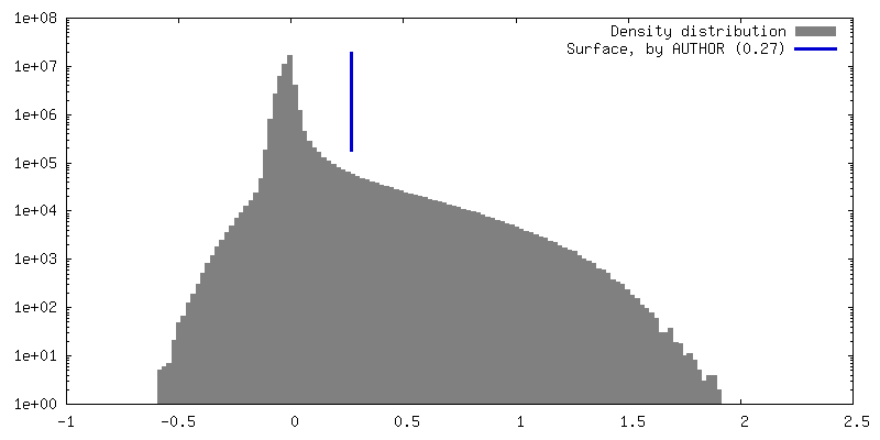
| Density |
| ||||||||||||||||||||||||||||||||||||
| Symmetry | Space group: 1 | ||||||||||||||||||||||||||||||||||||
| Details | EMDB XML:
|
 Z (Sec.)
 Y (Row.)
 X (Col.)


-Supplemental data
-Mask #1
| File | emd_72255_msk_1.map | ||||||||||||
|---|---|---|---|---|---|---|---|---|---|---|---|---|---|
| Projections & Slices |
| ||||||||||||
| Density Histograms |


-Half map: #1
| File | emd_72255_half_map_1.map | ||||||||||||
|---|---|---|---|---|---|---|---|---|---|---|---|---|---|
| Projections & Slices |
| ||||||||||||
| Density Histograms |


-Half map: #2
| File | emd_72255_half_map_2.map | ||||||||||||
|---|---|---|---|---|---|---|---|---|---|---|---|---|---|
| Projections & Slices |
| ||||||||||||
| Density Histograms |


- Sample components
-Entire : Crude cell extract
| Entire | Name: Crude cell extract |
|---|---|
| Components |
|
-Supramolecule #1: Crude cell extract
| Supramolecule | Name: Crude cell extract / type: organelle_or_cellular_component / ID: 1 / Parent: 0 / Macromolecule list: #1-#2 Details: Frozen cell pellets were lysed under cryogenic conditions using a cryomill. Frozen lysate was resuspended and clarified via centrifugation at 21,000g. DNase I (33 U/mL) was added and crude ...Details: Frozen cell pellets were lysed under cryogenic conditions using a cryomill. Frozen lysate was resuspended and clarified via centrifugation at 21,000g. DNase I (33 U/mL) was added and crude cell extract was plunge-frozen. |
|---|---|
| Source (natural) | Organism: |
-Experimental details
-Structure determination
| Method | cryo EM |
|---|---|
 Processing Processing | single particle reconstruction |
| Aggregation state | particle |
-Sample preparation
| Buffer | pH: 7.5 Component:
Details: 33 U/mL DNase I (New England Biolabs #M0303L) was added in addition. | ||||||||||||||||||
|---|---|---|---|---|---|---|---|---|---|---|---|---|---|---|---|---|---|---|---|
| Grid | Model: Quantifoil R2/1 / Material: COPPER / Mesh: 300 / Support film - #0 - Film type ID: 1 / Support film - #0 - Material: CARBON / Support film - #0 - topology: HOLEY / Support film - #1 - Film type ID: 2 / Support film - #1 - Material: GRAPHENE / Support film - #1 - topology: CONTINUOUS Details: Grid was coated with monolayer graphene following (Grassetti et al, JOVE 2023) and treated with UV/ozone for 10 minutes using a Bioforce PC440 UV/ozone cleaner | ||||||||||||||||||
| Vitrification | Cryogen name: ETHANE / Chamber humidity: 95 % / Chamber temperature: 283 K / Instrument: FEI VITROBOT MARK IV |
- Electron microscopy
| Microscope | TFS KRIOS |
|---|---|
| Image recording | Film or detector model: GATAN K3 BIOQUANTUM (6k x 4k) / Average electron dose: 45.1838 e/Å2 |
| Electron beam | Acceleration voltage: 300 kV / Electron source:  FIELD EMISSION GUN FIELD EMISSION GUN |
| Electron optics | Illumination mode: FLOOD BEAM / Imaging mode: BRIGHT FIELD / Nominal defocus max: 2.0 µm / Nominal defocus min: 0.03 µm |
| Experimental equipment |  Model: Titan Krios / Image courtesy: FEI Company |

