Movie
Movie Controller

[English] 日本語
 Yorodumi
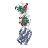
- EMDB-71791: Human 19S proteasome regulatory particle (RP) without observing T... -
+ Open data

- Basic information
| Entry |  | |||||||||
|---|---|---|---|---|---|---|---|---|---|---|
| Title | Human 19S proteasome regulatory particle (RP) without observing TXNL1 PITH domain bound | |||||||||
 Map data Map data | ||||||||||
 Sample Sample |
| |||||||||
 Keywords Keywords | Proteasome / 19S / RP / HYDROLASE | |||||||||
| Biological species |  Homo sapiens (human) Homo sapiens (human) | |||||||||
| Method | single particle reconstruction / cryo EM / Resolution: 3.92 Å | |||||||||
 Authors Authors | Chen X / Negi H / Walters KJ | |||||||||
| Funding support |  United States, 1 items United States, 1 items
| |||||||||
 Citation Citation | Journal: Mol Cell / Year: 2026 Title: Structures of dynamic interactors at native proteasomes by PhIX-MS and cryo-electron microscopy. Authors: Kitaik Lee / Hitendra Negi / Xiang Chen / Katerina Atallah-Yunes / Sophia Truslow / Rithik E Castelino / Mary R Guest / Anthony M Ciancone / Xiuxiu Lu / Sergey G Tarasov / Raj Chari / Kylie ...Authors: Kitaik Lee / Hitendra Negi / Xiang Chen / Katerina Atallah-Yunes / Sophia Truslow / Rithik E Castelino / Mary R Guest / Anthony M Ciancone / Xiuxiu Lu / Sergey G Tarasov / Raj Chari / Kylie J Walters / Francis J O'Reilly / Abstract: Molecular machines rely on dynamic, low-affinity interactions to perform their functional roles. We developed PhIX-MS (photo-induced in situ crosslinking-mass spectrometry), a structural proteomics ...Molecular machines rely on dynamic, low-affinity interactions to perform their functional roles. We developed PhIX-MS (photo-induced in situ crosslinking-mass spectrometry), a structural proteomics workflow to capture topological information for such transient interactions in cells by UV-activated crosslinking. Applying PhIX-MS with cryo-electron microscopy (cryo-EM) to proteasomes, we mapped the redox sensor TXNL1 at the proteasome regulatory particle (RP), including its dynamic thioredoxin-like domain near RPN2/PSMD1 and RPN13/ADRM1, where it is ideal for reducing substrates prior to proteolysis. RPs without the proteolytic core particle (CP) were structurally resolved while bound to TXNL1 and/or the chaperone PSMD5/S5b, which inserts its C terminus into the ATPase pore, causing extensive structural rearrangements. Additionally, PhIX-MS and AlphaFold identified the ubiquitin ligase UBE3C/Hul5 at RPN2, RPN3, and a dynamic RPN10 region, tethering UBE3C above the substrate entry channel. Our integrative approach enables the localization of native, low-affinity protein interactions and is broadly applicable to dynamic macromolecular assemblies. | |||||||||
| History |
|
- Structure visualization
| Supplemental images |
|---|

- Downloads & links
-EMDB archive
| Map data | emd_71791.map.gz | 152.2 MB |  EMDB map data format EMDB map data format | |
|---|---|---|---|---|
| Header (meta data) | emd-71791-v30.xmlemd-71791.xml | 24 KB 24 KB | Display Display | EMDB header |
| FSC (resolution estimation) | emd_71791_fsc.xml | 14.4 KB | Display | FSC data file |
| Images |  emd_71791.png emd_71791.png | 81.3 KB | ||
| Filedesc metadata | emd-71791.cif.gz | 5.1 KB | ||
| Others | emd_71791_additional_1.map.gzemd_71791_half_map_1.map.gzemd_71791_half_map_2.map.gz | 206.2 MB 285.2 MB 285.2 MB | ||
| Archive directory |  https://data.pdbj.org/pub/emdb/structures/EMD-71791ftp://data.pdbj.org/pub/emdb/structures/EMD-71791 https://data.pdbj.org/pub/emdb/structures/EMD-71791ftp://data.pdbj.org/pub/emdb/structures/EMD-71791 | HTTPS FTP |
-Related structure data
| Related structure data |  12bmC  9pmjC  9pmoC  9pmqC  9proC  9prtC  71736 C: citing same article ( |
|---|


-Links
| EMDB pages | EMDB (EBI/PDBe) / EMDataResource |
|---|
-Map
| File | Download / File: emd_71791.map.gz / Format: CCP4 / Size: 307.5 MB / Type: IMAGE STORED AS FLOATING POINT NUMBER (4 BYTES) | ||||||||||||||||||||||||||||||||||||
|---|---|---|---|---|---|---|---|---|---|---|---|---|---|---|---|---|---|---|---|---|---|---|---|---|---|---|---|---|---|---|---|---|---|---|---|---|---|
| Projections & slices | Image control
Images are generated by Spider. | ||||||||||||||||||||||||||||||||||||
| Voxel size | X=Y=Z: 0.81 Å | ||||||||||||||||||||||||||||||||||||
| Density |
| ||||||||||||||||||||||||||||||||||||
| Symmetry | Space group: 1 | ||||||||||||||||||||||||||||||||||||
| Details | EMDB XML:
|
 Z (Sec.)
 Y (Row.)
 X (Col.)


-Supplemental data
-Additional map: #1
| File | emd_71791_additional_1.map | ||||||||||||
|---|---|---|---|---|---|---|---|---|---|---|---|---|---|
| Projections & Slices |
| ||||||||||||
| Density Histograms |


-Half map: #1
| File | emd_71791_half_map_1.map | ||||||||||||
|---|---|---|---|---|---|---|---|---|---|---|---|---|---|
| Projections & Slices |
| ||||||||||||
| Density Histograms |


-Half map: #2
| File | emd_71791_half_map_2.map | ||||||||||||
|---|---|---|---|---|---|---|---|---|---|---|---|---|---|
| Projections & Slices |
| ||||||||||||
| Density Histograms |


- Sample components
-Entire : Human proteasome purified from HCT116 cells
| Entire | Name: Human proteasome purified from HCT116 cells |
|---|---|
| Components |
|
-Supramolecule #1: Human proteasome purified from HCT116 cells
| Supramolecule | Name: Human proteasome purified from HCT116 cells / type: complex / ID: 1 / Parent: 0 / Macromolecule list: #1-#12 |
|---|---|
| Source (natural) | Organism: Homo sapiens (human) |
| Molecular weight | Theoretical: 1 MDa |
-Experimental details
-Structure determination
| Method | cryo EM |
|---|---|
 Processing Processing | single particle reconstruction |
| Aggregation state | particle |
-Sample preparation
| Concentration | 0.75 mg/mL | ||||||||||||||||||
|---|---|---|---|---|---|---|---|---|---|---|---|---|---|---|---|---|---|---|---|
| Buffer | pH: 7.5 Component:
Details: 50 mM Tris [pH7.5], 50 mM NaCl, 1.5 mM ATP-gamma-S, 5 mM MgCl2 and 2 mM DTT | ||||||||||||||||||
| Grid | Model: Quantifoil R1.2/1.3 / Material: COPPER / Mesh: 300 / Pretreatment - Type: GLOW DISCHARGE / Pretreatment - Time: 30 sec. / Pretreatment - Atmosphere: AIR / Details: 25 mA | ||||||||||||||||||
| Vitrification | Cryogen name: ETHANE / Chamber humidity: 85 % / Chamber temperature: 295.15 K / Instrument: LEICA EM GP | ||||||||||||||||||
| Details | Quantifoil copper R 1.2/1.3 holey carbon 300 mesh grids (#Q3100CR1.3; Electron Microscopy Sciences). |
- Electron microscopy
| Microscope | FEI TALOS ARCTICA |
|---|---|
| Specialist optics | Energy filter - Name: GIF Bioquantum |
| Image recording | Film or detector model: GATAN K3 (6k x 4k) / Digitization - Dimensions - Width: 5760 pixel / Digitization - Dimensions - Height: 4092 pixel / Number real images: 40 / Average exposure time: 2.5 sec. / Average electron dose: 55.6 e/Å2 |
| Electron beam | Acceleration voltage: 200 kV / Electron source:  FIELD EMISSION GUN FIELD EMISSION GUN |
| Electron optics | Illumination mode: FLOOD BEAM / Imaging mode: BRIGHT FIELD / Cs: 2.7 mm / Nominal defocus max: 2.0 µm / Nominal defocus min: 0.8 µm / Nominal magnification: 100000 |
| Sample stage | Specimen holder model: FEI TITAN KRIOS AUTOGRID HOLDER / Cooling holder cryogen: NITROGEN |
| Experimental equipment | 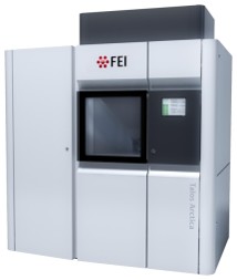 Model: Talos Arctica / Image courtesy: FEI Company |