 Movie
Movie Controller


+ Open data

- Basic information
| Entry |  | |||||||||
|---|---|---|---|---|---|---|---|---|---|---|
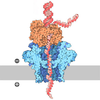
| Title | CsgG nanopore in complex with designed CsgX1C | |||||||||
 Map data Map data | ||||||||||
 Sample Sample |
| |||||||||
 Keywords Keywords | complex / de novo design / nanopore / MEMBRANE PROTEIN | |||||||||
| Function / homology | Curli production assembly/transport component CsgG / Curli production assembly/transport component CsgG / outer membrane-bounded periplasmic space / Prokaryotic membrane lipoprotein lipid attachment site profile. / identical protein binding / plasma membrane / Curli production assembly/transport component CsgG Function and homology information Function and homology information | |||||||||
| Biological species |   Escherichia coli (E. coli) / synthetic construct (others) Escherichia coli (E. coli) / synthetic construct (others) | |||||||||
| Method | single particle reconstruction / cryo EM / Resolution: 2.96 Å | |||||||||
 Authors Authors | Hatstat AK / Melo A / Tse E / Merz GE | |||||||||
| Funding support |  United States, 2 items United States, 2 items
| |||||||||
 Citation Citation | Journal: To Be Published Title: De novo design of semisynthetic protein nanopores Authors: Schnaider LS / Hatstat AK / Scott AJ / Tan S / Hambley R / Dawson W / Polizzi N / Wallace EJ / Merz GE / DeGrado WF | |||||||||
| History |
|
- Structure visualization
| Supplemental images |
|---|

- Downloads & links
-EMDB archive
| Map data | emd_71746.map.gz | 307.2 MB | EMDB map data format | |
|---|---|---|---|---|
| Header (meta data) | emd-71746-v30.xmlemd-71746.xml | 18.4 KB 18.4 KB | Display Display | EMDB header |
| FSC (resolution estimation) | emd_71746_fsc.xml | 14.6 KB | Display | FSC data file |
| Images |  emd_71746.png emd_71746.png | 75.6 KB | ||
| Filedesc metadata | emd-71746.cif.gz | 5.8 KB | ||
| Others | emd_71746_additional_1.map.gzemd_71746_half_map_1.map.gzemd_71746_half_map_2.map.gz | 162.2 MB 301.5 MB 301.5 MB | ||
| Archive directory |  https://data.pdbj.org/pub/emdb/structures/EMD-71746ftp://data.pdbj.org/pub/emdb/structures/EMD-71746 https://data.pdbj.org/pub/emdb/structures/EMD-71746ftp://data.pdbj.org/pub/emdb/structures/EMD-71746 | HTTPS FTP |
-Related structure data
| Related structure data |  9pn8MC  9pn9C  9pnaC  9pnbC M: atomic model generated by this map C: citing same article ( |
|---|---|
| Similar structure data |


-Links
| EMDB pages | EMDB (EBI/PDBe) / EMDataResource |
|---|---|
| Related items in Molecule of the Month |
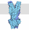
-Map
| File | Download / File: emd_71746.map.gz / Format: CCP4 / Size: 325 MB / Type: IMAGE STORED AS FLOATING POINT NUMBER (4 BYTES) | ||||||||||||||||||||||||||||||||||||
|---|---|---|---|---|---|---|---|---|---|---|---|---|---|---|---|---|---|---|---|---|---|---|---|---|---|---|---|---|---|---|---|---|---|---|---|---|---|
| Projections & slices | Image control
Images are generated by Spider. | ||||||||||||||||||||||||||||||||||||
| Voxel size | X=Y=Z: 0.834 Å | ||||||||||||||||||||||||||||||||||||
| Density |
| ||||||||||||||||||||||||||||||||||||
| Symmetry | Space group: 1 | ||||||||||||||||||||||||||||||||||||
| Details | EMDB XML:
|
 Z (Sec.)
 Y (Row.)
 X (Col.)


-Supplemental data
-Additional map: #1
| File | emd_71746_additional_1.map | ||||||||||||
|---|---|---|---|---|---|---|---|---|---|---|---|---|---|
| Projections & Slices |
| ||||||||||||
| Density Histograms |


-Half map: #2
| File | emd_71746_half_map_1.map | ||||||||||||
|---|---|---|---|---|---|---|---|---|---|---|---|---|---|
| Projections & Slices |
| ||||||||||||
| Density Histograms |


-Half map: #1
| File | emd_71746_half_map_2.map | ||||||||||||
|---|---|---|---|---|---|---|---|---|---|---|---|---|---|
| Projections & Slices |
| ||||||||||||
| Density Histograms |


- Sample components
-Entire : Nanopore assembly of CsgG nonamer with CsgX1C nonamer
| Entire | Name: Nanopore assembly of CsgG nonamer with CsgX1C nonamer |
|---|---|
| Components |
|
-Supramolecule #1: Nanopore assembly of CsgG nonamer with CsgX1C nonamer
| Supramolecule | Name: Nanopore assembly of CsgG nonamer with CsgX1C nonamer / type: complex / ID: 1 / Parent: 0 / Macromolecule list: all |
|---|---|
| Source (natural) | Organism: Escherichia coli (E. coli) |
-Macromolecule #1: CsgX1C
| Macromolecule | Name: CsgX1C / type: protein_or_peptide / ID: 1 / Number of copies: 9 / Enantiomer: LEVO |
|---|---|
| Source (natural) | Organism: synthetic construct (others) |
| Molecular weight | Theoretical: 6.210905 KDa |
| Sequence | String: GTMTFQFRNP NFGGNPNNGA FLLCSAQAQN AGILAAQLWN NGDYDRALSL FIAVVQSC |
-Macromolecule #2: Curli production assembly/transport component CsgG
| Macromolecule | Name: Curli production assembly/transport component CsgG / type: protein_or_peptide / ID: 2 / Number of copies: 9 / Enantiomer: LEVO |
|---|---|
| Source (natural) | Organism: Escherichia coli (E. coli) |
| Molecular weight | Theoretical: 30.091146 KDa |
| Recombinant expression | Organism: Escherichia coli (E. coli) |
| Sequence | String: CLTAPPKEAA RPTLMPRAQS YKDLTHLPAP TGKIFVSVYN IQDETGQFKP YPASNQSTAV PQSATAMLVT ALKDSRWFIP LERQGLQNL LNERKIIRAA QENGTVAINN RIPLQSLTAA NIMVEGSIIG YESNVKSGGV GARYFGIGAD TQYQLDQIAV N LRVVNVST ...String: CLTAPPKEAA RPTLMPRAQS YKDLTHLPAP TGKIFVSVYN IQDETGQFKP YPASNQSTAV PQSATAMLVT ALKDSRWFIP LERQGLQNL LNERKIIRAA QENGTVAINN RIPLQSLTAA NIMVEGSIIG YESNVKSGGV GARYFGIGAD TQYQLDQIAV N LRVVNVST GEILSSVNTS KTILSYEVQA GVFRFIDYQR LLEGEVGYTS NEPVMLCLMS AIETGVIFLI NDGIDRGLWD LQ NKAERQN DILVKYRHMS VPPESSAWSH PQFEK UniProtKB: Curli production assembly/transport component CsgG |
-Experimental details
-Structure determination
| Method | cryo EM |
|---|---|
 Processing Processing | single particle reconstruction |
| Aggregation state | particle |
-Sample preparation
| Concentration | 0.3 mg/mL |
|---|---|
| Buffer | pH: 7 |
| Grid | Model: Quantifoil / Material: GOLD / Mesh: 200 / Support film - Material: GRAPHENE OXIDE / Support film - topology: CONTINUOUS |
| Vitrification | Cryogen name: ETHANE / Chamber humidity: 100 % / Chamber temperature: 277 K / Instrument: FEI VITROBOT MARK IV |
- Electron microscopy
| Microscope | TFS KRIOS |
|---|---|
| Image recording | Film or detector model: GATAN K3 BIOCONTINUUM (6k x 4k) / Average electron dose: 46.0 e/Å2 |
| Electron beam | Acceleration voltage: 300 kV / Electron source:  FIELD EMISSION GUN FIELD EMISSION GUN |
| Electron optics | Illumination mode: FLOOD BEAM / Imaging mode: BRIGHT FIELD / Nominal defocus max: 1.8 µm / Nominal defocus min: 0.8 µm |
| Experimental equipment |  Model: Titan Krios / Image courtesy: FEI Company |

