 Movie
Movie Controller

[English] 日本語
 Yorodumi
- EMDB-71566: Structure of EBOV GP, MWAC-3634, and Nanosota-EB2 ternary complex -
+ Open data

- Basic information
| Entry |  | |||||||||
|---|---|---|---|---|---|---|---|---|---|---|
| Title | Structure of EBOV GP, MWAC-3634, and Nanosota-EB2 ternary complex | |||||||||
 Map data Map data | unsharpened map of EBOV GP in complex with compound 3634 | |||||||||
 Sample Sample |
| |||||||||
 Keywords Keywords | EBOV / glycoprotein / inhibitor / VIRAL PROTEIN / VIRAL PROTEIN-IMMUNE SYSTEM complex | |||||||||
| Function / homology |  Function and homology information Function and homology informationsymbiont-mediated killing of host cell / host cell endoplasmic reticulum / viral budding from plasma membrane / clathrin-dependent endocytosis of virus by host cell / symbiont-mediated-mediated suppression of host tetherin activity / host cell cytoplasm / entry receptor-mediated virion attachment to host cell / symbiont-mediated suppression of host innate immune response / membrane raft / fusion of virus membrane with host endosome membrane ...symbiont-mediated killing of host cell / host cell endoplasmic reticulum / viral budding from plasma membrane / clathrin-dependent endocytosis of virus by host cell / symbiont-mediated-mediated suppression of host tetherin activity / host cell cytoplasm / entry receptor-mediated virion attachment to host cell / symbiont-mediated suppression of host innate immune response / membrane raft / fusion of virus membrane with host endosome membrane / viral envelope / symbiont entry into host cell / lipid binding / host cell plasma membrane / virion membrane / extracellular region / identical protein binding Similarity search - Function | |||||||||
| Biological species |  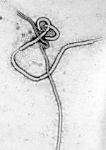 Ebola virus - Mayinga, Zaire, 1976 / Ebola virus - Mayinga, Zaire, 1976 /   Vicugna pacos (alpaca) / Ebola virus Vicugna pacos (alpaca) / Ebola virus | |||||||||
| Method | single particle reconstruction / cryo EM / Resolution: 2.54 Å | |||||||||
 Authors Authors | Bu F / Ye G / Li F | |||||||||
| Funding support |  United States, 1 items United States, 1 items
| |||||||||
 Citation Citation | Journal: To Be Published Title: Structure of EBOV GP, MWAC-3634, and Nanosota-EB2 ternary complex Authors: Bu F / Ye G / Li F | |||||||||
| History |
|
- Structure visualization
| Supplemental images |
|---|

- Downloads & links
-EMDB archive
| Map data | emd_71566.map.gz | 51.7 MB | EMDB map data format | |
|---|---|---|---|---|
| Header (meta data) | emd-71566-v30.xmlemd-71566.xml | 20.5 KB 20.5 KB | Display Display | EMDB header |
| FSC (resolution estimation) | emd_71566_fsc.xml | 9.9 KB | Display | FSC data file |
| Images |  emd_71566.png emd_71566.png | 42.9 KB | ||
| Filedesc metadata | emd-71566.cif.gz | 6.3 KB | ||
| Others | emd_71566_additional_1.map.gzemd_71566_half_map_1.map.gzemd_71566_half_map_2.map.gz | 97.1 MB 95.5 MB 95.5 MB | ||
| Archive directory |  http://ftp.pdbj.org/pub/emdb/structures/EMD-71566ftp://ftp.pdbj.org/pub/emdb/structures/EMD-71566 http://ftp.pdbj.org/pub/emdb/structures/EMD-71566ftp://ftp.pdbj.org/pub/emdb/structures/EMD-71566 | HTTPS FTP |
-Related structure data
| Related structure data |  9pelMC M: atomic model generated by this map C: citing same article ( |
|---|---|
| Similar structure data |
-Links
| EMDB pages | EMDB (EBI/PDBe) / EMDataResource |
|---|---|
| Related items in Molecule of the Month |

-Map
| File | Download / File: emd_71566.map.gz / Format: CCP4 / Size: 103 MB / Type: IMAGE STORED AS FLOATING POINT NUMBER (4 BYTES) | ||||||||||||||||||||||||||||||||||||
|---|---|---|---|---|---|---|---|---|---|---|---|---|---|---|---|---|---|---|---|---|---|---|---|---|---|---|---|---|---|---|---|---|---|---|---|---|---|
| Annotation | unsharpened map of EBOV GP in complex with compound 3634 | ||||||||||||||||||||||||||||||||||||
| Projections & slices | Image control
Images are generated by Spider. | ||||||||||||||||||||||||||||||||||||
| Voxel size | X=Y=Z: 0.88533 Å | ||||||||||||||||||||||||||||||||||||
| Density |
| ||||||||||||||||||||||||||||||||||||
| Symmetry | Space group: 1 | ||||||||||||||||||||||||||||||||||||
| Details | EMDB XML:
|
 Z (Sec.)
 Y (Row.)
 X (Col.)


-Supplemental data
-Additional map: sharpened map of EBOV GP in complex with compound 3634
| File | emd_71566_additional_1.map | ||||||||||||
|---|---|---|---|---|---|---|---|---|---|---|---|---|---|
| Annotation | sharpened map of EBOV GP in complex with compound 3634 | ||||||||||||
| Projections & Slices |
| ||||||||||||
| Density Histograms |


-Half map: half map A of EBOV GP in complex with compound 3634
| File | emd_71566_half_map_1.map | ||||||||||||
|---|---|---|---|---|---|---|---|---|---|---|---|---|---|
| Annotation | half map_A of EBOV GP in complex with compound 3634 | ||||||||||||
| Projections & Slices |
| ||||||||||||
| Density Histograms |


-Half map: half map B of EBOV GP in complex with compound 3634
| File | emd_71566_half_map_2.map | ||||||||||||
|---|---|---|---|---|---|---|---|---|---|---|---|---|---|
| Annotation | half map_B of EBOV GP in complex with compound 3634 | ||||||||||||
| Projections & Slices |
| ||||||||||||
| Density Histograms |


- Sample components
-Entire : EBOV GP, MWAC-3634, and Nanosota-EB2 ternary complex
| Entire | Name: EBOV GP, MWAC-3634, and Nanosota-EB2 ternary complex |
|---|---|
| Components |
|
-Supramolecule #1: EBOV GP, MWAC-3634, and Nanosota-EB2 ternary complex
| Supramolecule | Name: EBOV GP, MWAC-3634, and Nanosota-EB2 ternary complex / type: complex / ID: 1 / Parent: 0 / Macromolecule list: #1-#3 |
|---|
-Supramolecule #2: EBOV GP
| Supramolecule | Name: EBOV GP / type: complex / ID: 2 / Parent: 1 / Macromolecule list: #2-#3 |
|---|---|
| Source (natural) | Organism: Ebola virus - Mayinga, Zaire, 1976 |
-Supramolecule #3: Nanosota-EB2
| Supramolecule | Name: Nanosota-EB2 / type: complex / ID: 3 / Parent: 1 / Macromolecule list: #1 |
|---|---|
| Source (natural) | Organism: Vicugna pacos (alpaca) |
-Macromolecule #1: Nanosota-EB2
| Macromolecule | Name: Nanosota-EB2 / type: protein_or_peptide / ID: 1 / Number of copies: 3 / Enantiomer: LEVO |
|---|---|
| Source (natural) | Organism: Vicugna pacos (alpaca) |
| Molecular weight | Theoretical: 15.722139 KDa |
| Recombinant expression | Organism:   Escherichia coli (E. coli) Escherichia coli (E. coli) |
| Sequence | String: QVQLQESGGG LVQAGGSLRL SCVHSGRTFS NDAMAWFRQA PGKDREFAAG IDYNGGRTAY TDSVKGRFTI SRDNAKNTVY LQMNSLKPE DTAVYSCAAR PWSIANLAYT YDSWGQGTQV TVSSGGQHHH HHHGAYPYDV PDYAS |
-Macromolecule #2: Envelope glycoprotein GP1
| Macromolecule | Name: Envelope glycoprotein GP1 / type: protein_or_peptide / ID: 2 / Number of copies: 3 / Enantiomer: LEVO |
|---|---|
| Source (natural) | Organism: Ebola virus / Strain: Mayinga, Zaire, 1976 |
| Molecular weight | Theoretical: 37.6645 KDa |
| Recombinant expression | Organism:  Homo sapiens (human) Homo sapiens (human) |
| Sequence | String: MDAMKRGLCC VLLLCGAVFV SASSIPLGVI HNSTLQVSDV DKLVCRDKLS STNQLRSVGL NLEGNGVATD VPSATKRWGF RSGVPPKVV NYEAGEWAEN CYNLEIKKPD GSECLPAAPD GIRGFPRCRY VHKVSGTGPC AGDFAFHKEG AFFLYDRLAS T VIYRGTTF ...String: MDAMKRGLCC VLLLCGAVFV SASSIPLGVI HNSTLQVSDV DKLVCRDKLS STNQLRSVGL NLEGNGVATD VPSATKRWGF RSGVPPKVV NYEAGEWAEN CYNLEIKKPD GSECLPAAPD GIRGFPRCRY VHKVSGTGPC AGDFAFHKEG AFFLYDRLAS T VIYRGTTF AEGVVAFLIL PQAKKDFFSS HPLREPVNAT EDPSSGYYST TIRYQATGFG TNETEYLFEV DNLTYVQLES RF TPQFLLQ LNETIYTSGK RSNTTGKLIW KVNPEIDTTI GEWAFWETKK NLTRKIRSEE LSFTVVSTHH QDTGEESASS GKL GLITNT IAGVAGLITG GRRTRR UniProtKB: Envelope glycoprotein, Envelope glycoprotein |
-Macromolecule #3: Envelope glycoprotein GP2
| Macromolecule | Name: Envelope glycoprotein GP2 / type: protein_or_peptide / ID: 3 / Number of copies: 3 / Enantiomer: LEVO |
|---|---|
| Source (natural) | Organism: Ebola virus / Strain: Mayinga, Zaire, 1976 |
| Molecular weight | Theoretical: 18.888287 KDa |
| Recombinant expression | Organism: Homo sapiens (human) |
| Sequence | String: EAIVNAQPKC NPNLHYWTTQ DEGAAIGLAW IPYFGPAAEG IYIEGLMHNQ DGLICGLRQL ANETTQALQL FLRATTELRT FSILNRKAI DFLLQRWGGT CHILGPDCCI EPHDWTKNIT DKIDQIIHDF VDGSGYIPEA PRDGQAYVRK DGEWVLLSTF L GHHHHHH UniProtKB: Envelope glycoprotein |
-Macromolecule #7: (3S,4R)-1-{(2P)-2-(6-chloroquinolin-4-yl)-1-[(1R)-1-(naphthalen-1...
| Macromolecule | Name: (3S,4R)-1-{(2P)-2-(6-chloroquinolin-4-yl)-1-[(1R)-1-(naphthalen-1-yl)ethyl]-1H-1,3-benzimidazole-6-carbonyl}-N-methyl-4-[3-(trifluoromethyl)phenyl]pyrrolidine-3-carboxamide type: ligand / ID: 7 / Number of copies: 3 / Formula: A1CIK |
|---|---|
| Molecular weight | Theoretical: 732.192 Da |
-Experimental details
-Structure determination
| Method | cryo EM |
|---|---|
 Processing Processing | single particle reconstruction |
| Aggregation state | particle |
-Sample preparation
| Buffer | pH: 7.4 |
|---|---|
| Vitrification | Cryogen name: ETHANE |
- Electron microscopy
| Microscope | TFS KRIOS |
|---|---|
| Image recording | Film or detector model: GATAN K3 (6k x 4k) / Number real images: 6106 / Average electron dose: 54.5 e/Å2 |
| Electron beam | Acceleration voltage: 300 kV / Electron source:  FIELD EMISSION GUN FIELD EMISSION GUN |
| Electron optics | Illumination mode: FLOOD BEAM / Imaging mode: BRIGHT FIELD / Nominal defocus max: 2.0 µm / Nominal defocus min: 1.0 µm |
| Experimental equipment |  Model: Titan Krios / Image courtesy: FEI Company |


