 Movie
Movie Controller


+ Open data

- Basic information
| Entry | Database: EMDB / ID: EMD-7153 | |||||||||
|---|---|---|---|---|---|---|---|---|---|---|
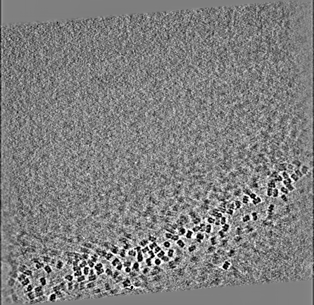
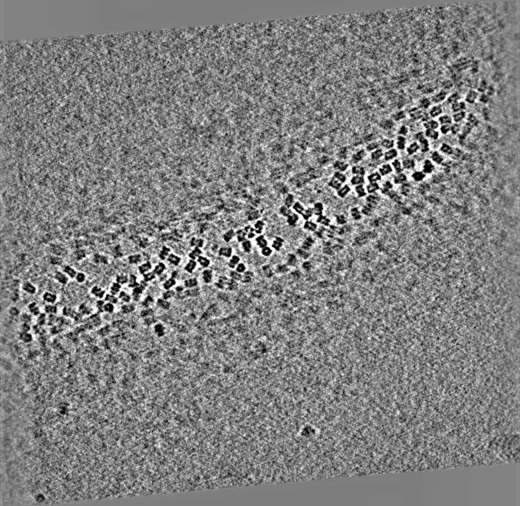
| Title | T20S proteasome on a gold Quantifoil grid | |||||||||
 Map data Map data | Tomogram of T20S proteasome single particle | |||||||||
 Sample Sample |
| |||||||||
| Biological species | unidentified (others) | |||||||||
| Method | electron tomography / cryo EM | |||||||||
 Authors Authors | Noble AN / Dandey VP / Wei H / Brasch J / Chase J / Acharya P / Tan Y / Zhang Z / Kim LY / Scapin G ...Noble AN / Dandey VP / Wei H / Brasch J / Chase J / Acharya P / Tan Y / Zhang Z / Kim LY / Scapin G / Rapp M / Eng ET / Rice WJ / Cheng A / Negro CJ / Shapiro L / Kwong PD / Jeruzalmi D / des Georges A / Potter CS / Carragher B | |||||||||
 Citation Citation | Journal: Elife / Year: 2018 Title: Routine single particle CryoEM sample and grid characterization by tomography. Authors: Alex J Noble / Venkata P Dandey / Hui Wei / Julia Brasch / Jillian Chase / Priyamvada Acharya / Yong Zi Tan / Zhening Zhang / Laura Y Kim / Giovanna Scapin / Micah Rapp / Edward T Eng / ...Authors: Alex J Noble / Venkata P Dandey / Hui Wei / Julia Brasch / Jillian Chase / Priyamvada Acharya / Yong Zi Tan / Zhening Zhang / Laura Y Kim / Giovanna Scapin / Micah Rapp / Edward T Eng / William J Rice / Anchi Cheng / Carl J Negro / Lawrence Shapiro / Peter D Kwong / David Jeruzalmi / Amedee des Georges / Clinton S Potter / Bridget Carragher /  Abstract: Single particle cryo-electron microscopy (cryoEM) is often performed under the assumption that particles are not adsorbed to the air-water interfaces and in thin, vitreous ice. In this study, we ...Single particle cryo-electron microscopy (cryoEM) is often performed under the assumption that particles are not adsorbed to the air-water interfaces and in thin, vitreous ice. In this study, we performed fiducial-less tomography on over 50 different cryoEM grid/sample preparations to determine the particle distribution within the ice and the overall geometry of the ice in grid holes. Surprisingly, by studying particles in holes in 3D from over 1000 tomograms, we have determined that the vast majority of particles (approximately 90%) are adsorbed to an air-water interface. The implications of this observation are wide-ranging, with potential ramifications regarding protein denaturation, conformational change, and preferred orientation. We also show that fiducial-less cryo-electron tomography on single particle grids may be used to determine ice thickness, optimal single particle collection areas and strategies, particle heterogeneity, and de novo models for template picking and single particle alignment. | |||||||||
| History |
|
- Structure visualization
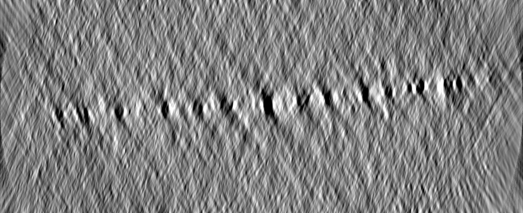
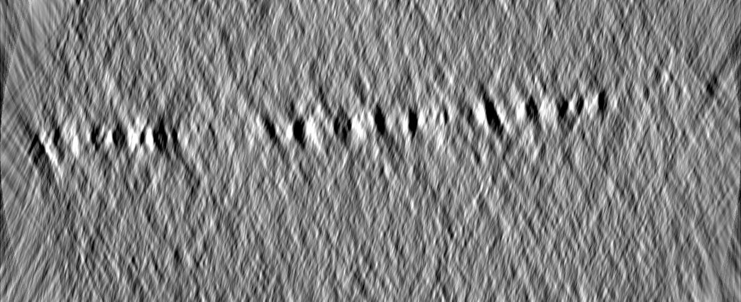
| Movie |
 Movie viewer Movie viewer |
|---|---|
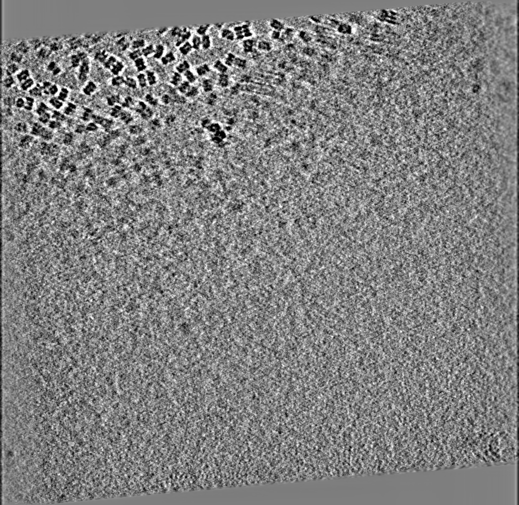
| Supplemental images |

- Downloads & links
-EMDB archive
| Map data | emd_7153.map.gz | 1.5 GB | EMDB map data format | |
|---|---|---|---|---|
| Header (meta data) | emd-7153-v30.xmlemd-7153.xml | 8.4 KB 8.4 KB | Display Display | EMDB header |
| Images | 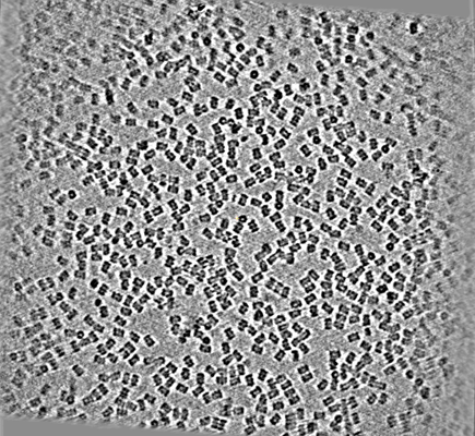 emd_7153.png emd_7153.png | 272.7 KB | ||
| Archive directory |  http://ftp.pdbj.org/pub/emdb/structures/EMD-7153ftp://ftp.pdbj.org/pub/emdb/structures/EMD-7153 http://ftp.pdbj.org/pub/emdb/structures/EMD-7153ftp://ftp.pdbj.org/pub/emdb/structures/EMD-7153 | HTTPS FTP |
-Related structure data
| Related structure data |  7135C  7138C  7139C  7140C  7141C  7142C  7143C  7144C  7145C  7146C  7147C  7148C  7149C  7150C  7151C  7152C  7154C C: citing same article ( |
|---|---|
| EM raw data | EMPIAR-10144 (Title: CryoET of T20S proteasome single particle / Data size: 12.4 Data #1: Tilt images along with all other magnification images from the collection [micrographs - single frame] Data #2: Appion-Protomo tilt-series alignments [tilt series]) EMPIAR-10188 (Title: T20S proteasome single particle / Data size: 116.1 Data #1: Unaligned, frame aligned, and frame aligned+dose weighted micrographs [micrographs - single frame]) |
-Links
| EMDB pages | EMDB (EBI/PDBe) / EMDataResource |
|---|
-Map
| File | Download / File: emd_7153.map.gz / Format: CCP4 / Size: 1.7 GB / Type: IMAGE STORED AS FLOATING POINT NUMBER (4 BYTES) | ||||||||||||||||||||||||||||||||||||||||||||||||||||||||||||
|---|---|---|---|---|---|---|---|---|---|---|---|---|---|---|---|---|---|---|---|---|---|---|---|---|---|---|---|---|---|---|---|---|---|---|---|---|---|---|---|---|---|---|---|---|---|---|---|---|---|---|---|---|---|---|---|---|---|---|---|---|---|
| Annotation | Tomogram of T20S proteasome single particle | ||||||||||||||||||||||||||||||||||||||||||||||||||||||||||||
| Projections & slices | Image control
Images are generated by Spider. generated in cubic-lattice coordinate | ||||||||||||||||||||||||||||||||||||||||||||||||||||||||||||
| Voxel size | X=Y=Z: 1 Å | ||||||||||||||||||||||||||||||||||||||||||||||||||||||||||||
| Density |
| ||||||||||||||||||||||||||||||||||||||||||||||||||||||||||||
| Symmetry | Space group: 1 | ||||||||||||||||||||||||||||||||||||||||||||||||||||||||||||
| Details | EMDB XML:
CCP4 map header:
| ||||||||||||||||||||||||||||||||||||||||||||||||||||||||||||
 Z (Sec.)
 Y (Row.)
 X (Col.)


-Supplemental data
- Sample components
-Entire : T20S proteasome
| Entire | Name: T20S proteasome |
|---|---|
| Components |
|
-Supramolecule #1: T20S proteasome
| Supramolecule | Name: T20S proteasome / type: complex / ID: 1 / Parent: 0 / Details: Tomography on single particle sample |
|---|---|
| Source (natural) | Organism: unidentified (others) |
-Experimental details
-Structure determination
| Method | cryo EM |
|---|---|
 Processing Processing | electron tomography |
| Aggregation state | particle |
-Sample preparation
| Vitrification | Cryogen name: ETHANE |
|---|---|
| Sectioning | Other: NO SECTIONING |
- Electron microscopy
| Microscope | FEI TITAN KRIOS |
|---|---|
| Image recording | Film or detector model: GATAN K2 SUMMIT (4k x 4k) / Detector mode: COUNTING / Average electron dose: 2.0 e/Å2 |
| Electron beam | Acceleration voltage: 300 kV / Electron source:  FIELD EMISSION GUN FIELD EMISSION GUN |
| Electron optics | Illumination mode: FLOOD BEAM / Imaging mode: BRIGHT FIELD / Cs: 2.7 mm |
| Experimental equipment |  Model: Titan Krios / Image courtesy: FEI Company |
-Image processing
| Details | Appion-Protomo fiducial-less tilt-series alignment |
|---|---|
| Final reconstruction | Algorithm: SIMULTANEOUS ITERATIVE (SIRT) / Software - Name: TOMO3D / Number images used: 28 |
