 Movie
Movie Controller


+ Open data

- Basic information
| Entry |  | ||||||||||||
|---|---|---|---|---|---|---|---|---|---|---|---|---|---|
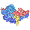
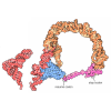
| Title | In situ human A/P-P/E state 80S ribosome | ||||||||||||
 Map data Map data | |||||||||||||
 Sample Sample |
| ||||||||||||
 Keywords Keywords | In situ / Ribosome | ||||||||||||
| Function / homology |  Function and homology information Function and homology informationnegative regulation of protein localization to endoplasmic reticulum / nascent polypeptide-associated complex / endothelial cell differentiation / male meiosis I / response to insecticide / negative regulation of endoplasmic reticulum unfolded protein response / eukaryotic 80S initiation complex / ribosomal protein import into nucleus / regulation of G1 to G0 transition / positive regulation of DNA binding ...negative regulation of protein localization to endoplasmic reticulum / nascent polypeptide-associated complex / endothelial cell differentiation / male meiosis I / response to insecticide / negative regulation of endoplasmic reticulum unfolded protein response / eukaryotic 80S initiation complex / ribosomal protein import into nucleus / regulation of G1 to G0 transition / positive regulation of DNA binding / positive regulation of respiratory burst involved in inflammatory response / positive regulation of gastrulation / regulation of adenylate cyclase-activating G protein-coupled receptor signaling pathway / protein tyrosine kinase inhibitor activity / IRE1-RACK1-PP2A complex / TNFR1-mediated ceramide production / positive regulation of Golgi to plasma membrane protein transport / G1 to G0 transition / negative regulation of formation of translation preinitiation complex / nucleolus organization / positive regulation of ubiquitin-protein transferase activity / negative regulation of RNA splicing / GAIT complex / TORC2 complex binding / erythrocyte homeostasis / regulation of establishment of cell polarity / rRNA modification in the nucleus and cytosol / negative regulation of intrinsic apoptotic signaling pathway in response to hydrogen peroxide / negative regulation of phagocytosis / cytoplasmic side of rough endoplasmic reticulum membrane / regulation of translation involved in cellular response to UV / A band / Formation of the ternary complex, and subsequently, the 43S complex / laminin receptor activity / ion channel inhibitor activity / positive regulation of intrinsic apoptotic signaling pathway in response to DNA damage by p53 class mediator / response to aldosterone / negative regulation of myoblast fusion / protein-DNA complex disassembly / Ribosomal scanning and start codon recognition / Translation initiation complex formation / TFIID-class transcription factor complex binding / negative regulation of Wnt signaling pathway / positive regulation of DNA damage response, signal transduction by p53 class mediator / fibroblast growth factor binding / BH3 domain binding / Protein hydroxylation / TOR signaling / mTORC1-mediated signalling / negative regulation of translational frameshifting / monocyte chemotaxis / PELO:HBS1L and ABCE1 dissociate a ribosome on a non-stop mRNA / SRC activates STAT3 in a quantitative manner, through Cadherin-11 (CDH11), RAC1 and gp130 (IL6ST) / regulation of lipid metabolic process / SARS-CoV-1 modulates host translation machinery / regulation of cell division / positive regulation of GTPase activity / protein localization to nucleus / Peptide chain elongation / Selenocysteine synthesis / negative regulation of protein binding / Formation of a pool of free 40S subunits / negative regulation of respiratory burst involved in inflammatory response / positive regulation of intrinsic apoptotic signaling pathway by p53 class mediator / protein targeting / protein serine/threonine kinase inhibitor activity / Eukaryotic Translation Termination / Dengue Virus Attachment and Entry / SRP-dependent cotranslational protein targeting to membrane / Response of EIF2AK4 (GCN2) to amino acid deficiency / ubiquitin ligase inhibitor activity / Viral mRNA Translation / endonucleolytic cleavage to generate mature 3'-end of SSU-rRNA from (SSU-rRNA, 5.8S rRNA, LSU-rRNA) / Nonsense Mediated Decay (NMD) independent of the Exon Junction Complex (EJC) / positive regulation of signal transduction by p53 class mediator / GTP hydrolysis and joining of the 60S ribosomal subunit / embryo implantation / L13a-mediated translational silencing of Ceruloplasmin expression / cleavage in ITS2 between 5.8S rRNA and LSU-rRNA of tricistronic rRNA transcript (SSU-rRNA, 5.8S rRNA, LSU-rRNA) / regulation of translational fidelity / Major pathway of rRNA processing in the nucleolus and cytosol / phagocytic cup / Nonsense Mediated Decay (NMD) enhanced by the Exon Junction Complex (EJC) / maturation of LSU-rRNA / negative regulation of ubiquitin-dependent protein catabolic process / Protein methylation / positive regulation of cell cycle / endonucleolytic cleavage in ITS1 to separate SSU-rRNA from 5.8S rRNA and LSU-rRNA from tricistronic rRNA transcript (SSU-rRNA, 5.8S rRNA, LSU-rRNA) / translation regulator activity / Nuclear events stimulated by ALK signaling in cancer / ribosomal small subunit export from nucleus / Amplification of signal from unattached kinetochores via a MAD2 inhibitory signal / rough endoplasmic reticulum / laminin binding / SH2 domain binding / signaling adaptor activity / negative regulation of proteasomal ubiquitin-dependent protein catabolic process / Gene and protein expression by JAK-STAT signaling after Interleukin-12 stimulation / Maturation of protein E / MDM2/MDM4 family protein binding Similarity search - Function | ||||||||||||
| Biological species |  Homo sapiens (human) Homo sapiens (human) | ||||||||||||
| Method | single particle reconstruction / cryo EM / Resolution: 2.98 Å | ||||||||||||
 Authors Authors | Zheng W / Xiong Y | ||||||||||||
| Funding support | 1 items
| ||||||||||||
 Citation Citation | Journal: Nat Commun / Year: 2025 Title: Visualizing the translation landscape in human cells at high resolution. Authors: Wei Zheng / Yuekang Zhang / Jimin Wang / Shuhui Wang / Pengxin Chai / Elizabeth J Bailey / Chenghao Zhu / Wangbiao Guo / Swapnil C Devarkar / Shenping Wu / Jianfeng Lin / Kai Zhang / Jun Liu ...Authors: Wei Zheng / Yuekang Zhang / Jimin Wang / Shuhui Wang / Pengxin Chai / Elizabeth J Bailey / Chenghao Zhu / Wangbiao Guo / Swapnil C Devarkar / Shenping Wu / Jianfeng Lin / Kai Zhang / Jun Liu / Ivan B Lomakin / Yong Xiong /  Abstract: Comprehensive in situ structures of macromolecules can transform our understanding of biology and advance human health. Here, we map protein synthesis inside human cells in detail by combining ...Comprehensive in situ structures of macromolecules can transform our understanding of biology and advance human health. Here, we map protein synthesis inside human cells in detail by combining automated cryo-focused ion beam (FIB) milling and in situ single-particle cryo electron microscopy (cryo-EM). With this in situ cryo-EM approach, we resolved a 2.2 Å consensus structure of the human 80S ribosome and unveiled 23 functional states, nearly all better than 3 Å resolution. Compared to in vitro studies, we observed variations in ribosome structures, distinct environments of ion and polyamine binding, and associated proteins such as EDF1 and NACβ that are typically not enriched with purified ribosomes. We also detected additional peptide-related density features on the ribosome and visualized ribosome-ribosome interactions in helical polysomes. Finally, high-resolution structures from cells treated with homoharringtonine and cycloheximide revealed a distinct translational landscape and a spermidine that interacts with cycloheximide at the E site, one of the numerous polyamines that also bind native ribosomes. These results underscore the value of high-resolution in situ studies in the native environment. #1: Journal: Acta Crystallogr., Sect. D: Biol. Crystallogr. / Year: 2018Title: Real-space refinement in PHENIX for cryo-EM and crystallography Authors: Wei Z | ||||||||||||
| History |
|
- Structure visualization
| Supplemental images |
|---|

- Downloads & links
-EMDB archive
| Map data | emd_71377.map.gz | 257.6 MB | EMDB map data format | |
|---|---|---|---|---|
| Header (meta data) | emd-71377-v30.xmlemd-71377.xml | 106.7 KB 106.7 KB | Display Display | EMDB header |
| FSC (resolution estimation) | emd_71377_fsc.xml | 16.9 KB | Display | FSC data file |
| Images |  emd_71377.png emd_71377.png | 13.1 KB | ||
| Filedesc metadata | emd-71377.cif.gz | 20.5 KB | ||
| Others | emd_71377_half_map_1.map.gzemd_71377_half_map_2.map.gz | 475.7 MB 475.7 MB | ||
| Archive directory |  https://data.pdbj.org/pub/emdb/structures/EMD-71377ftp://data.pdbj.org/pub/emdb/structures/EMD-71377 https://data.pdbj.org/pub/emdb/structures/EMD-71377ftp://data.pdbj.org/pub/emdb/structures/EMD-71377 | HTTPS FTP |
-Related structure data
| Related structure data |  9p8hMC  9p6zC  9p72C  9p73C  9p76C  9p78C  9p79C  9p7aC  9p7cC  9p7dC  9p7eC  9p7fC  9p7gC  9p7hC  9p7iC  9p7jC  9p7kC  9p7lC  9p7nC  9p7oC  9p7wC  9p7xC  9p7yC  9p8bC  9p8cC  9p8iC  9p9hC  9p9iC  9p9jC  9p9kC  9pa7C  9pbeC  9pkgC M: atomic model generated by this map C: citing same article ( |
|---|---|
| Similar structure data |


-Links
| EMDB pages | EMDB (EBI/PDBe) / EMDataResource |
|---|---|
| Related items in Molecule of the Month |

















-Map
| File | Download / File: emd_71377.map.gz / Format: CCP4 / Size: 512 MB / Type: IMAGE STORED AS FLOATING POINT NUMBER (4 BYTES) | ||||||||||||||||||||||||||||||||||||
|---|---|---|---|---|---|---|---|---|---|---|---|---|---|---|---|---|---|---|---|---|---|---|---|---|---|---|---|---|---|---|---|---|---|---|---|---|---|
| Projections & slices | Image control
Images are generated by Spider. | ||||||||||||||||||||||||||||||||||||
| Voxel size | X=Y=Z: 1.068 Å | ||||||||||||||||||||||||||||||||||||
| Density |
| ||||||||||||||||||||||||||||||||||||
| Symmetry | Space group: 1 | ||||||||||||||||||||||||||||||||||||
| Details | EMDB XML:
|
 Z (Sec.)
 Y (Row.)
 X (Col.)


-Supplemental data
-Half map: #2
| File | emd_71377_half_map_1.map | ||||||||||||
|---|---|---|---|---|---|---|---|---|---|---|---|---|---|
| Projections & Slices |
| ||||||||||||
| Density Histograms |


-Half map: #1
| File | emd_71377_half_map_2.map | ||||||||||||
|---|---|---|---|---|---|---|---|---|---|---|---|---|---|
| Projections & Slices |
| ||||||||||||
| Density Histograms |


- Sample components
+Entire : In situ human A/P-P/E state 80S ribosome
+Supramolecule #1: In situ human A/P-P/E state 80S ribosome
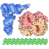
+Macromolecule #1: A/P site tRNA
+Macromolecule #2: P/E site tRNA
+Macromolecule #35: 18S rRNA
+Macromolecule #39: 28S rRNA
+Macromolecule #40: 5S rRNA
+Macromolecule #41: 5.8S rRNA
+Macromolecule #3: 40S ribosomal protein S3
+Macromolecule #4: 40S ribosomal protein S5
+Macromolecule #5: 40S ribosomal protein S10
+Macromolecule #6: Small ribosomal subunit protein eS12
+Macromolecule #7: 40S ribosomal protein S15
+Macromolecule #8: Small ribosomal subunit protein uS9
+Macromolecule #9: Small ribosomal subunit protein eS17
+Macromolecule #10: 40S ribosomal protein S18
+Macromolecule #11: 40S ribosomal protein S19
+Macromolecule #12: 40S ribosomal protein S20
+Macromolecule #13: Small ribosomal subunit protein eS25
+Macromolecule #14: 40S ribosomal protein S28
+Macromolecule #15: 40S ribosomal protein S29
+Macromolecule #16: Ubiquitin-40S ribosomal protein S27a
+Macromolecule #17: Receptor of activated protein C kinase 1
+Macromolecule #18: 40S ribosomal protein SA
+Macromolecule #19: 40S ribosomal protein S3a
+Macromolecule #20: 40S ribosomal protein S2
+Macromolecule #21: Small ribosomal subunit protein eS4, X isoform
+Macromolecule #22: 40S ribosomal protein S6
+Macromolecule #23: Small ribosomal subunit protein eS7
+Macromolecule #24: 40S ribosomal protein S8
+Macromolecule #25: 40S ribosomal protein S9
+Macromolecule #26: 40S ribosomal protein S11
+Macromolecule #27: 40S ribosomal protein S13
+Macromolecule #28: Small ribosomal subunit protein uS11
+Macromolecule #29: Small ribosomal subunit protein eS21
+Macromolecule #30: 40S ribosomal protein S15a
+Macromolecule #31: 40S ribosomal protein S23
+Macromolecule #32: 40S ribosomal protein S24
+Macromolecule #33: 40S ribosomal protein S26
+Macromolecule #34: Small ribosomal subunit protein eS27
+Macromolecule #36: 60S ribosomal protein L19
+Macromolecule #37: Ribosomal protein L24
+Macromolecule #38: Transcription factor BTF3
+Macromolecule #42: 60S ribosomal protein L8
+Macromolecule #43: Large ribosomal subunit protein uL3
+Macromolecule #44: 60S ribosomal protein L4
+Macromolecule #45: Large ribosomal subunit protein uL18
+Macromolecule #46: Large ribosomal subunit protein eL6
+Macromolecule #47: 60S ribosomal protein L7
+Macromolecule #48: 60S ribosomal protein L7a
+Macromolecule #49: 60S ribosomal protein L9
+Macromolecule #50: Ribosomal protein uL16-like
+Macromolecule #51: 60S ribosomal protein L11
+Macromolecule #52: Large ribosomal subunit protein eL13
+Macromolecule #53: 60S ribosomal protein L14
+Macromolecule #54: 60S ribosomal protein L15
+Macromolecule #55: 60S ribosomal protein L13a
+Macromolecule #56: 60S ribosomal protein L17
+Macromolecule #57: 60S ribosomal protein L18
+Macromolecule #58: 60S ribosomal protein L18a
+Macromolecule #59: 60S ribosomal protein L21
+Macromolecule #60: Heparin-binding protein HBp15
+Macromolecule #61: 60S ribosomal protein L23
+Macromolecule #62: 60S ribosomal protein L23a
+Macromolecule #63: 60S ribosomal protein L26
+Macromolecule #64: 60S ribosomal protein L27
+Macromolecule #65: 60S ribosomal protein L27a
+Macromolecule #66: Large ribosomal subunit protein eL29
+Macromolecule #67: 60S ribosomal protein L30
+Macromolecule #68: 60S ribosomal protein L31
+Macromolecule #69: 60S ribosomal protein L32
+Macromolecule #70: 60S ribosomal protein L35a
+Macromolecule #71: 60S ribosomal protein L34
+Macromolecule #72: 60S ribosomal protein L35
+Macromolecule #73: 60S ribosomal protein L36
+Macromolecule #74: 60S ribosomal protein L37
+Macromolecule #75: 60S ribosomal protein L38
+Macromolecule #76: 60S ribosomal protein L39
+Macromolecule #77: Large ribosomal subunit protein eL40
+Macromolecule #78: 60S ribosomal protein L41
+Macromolecule #79: 60S ribosomal protein L36a
+Macromolecule #80: 60S ribosomal protein L37a
+Macromolecule #81: 60S ribosomal protein L28
+Macromolecule #82: 60S acidic ribosomal protein P0
+Macromolecule #83: Large ribosomal subunit protein uL11
+Macromolecule #84: Endothelial differentiation-related factor 1
+Macromolecule #85: MAGNESIUM ION
+Macromolecule #86: ZINC ION
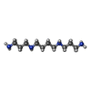
+Macromolecule #87: SPERMINE
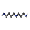
+Macromolecule #88: SPERMIDINE
-Experimental details
-Structure determination
| Method | cryo EM |
|---|---|
 Processing Processing | single particle reconstruction |
| Aggregation state | cell |
-Sample preparation
| Buffer | pH: 7.4 |
|---|---|
| Vitrification | Cryogen name: ETHANE |
- Electron microscopy
| Microscope | FEI TECNAI F30 |
|---|---|
| Image recording | Film or detector model: GATAN K3 (6k x 4k) / Average electron dose: 50.0 e/Å2 |
| Electron beam | Acceleration voltage: 300 kV / Electron source:  FIELD EMISSION GUN FIELD EMISSION GUN |
| Electron optics | Illumination mode: FLOOD BEAM / Imaging mode: BRIGHT FIELD / Nominal defocus max: 2.0 µm / Nominal defocus min: 1.2 µm |
| Experimental equipment | 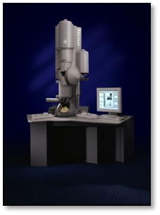 Model: Tecnai F30 / Image courtesy: FEI Company |

