ムービー
ムービー コントローラー


+ データを開く

- 基本情報
基本情報
| 登録情報 |  | |||||||||
|---|---|---|---|---|---|---|---|---|---|---|
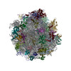
| タイトル | Reconstruction of the yeast 60S ribosomal subunit from CEMOVIS sections | |||||||||
 マップデータ マップデータ | 3D reconstruction calculated from 2DTM results, where the yeast 60S subunit was matched in micrographs from vitreous sections of Saccharomyces cerevisiae cells | |||||||||
 試料 試料 |
| |||||||||
 キーワード キーワード | Ribosome CEMOVIS Yeast / TRANSLATION | |||||||||
| 生物種 |  | |||||||||
| 手法 | 単粒子再構成法 / クライオ電子顕微鏡法 / 解像度: 3.5 Å | |||||||||
 データ登録者 データ登録者 | Elferich J / Kaminek M / Kong L / Odriozola A / Kukulski W / Zuber B / Grigorieff N | |||||||||
| 資金援助 |  米国, 米国,  スイス, 2件 スイス, 2件
| |||||||||
 引用 引用 | ジャーナル: IUCrJ / 年: 2025 タイトル: In situ high-resolution cryo-EM reconstructions from CEMOVIS. 著者: Johannes Elferich / Marek Kaminek / Lingli Kong / Adolfo Odriozola / Wanda Kukulski / Benoît Zuber / Nikolaus Grigorieff / 要旨: Cryo-electron microscopy can be used to image cells and tissue at high resolution. To ensure electron transparency, the sample thickness must not exceed 500 nm. Focused-ion-beam (FIB) milling has ...Cryo-electron microscopy can be used to image cells and tissue at high resolution. To ensure electron transparency, the sample thickness must not exceed 500 nm. Focused-ion-beam (FIB) milling has become the standard method for preparing thin samples (lamellae); however, the material removed by the milling process is lost, the imageable area is usually limited to a few square micrometres and the surface layers sustain damage from the ion beam. We have examined cryo-electron microscopy of vitreous sections (CEMOVIS), a technique based on cutting thin sections with a knife, as an alternative to FIB milling. Vitreous sections also sustain damage, including compression, shearing and cracks. However, samples can be sectioned in series, producing many orders of magnitude more imageable area compared to lamellae, making CEMOVIS an alternative to FIB milling with distinct advantages. Using two-dimensional template matching on images of vitreous sections of Saccharomyces cerevisiae cells, we reconstructed the 60S ribosomal subunit at near-atomic resolution, demonstrating that, in many regions of the sections, the molecular structure of these subunits is largely intact, comparable to FIB-milled lamellae. | |||||||||
| 履歴 |
|
- 構造の表示
構造の表示
| 添付画像 |
|---|

- ダウンロードとリンク
-EMDBアーカイブ
| マップデータ | emd_71068.map.gz | 474.5 MB |  EMDBマップデータ形式 EMDBマップデータ形式 | |
|---|---|---|---|---|
| ヘッダ (付随情報) | emd-71068-v30.xmlemd-71068.xml | 15.6 KB 15.6 KB | 表示 表示 | EMDBヘッダ |
| 画像 |  emd_71068.png emd_71068.png | 155.5 KB | ||
| Filedesc metadata | emd-71068.cif.gz | 4.3 KB | ||
| その他 | emd_71068_half_map_1.map.gzemd_71068_half_map_2.map.gz | 474.2 MB 474.2 MB | ||
| アーカイブディレクトリ |  http://ftp.pdbj.org/pub/emdb/structures/EMD-71068ftp://ftp.pdbj.org/pub/emdb/structures/EMD-71068 http://ftp.pdbj.org/pub/emdb/structures/EMD-71068ftp://ftp.pdbj.org/pub/emdb/structures/EMD-71068 | HTTPS FTP |
-検証レポート
| 文書・要旨 | emd_71068_validation.pdf.gz | 1.3 MB | 表示 | EMDB検証レポート |
|---|---|---|---|---|
| 文書・詳細版 | emd_71068_full_validation.pdf.gz | 1.3 MB | 表示 | |
| XML形式データ | emd_71068_validation.xml.gz | 18.8 KB | 表示 | |
| CIF形式データ | emd_71068_validation.cif.gz | 22.5 KB | 表示 | |
| アーカイブディレクトリ | https://ftp.pdbj.org/pub/emdb/validation_reports/EMD-71068ftp://ftp.pdbj.org/pub/emdb/validation_reports/EMD-71068 | HTTPS FTP |
-リンク
| EMDBのページ | EMDB (EBI/PDBe) / EMDataResource |
|---|
-マップ
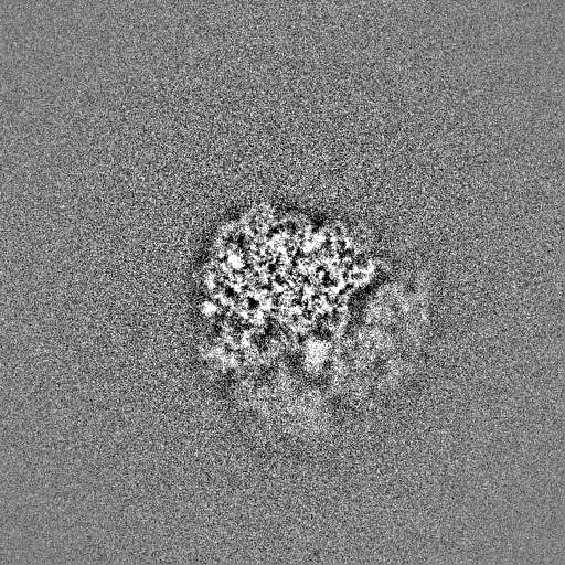
| ファイル | ダウンロード / ファイル: emd_71068.map.gz / 形式: CCP4 / 大きさ: 512 MB / タイプ: IMAGE STORED AS FLOATING POINT NUMBER (4 BYTES) | ||||||||||||||||||||||||||||||||||||
|---|---|---|---|---|---|---|---|---|---|---|---|---|---|---|---|---|---|---|---|---|---|---|---|---|---|---|---|---|---|---|---|---|---|---|---|---|---|
| 注釈 | 3D reconstruction calculated from 2DTM results, where the yeast 60S subunit was matched in micrographs from vitreous sections of Saccharomyces cerevisiae cells | ||||||||||||||||||||||||||||||||||||
| 投影像・断面図 | 画像のコントロール
画像は Spider により作成 | ||||||||||||||||||||||||||||||||||||
| ボクセルのサイズ | X=Y=Z: 1.17 Å | ||||||||||||||||||||||||||||||||||||
| 密度 |
| ||||||||||||||||||||||||||||||||||||
| 対称性 | 空間群: 1 | ||||||||||||||||||||||||||||||||||||
| 詳細 | EMDB XML:
|
 Z (Sec.)
 Y (Row.)
 X (Col.)


-添付データ
-ハーフマップ: Half-Map 2 of 3D reconstruction calculated from 2DTM...
| ファイル | emd_71068_half_map_1.map | ||||||||||||
|---|---|---|---|---|---|---|---|---|---|---|---|---|---|
| 注釈 | Half-Map 2 of 3D reconstruction calculated from 2DTM results, where the yeast 60S subunit was matched in micrographs from vitreous sections of Saccharomyces cerevisiae cells | ||||||||||||
| 投影像・断面図 |
| ||||||||||||
| 密度ヒストグラム |


-ハーフマップ: Half-Map 1 of 3D reconstruction calculated from 2DTM...
| ファイル | emd_71068_half_map_2.map | ||||||||||||
|---|---|---|---|---|---|---|---|---|---|---|---|---|---|
| 注釈 | Half-Map 1 of 3D reconstruction calculated from 2DTM results, where the yeast 60S subunit was matched in micrographs from vitreous sections of Saccharomyces cerevisiae cells | ||||||||||||
| 投影像・断面図 |
| ||||||||||||
| 密度ヒストグラム |


- 試料の構成要素
-全体 : Yeast cells sectioned using CEMOVIS
| 全体 | 名称: Yeast cells sectioned using CEMOVIS |
|---|---|
| 要素 |
|
-超分子 #1: Yeast cells sectioned using CEMOVIS
| 超分子 | 名称: Yeast cells sectioned using CEMOVIS / タイプ: cell / ID: 1 / 親要素: 0 |
|---|---|
| 由来(天然) | 生物種: |
-超分子 #2: 60S Ribosomal subunit
| 超分子 | 名称: 60S Ribosomal subunit / タイプ: complex / ID: 2 / 親要素: 1 |
|---|---|
| 由来(天然) | 生物種: |
| 分子量 | 理論値: 1.7 MDa |
-実験情報
-構造解析
| 手法 | クライオ電子顕微鏡法 |
|---|---|
 解析 解析 | 単粒子再構成法 |
| 試料の集合状態 | cell |
-試料調製
| 緩衝液 | pH: 7.4 |
|---|---|
| グリッド | モデル: Quantifoil R3.5/1 / 材質: COPPER / メッシュ: 200 / 支持フィルム - #0 - Film type ID: 1 / 支持フィルム - #0 - 材質: CARBON / 支持フィルム - #0 - トポロジー: HOLEY / 支持フィルム - #1 - Film type ID: 2 / 支持フィルム - #1 - トポロジー: HOLEY / 支持フィルム - #1 - Film thickness: 10 |
| 凍結 | 凍結剤: NITROGEN / 詳細: Leica EM PACT2 High Pressure Freezer. |
- 電子顕微鏡法
| 顕微鏡 | TFS KRIOS |
|---|---|
| 特殊光学系 | エネルギーフィルター - 名称: TFS Selectris / エネルギーフィルター - スリット幅: 20 eV |
| 撮影 | フィルム・検出器のモデル: TFS FALCON 4i (4k x 4k) 撮影したグリッド数: 2 / 実像数: 933 / 平均電子線量: 40.0 e/Å2 |
| 電子線 | 加速電圧: 300 kV / 電子線源:  FIELD EMISSION GUN FIELD EMISSION GUN |
| 電子光学系 | 照射モード: FLOOD BEAM / 撮影モード: BRIGHT FIELD / 最大 デフォーカス(公称値): 0.5 µm / 最小 デフォーカス(公称値): 0.5 µm |
| 試料ステージ | ホルダー冷却材: NITROGEN |
| 実験機器 |  モデル: Titan Krios / 画像提供: FEI Company |