Movie
Movie Controller

[English] 日本語
 Yorodumi
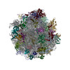
- EMDB-71068: Reconstruction of the yeast 60S ribosomal subunit from CEMOVIS se... -
+ Open data

- Basic information
| Entry |  | |||||||||
|---|---|---|---|---|---|---|---|---|---|---|
| Title | Reconstruction of the yeast 60S ribosomal subunit from CEMOVIS sections | |||||||||
 Map data Map data | 3D reconstruction calculated from 2DTM results, where the yeast 60S subunit was matched in micrographs from vitreous sections of Saccharomyces cerevisiae cells | |||||||||
 Sample Sample |
| |||||||||
 Keywords Keywords | Ribosome CEMOVIS Yeast / TRANSLATION | |||||||||
| Biological species |  | |||||||||
| Method | single particle reconstruction / cryo EM / Resolution: 3.5 Å | |||||||||
 Authors Authors | Elferich J / Kaminek M / Kong L / Odriozola A / Kukulski W / Zuber B / Grigorieff N | |||||||||
| Funding support |  United States, United States,  Switzerland, 2 items Switzerland, 2 items
| |||||||||
 Citation Citation | Journal: IUCrJ / Year: 2025 Title: In situ high-resolution cryo-EM reconstructions from CEMOVIS. Authors: Johannes Elferich / Marek Kaminek / Lingli Kong / Adolfo Odriozola / Wanda Kukulski / Benoît Zuber / Nikolaus Grigorieff / Abstract: Cryo-electron microscopy can be used to image cells and tissue at high resolution. To ensure electron transparency, the sample thickness must not exceed 500 nm. Focused-ion-beam (FIB) milling has ...Cryo-electron microscopy can be used to image cells and tissue at high resolution. To ensure electron transparency, the sample thickness must not exceed 500 nm. Focused-ion-beam (FIB) milling has become the standard method for preparing thin samples (lamellae); however, the material removed by the milling process is lost, the imageable area is usually limited to a few square micrometres and the surface layers sustain damage from the ion beam. We have examined cryo-electron microscopy of vitreous sections (CEMOVIS), a technique based on cutting thin sections with a knife, as an alternative to FIB milling. Vitreous sections also sustain damage, including compression, shearing and cracks. However, samples can be sectioned in series, producing many orders of magnitude more imageable area compared to lamellae, making CEMOVIS an alternative to FIB milling with distinct advantages. Using two-dimensional template matching on images of vitreous sections of Saccharomyces cerevisiae cells, we reconstructed the 60S ribosomal subunit at near-atomic resolution, demonstrating that, in many regions of the sections, the molecular structure of these subunits is largely intact, comparable to FIB-milled lamellae. | |||||||||
| History |
|
- Structure visualization
| Supplemental images |
|---|

- Downloads & links
-EMDB archive
| Map data | emd_71068.map.gz | 474.5 MB |  EMDB map data format EMDB map data format | |
|---|---|---|---|---|
| Header (meta data) | emd-71068-v30.xmlemd-71068.xml | 15.7 KB 15.7 KB | Display Display | EMDB header |
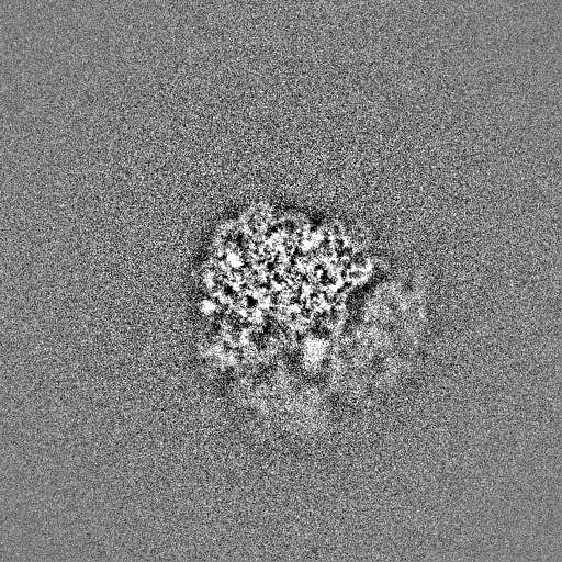
| Images |  emd_71068.png emd_71068.png | 155.5 KB | ||
| Filedesc metadata | emd-71068.cif.gz | 4.3 KB | ||
| Others | emd_71068_half_map_1.map.gzemd_71068_half_map_2.map.gz | 474.2 MB 474.2 MB | ||
| Archive directory |  http://ftp.pdbj.org/pub/emdb/structures/EMD-71068ftp://ftp.pdbj.org/pub/emdb/structures/EMD-71068 http://ftp.pdbj.org/pub/emdb/structures/EMD-71068ftp://ftp.pdbj.org/pub/emdb/structures/EMD-71068 | HTTPS FTP |
-Links
| EMDB pages | EMDB (EBI/PDBe) / EMDataResource |
|---|
-Map
| File | Download / File: emd_71068.map.gz / Format: CCP4 / Size: 512 MB / Type: IMAGE STORED AS FLOATING POINT NUMBER (4 BYTES) | ||||||||||||||||||||||||||||||||||||
|---|---|---|---|---|---|---|---|---|---|---|---|---|---|---|---|---|---|---|---|---|---|---|---|---|---|---|---|---|---|---|---|---|---|---|---|---|---|
| Annotation | 3D reconstruction calculated from 2DTM results, where the yeast 60S subunit was matched in micrographs from vitreous sections of Saccharomyces cerevisiae cells | ||||||||||||||||||||||||||||||||||||
| Projections & slices | Image control
Images are generated by Spider. | ||||||||||||||||||||||||||||||||||||
| Voxel size | X=Y=Z: 1.17 Å | ||||||||||||||||||||||||||||||||||||
| Density |
| ||||||||||||||||||||||||||||||||||||
| Symmetry | Space group: 1 | ||||||||||||||||||||||||||||||||||||
| Details | EMDB XML:
|
 Z (Sec.)
 Y (Row.)
 X (Col.)


-Supplemental data
-Half map: Half-Map 2 of 3D reconstruction calculated from 2DTM...
| File | emd_71068_half_map_1.map | ||||||||||||
|---|---|---|---|---|---|---|---|---|---|---|---|---|---|
| Annotation | Half-Map 2 of 3D reconstruction calculated from 2DTM results, where the yeast 60S subunit was matched in micrographs from vitreous sections of Saccharomyces cerevisiae cells | ||||||||||||
| Projections & Slices |
| ||||||||||||
| Density Histograms |


-Half map: Half-Map 1 of 3D reconstruction calculated from 2DTM...
| File | emd_71068_half_map_2.map | ||||||||||||
|---|---|---|---|---|---|---|---|---|---|---|---|---|---|
| Annotation | Half-Map 1 of 3D reconstruction calculated from 2DTM results, where the yeast 60S subunit was matched in micrographs from vitreous sections of Saccharomyces cerevisiae cells | ||||||||||||
| Projections & Slices |
| ||||||||||||
| Density Histograms |


- Sample components
-Entire : Yeast cells sectioned using CEMOVIS
| Entire | Name: Yeast cells sectioned using CEMOVIS |
|---|---|
| Components |
|
-Supramolecule #1: Yeast cells sectioned using CEMOVIS
| Supramolecule | Name: Yeast cells sectioned using CEMOVIS / type: cell / ID: 1 / Parent: 0 |
|---|---|
| Source (natural) | Organism: |
-Supramolecule #2: 60S Ribosomal subunit
| Supramolecule | Name: 60S Ribosomal subunit / type: complex / ID: 2 / Parent: 1 |
|---|---|
| Source (natural) | Organism: |
| Molecular weight | Theoretical: 1.7 MDa |
-Experimental details
-Structure determination
| Method | cryo EM |
|---|---|
 Processing Processing | single particle reconstruction |
| Aggregation state | cell |
-Sample preparation
| Buffer | pH: 7.4 |
|---|---|
| Grid | Model: Quantifoil R3.5/1 / Material: COPPER / Mesh: 200 / Support film - #0 - Film type ID: 1 / Support film - #0 - Material: CARBON / Support film - #0 - topology: HOLEY / Support film - #1 - Film type ID: 2 / Support film - #1 - topology: HOLEY / Support film - #1 - Film thickness: 10 |
| Vitrification | Cryogen name: NITROGEN / Details: Leica EM PACT2 High Pressure Freezer. |
- Electron microscopy
| Microscope | TFS KRIOS |
|---|---|
| Specialist optics | Energy filter - Name: TFS Selectris / Energy filter - Slit width: 20 eV |
| Image recording | Film or detector model: TFS FALCON 4i (4k x 4k) / Number grids imaged: 2 / Number real images: 933 / Average electron dose: 40.0 e/Å2 |
| Electron beam | Acceleration voltage: 300 kV / Electron source:  FIELD EMISSION GUN FIELD EMISSION GUN |
| Electron optics | Illumination mode: FLOOD BEAM / Imaging mode: BRIGHT FIELD / Nominal defocus max: 0.5 µm / Nominal defocus min: 0.5 µm |
| Sample stage | Cooling holder cryogen: NITROGEN |
| Experimental equipment |  Model: Titan Krios / Image courtesy: FEI Company |