National Institutes of Health/National Institute Of Allergy and Infectious Diseases (NIH/NIAID)
AI151055
United States
National Institutes of Health/National Institute of General Medical Sciences (NIH/NIGMS)
GM071940
United States
Citation
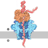
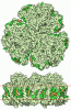
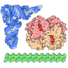
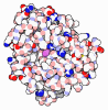
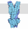
Journal: PLoS Pathog / Year: 2026 Title: Discovery and cryoEM structure of FPM13, a periplasmic metalloprotein unique to Francisella. Authors: Daniel L Clemens / Bai-Yu Lee / Xiaoyu Liu / Z Hong Zhou / Marcus A Horwitz / Abstract: We report the identification and cryoEM structure of the Francisella protein FTN_1118, a previously uncharacterized 13 kDa periplasmic protein unique to the Francisella genus. The protein was ...We report the identification and cryoEM structure of the Francisella protein FTN_1118, a previously uncharacterized 13 kDa periplasmic protein unique to the Francisella genus. The protein was serendipitously discovered during purification of Francisella type VI secretion system (T6SS) effector proteins and is hereby designated as FPM13 (Francisella Periplasmic Metalloprotein, 13 kDa) based on its cellular and biochemical properties. Identified by the cryoID approach based on our cryoEM density map, FPM13 exists naturally as a cylindrical 18-mer complex with 9-fold dihedral symmetry, formed by stacking two donut-shaped nonamers head-to-head. Measuring ~8 nm in height and outer diameter with a 3.5 nm central channel, the complex features a double-layered wall comprising an inner β-sheet core and an outer α-helical shell. Each FPM13 monomer adopts a compact fold comprising an N-terminus β-strand, an α-helix and two additional β strands at the C-terminus. Inter-ring loop interactions, hydrophobic contacts, and electrostatic interactions between adjacent subunits stabilize the assembly. Biochemical analyses, including APEX-biotinylation and Triton X-114 phase partitioning, confirmed FPM13 as a soluble periplasmic protein. Inductively coupled plasma mass spectrometry (ICP-MS) revealed FPM13 binds iron, copper, and zinc, with alanine substitution of predicted metal-binding cysteine and histidine residues abolishing this capability. Biochemical assays further revealed that wild-type FPM13 catalyzes disulfide bond formation and rescues alkaline phosphatase from reductive inactivation, indicating a role in maintaining periplasmic disulfide bonds. The metal-binding disruption mutant loses this oxidation activity. Deletion of FPM13 in Francisella novicida caused no growth defects in vitro, in macrophages, or in mice under tested conditions, suggesting functional redundancy may compensate for its absence. This study unveils a novel metalloprotein and demonstrates the power of cryoID in identifying uncharacterized proteins directly from structural data, offering new insights into Francisella biology.
In the structure databanks used in Yorodumi, some data are registered as the other names, "COVID-19 virus" and "2019-nCoV". Here are the details of the virus and the list of structure data.
Jan 31, 2019. EMDB accession codes are about to change! (news from PDBe EMDB page)
EMDB accession codes are about to change! (news from PDBe EMDB page)
The allocation of 4 digits for EMDB accession codes will soon come to an end. Whilst these codes will remain in use, new EMDB accession codes will include an additional digit and will expand incrementally as the available range of codes is exhausted. The current 4-digit format prefixed with “EMD-” (i.e. EMD-XXXX) will advance to a 5-digit format (i.e. EMD-XXXXX), and so on. It is currently estimated that the 4-digit codes will be depleted around Spring 2019, at which point the 5-digit format will come into force.
The EM Navigator/Yorodumi systems omit the EMD- prefix.
Related info.:Q: What is EMD? / ID/Accession-code notation in Yorodumi/EM Navigator
Yorodumi is a browser for structure data from EMDB, PDB, SASBDB, etc.
This page is also the successor to EM Navigator detail page, and also detail information page/front-end page for Omokage search.
The word "yorodu" (or yorozu) is an old Japanese word meaning "ten thousand". "mi" (miru) is to see.
Related info.:EMDB / PDB / SASBDB / Comparison of 3 databanks / Yorodumi Search / Aug 31, 2016. New EM Navigator & Yorodumi / Yorodumi Papers / Jmol/JSmol / Function and homology information / Changes in new EM Navigator and Yorodumi
 Movie
Movie Controller


 Open data
 Basic information
 Map data
 Sample
 Keywords
 Function and homology information
 Francisella tularensis subsp. novicida (bacteria) /
  Authors
 United States, 2 items
  Citation
 Structure visualization
 Downloads & links
 emd_70611.png
 http://ftp.pdbj.org/pub/emdb/structures/EMD-70611
 Z (Sec.)
 Y (Row.)
 X (Col.)


 Sample components
 Processing
 Electron microscopy
 FIELD EMISSION GUN
