 Movie
Movie Controller


+ Open data

- Basic information
| Entry |  | |||||||||
|---|---|---|---|---|---|---|---|---|---|---|
| Title | Extended conformation of dusk state KaiC | |||||||||
 Map data Map data | sharpened map used for model building | |||||||||
 Sample Sample |
| |||||||||
 Keywords Keywords | AAA ATPase / circadian oscillator / kinase / phosphatase / CIRCADIAN CLOCK PROTEIN | |||||||||
| Function / homology |  Function and homology information Function and homology informationregulation of phosphorelay signal transduction system / negative regulation of circadian rhythm / entrainment of circadian clock / protein serine/threonine/tyrosine kinase activity / circadian rhythm / Hydrolases; Acting on acid anhydrides; Acting on acid anhydrides to facilitate cellular and subcellular movement / non-specific serine/threonine protein kinase / protein serine kinase activity / protein serine/threonine kinase activity / regulation of DNA-templated transcription ...regulation of phosphorelay signal transduction system / negative regulation of circadian rhythm / entrainment of circadian clock / protein serine/threonine/tyrosine kinase activity / circadian rhythm / Hydrolases; Acting on acid anhydrides; Acting on acid anhydrides to facilitate cellular and subcellular movement / non-specific serine/threonine protein kinase / protein serine kinase activity / protein serine/threonine kinase activity / regulation of DNA-templated transcription / magnesium ion binding / ATP hydrolysis activity / DNA binding / ATP binding / identical protein binding Similarity search - Function | |||||||||
| Biological species |  Synechococcus elongatus (bacteria) Synechococcus elongatus (bacteria) | |||||||||
| Method | single particle reconstruction / cryo EM / Resolution: 2.59 Å | |||||||||
 Authors Authors | Dzimianski JV / Crosby P / Balasco Serrao VH / Partch CL | |||||||||
| Funding support |  United States, 2 items United States, 2 items
| |||||||||
 Citation Citation | Journal: To Be Published Title: Extended conformation of dusk state KaiC Authors: Dzimianski JV / Crosby P / Balasco Serrao VH / Partch CL | |||||||||
| History |
|
- Structure visualization
| Supplemental images |
|---|

- Downloads & links
-EMDB archive
| Map data | emd_70432.map.gz | 1.2 GB | EMDB map data format | |
|---|---|---|---|---|
| Header (meta data) | emd-70432-v30.xmlemd-70432.xml | 21.5 KB 21.5 KB | Display Display | EMDB header |
| FSC (resolution estimation) | emd_70432_fsc.xml | 23.3 KB | Display | FSC data file |
| Images |  emd_70432.png emd_70432.png | 92.3 KB | ||
| Masks | emd_70432_msk_1.map | 1.3 GB | Mask map | |
| Filedesc metadata | emd-70432.cif.gz | 6.5 KB | ||
| Others | emd_70432_additional_1.map.gzemd_70432_half_map_1.map.gzemd_70432_half_map_2.map.gz | 621.4 MB 1.2 GB 1.2 GB | ||
| Archive directory |  http://ftp.pdbj.org/pub/emdb/structures/EMD-70432ftp://ftp.pdbj.org/pub/emdb/structures/EMD-70432 http://ftp.pdbj.org/pub/emdb/structures/EMD-70432ftp://ftp.pdbj.org/pub/emdb/structures/EMD-70432 | HTTPS FTP |
-Related structure data
| Related structure data | 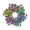 9ofhMC M: atomic model generated by this map C: citing same article ( |
|---|---|
| Similar structure data |
-Links
| EMDB pages | EMDB (EBI/PDBe) / EMDataResource |
|---|---|
| Related items in Molecule of the Month |

-Map
| File | Download / File: emd_70432.map.gz / Format: CCP4 / Size: 1.3 GB / Type: IMAGE STORED AS FLOATING POINT NUMBER (4 BYTES) | ||||||||||||||||||||||||||||||||||||
|---|---|---|---|---|---|---|---|---|---|---|---|---|---|---|---|---|---|---|---|---|---|---|---|---|---|---|---|---|---|---|---|---|---|---|---|---|---|
| Annotation | sharpened map used for model building | ||||||||||||||||||||||||||||||||||||
| Projections & slices | Image control
Images are generated by Spider. | ||||||||||||||||||||||||||||||||||||
| Voxel size | X=Y=Z: 0.3241 Å | ||||||||||||||||||||||||||||||||||||
| Density |
| ||||||||||||||||||||||||||||||||||||
| Symmetry | Space group: 1 | ||||||||||||||||||||||||||||||||||||
| Details | EMDB XML:
|
 Z (Sec.)
 Y (Row.)
 X (Col.)


-Supplemental data
-Mask #1
| File | emd_70432_msk_1.map | ||||||||||||
|---|---|---|---|---|---|---|---|---|---|---|---|---|---|
| Projections & Slices |
| ||||||||||||
| Density Histograms |


-Additional map: unsharpened map
| File | emd_70432_additional_1.map | ||||||||||||
|---|---|---|---|---|---|---|---|---|---|---|---|---|---|
| Annotation | unsharpened map | ||||||||||||
| Projections & Slices |
| ||||||||||||
| Density Histograms |


-Half map: half map A
| File | emd_70432_half_map_1.map | ||||||||||||
|---|---|---|---|---|---|---|---|---|---|---|---|---|---|
| Annotation | half map A | ||||||||||||
| Projections & Slices |
| ||||||||||||
| Density Histograms |


-Half map: half map B
| File | emd_70432_half_map_2.map | ||||||||||||
|---|---|---|---|---|---|---|---|---|---|---|---|---|---|
| Annotation | half map B | ||||||||||||
| Projections & Slices |
| ||||||||||||
| Density Histograms |


- Sample components
-Entire : KaiC hexamer
| Entire | Name: KaiC hexamer |
|---|---|
| Components |
|
-Supramolecule #1: KaiC hexamer
| Supramolecule | Name: KaiC hexamer / type: complex / ID: 1 / Parent: 0 / Macromolecule list: #1 |
|---|---|
| Source (natural) | Organism: Synechococcus elongatus (bacteria) |
-Macromolecule #1: Circadian clock oscillator protein KaiC
| Macromolecule | Name: Circadian clock oscillator protein KaiC / type: protein_or_peptide / ID: 1 / Number of copies: 6 / Enantiomer: LEVO / EC number: non-specific serine/threonine protein kinase |
|---|---|
| Source (natural) | Organism: Synechococcus elongatus (bacteria) |
| Molecular weight | Theoretical: 59.139801 KDa |
| Recombinant expression | Organism: |
| Sequence | String: DYKDDDDKMT SAEMTSPNNN SEHQAIAKMR TMIEGFDDIS HGGLPIGRST LVSGTSGTGK TLFSIQFLYN GIIEFDEPGV FVTFEETPQ DIIKNARSFG WDLAKLVDEG KLFILDASPD PEGQEVVGGF DLSALIERIN YAIQKYRARR VSIDSVTSVF Q QYDASSVV ...String: DYKDDDDKMT SAEMTSPNNN SEHQAIAKMR TMIEGFDDIS HGGLPIGRST LVSGTSGTGK TLFSIQFLYN GIIEFDEPGV FVTFEETPQ DIIKNARSFG WDLAKLVDEG KLFILDASPD PEGQEVVGGF DLSALIERIN YAIQKYRARR VSIDSVTSVF Q QYDASSVV RRELFRLVAR LKQIGATTVM TTERIEEYGP IARYGVEEFV SDNVVILRNV LEGERRRRTL EILKLRGTSH MK GEYPFTI TDHGINIFPL GAMRLTQRSS NVRVSSGVVR LDEMCGGGFF KDSIILATGA TGTGKTLLVS RFVENACANK ERA ILFAYE ESRAQLLRNA YSWGMDFEEM ERQNLLKIVC AYPESAGLED HLQIIKSEIN DFKPARIAID SLSALARGVS NNAF RQFVI GVTGYAKQEE ITGLFTNTSD QFMGAHSITD SHIEEITDTI ILLQYVEIRG EMSRAINVFK MRGSWHDKAI REFMI SDKG PDIKDSFRNF ERIISGSPTR ITVDEKSELS RIVRGVQEKG PES UniProtKB: Circadian clock oscillator protein KaiC |
-Macromolecule #2: ADENOSINE-5'-TRIPHOSPHATE
| Macromolecule | Name: ADENOSINE-5'-TRIPHOSPHATE / type: ligand / ID: 2 / Number of copies: 12 / Formula: ATP |
|---|---|
| Molecular weight | Theoretical: 507.181 Da |
| Chemical component information |  ChemComp-ATP: |
-Macromolecule #3: MAGNESIUM ION
| Macromolecule | Name: MAGNESIUM ION / type: ligand / ID: 3 / Number of copies: 12 / Formula: MG |
|---|---|
| Molecular weight | Theoretical: 24.305 Da |
-Experimental details
-Structure determination
| Method | cryo EM |
|---|---|
 Processing Processing | single particle reconstruction |
| Aggregation state | particle |
-Sample preparation
| Buffer | pH: 7.4 |
|---|---|
| Grid | Model: Quantifoil R2/2 / Material: COPPER / Mesh: 200 / Pretreatment - Type: GLOW DISCHARGE / Pretreatment - Time: 15 sec. |
| Vitrification | Cryogen name: ETHANE / Chamber humidity: 100 % / Chamber temperature: 298 K / Instrument: FEI VITROBOT MARK IV |
- Electron microscopy
| Microscope | TFS KRIOS |
|---|---|
| Image recording | Film or detector model: GATAN K3 BIOCONTINUUM (6k x 4k) / Number grids imaged: 2 / Number real images: 15001 / Average electron dose: 50.0 e/Å2 |
| Electron beam | Acceleration voltage: 300 kV / Electron source:  FIELD EMISSION GUN FIELD EMISSION GUN |
| Electron optics | Illumination mode: FLOOD BEAM / Imaging mode: BRIGHT FIELD / Nominal defocus max: 2.2 µm / Nominal defocus min: 0.5 µm / Nominal magnification: 130000 |
| Sample stage | Specimen holder model: FEI TITAN KRIOS AUTOGRID HOLDER / Cooling holder cryogen: NITROGEN |
| Experimental equipment |  Model: Titan Krios / Image courtesy: FEI Company |


