Movie
Movie Controller

[English] 日本語
 Yorodumi
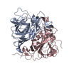
- EMDB-70397: Structure of the human astrovirus VA1 capsid spike bound to antib... -
+ Open data

- Basic information
| Entry |  | |||||||||
|---|---|---|---|---|---|---|---|---|---|---|
| Title | Structure of the human astrovirus VA1 capsid spike bound to antibody 7C8 | |||||||||
 Map data Map data | Main refined and sharpened map | |||||||||
 Sample Sample |
| |||||||||
 Keywords Keywords | Astrovirus / neutralizing antibody / epitope mapping / neurovirulent. / VIRAL PROTEIN | |||||||||
| Function / homology | Capsid, astroviral / Astrovirus capsid protein nucleoplasmin-like domain / T=3 icosahedral viral capsid / Viral coat protein subunit / clathrin-dependent endocytosis of virus by host cell / Capsid polyprotein VP86 Function and homology information Function and homology information | |||||||||
| Biological species |  Astrovirus VA1 / Astrovirus VA1 /   Mus musculus (house mouse) Mus musculus (house mouse) | |||||||||
| Method | single particle reconstruction / cryo EM / Resolution: 2.69 Å | |||||||||
 Authors Authors | Ghosh A / Dzimianski JV / Balasco Serrao VH / DuBois RM | |||||||||
| Funding support |  United States, 1 items United States, 1 items
| |||||||||
 Citation Citation | Journal: To Be Published Title: Cryo-EM Structures of the Human Astrovirus VA1 Capsid Spike Bound to Neutralizing Antibodies: Implications for Therapeutic Strategies Authors: Ghosh A / Dzimianski JV / Lopez T / Espinosa R / Balasco Serrao VH / Arias CF / DuBois RM | |||||||||
| History |
|
- Structure visualization
| Supplemental images |
|---|

- Downloads & links
-EMDB archive
| Map data | emd_70397.map.gz | 97.2 MB | EMDB map data format | |
|---|---|---|---|---|
| Header (meta data) | emd-70397-v30.xmlemd-70397.xml | 32.2 KB 32.2 KB | Display Display | EMDB header |
| FSC (resolution estimation) | emd_70397_fsc.xml | 9.9 KB | Display | FSC data file |
| Images |  emd_70397.png emd_70397.png | 67.4 KB | ||
| Masks | emd_70397_msk_1.map | 103 MB | Mask map | |
| Filedesc metadata | emd-70397.cif.gz | 7.1 KB | ||
| Others | emd_70397_additional_1.map.gzemd_70397_additional_2.map.gzemd_70397_additional_3.map.gzemd_70397_additional_4.map.gzemd_70397_half_map_1.map.gzemd_70397_half_map_2.map.gz | 95.6 MB 95.6 MB 51.6 MB 51.6 MB 95.5 MB 95.5 MB | ||
| Archive directory |  http://ftp.pdbj.org/pub/emdb/structures/EMD-70397ftp://ftp.pdbj.org/pub/emdb/structures/EMD-70397 http://ftp.pdbj.org/pub/emdb/structures/EMD-70397ftp://ftp.pdbj.org/pub/emdb/structures/EMD-70397 | HTTPS FTP |
-Related structure data
| Related structure data |  9oefMC  9oegC M: atomic model generated by this map C: citing same article ( |
|---|---|
| Similar structure data |

-Links
| EMDB pages | EMDB (EBI/PDBe) / EMDataResource |
|---|---|
| Related items in Molecule of the Month |
-Map
| File | Download / File: emd_70397.map.gz / Format: CCP4 / Size: 103 MB / Type: IMAGE STORED AS FLOATING POINT NUMBER (4 BYTES) | ||||||||||||||||||||||||||||||||||||
|---|---|---|---|---|---|---|---|---|---|---|---|---|---|---|---|---|---|---|---|---|---|---|---|---|---|---|---|---|---|---|---|---|---|---|---|---|---|
| Annotation | Main refined and sharpened map | ||||||||||||||||||||||||||||||||||||
| Projections & slices | Image control
Images are generated by Spider. | ||||||||||||||||||||||||||||||||||||
| Voxel size | X=Y=Z: 0.83 Å | ||||||||||||||||||||||||||||||||||||
| Density |
| ||||||||||||||||||||||||||||||||||||
| Symmetry | Space group: 1 | ||||||||||||||||||||||||||||||||||||
| Details | EMDB XML:
|
 Z (Sec.)
 Y (Row.)
 X (Col.)


-Supplemental data
-Mask #1
| File | emd_70397_msk_1.map | ||||||||||||
|---|---|---|---|---|---|---|---|---|---|---|---|---|---|
| Projections & Slices |
| ||||||||||||
| Density Histograms |


-Additional map: Original half map B before particle subtraction
| File | emd_70397_additional_1.map | ||||||||||||
|---|---|---|---|---|---|---|---|---|---|---|---|---|---|
| Annotation | Original half map B before particle subtraction | ||||||||||||
| Projections & Slices |
| ||||||||||||
| Density Histograms |


-Additional map: Original half map A before particle subtraction
| File | emd_70397_additional_2.map | ||||||||||||
|---|---|---|---|---|---|---|---|---|---|---|---|---|---|
| Annotation | Original half map A before particle subtraction | ||||||||||||
| Projections & Slices |
| ||||||||||||
| Density Histograms |


-Additional map: Unsharpened map
| File | emd_70397_additional_3.map | ||||||||||||
|---|---|---|---|---|---|---|---|---|---|---|---|---|---|
| Annotation | Unsharpened map | ||||||||||||
| Projections & Slices |
| ||||||||||||
| Density Histograms |


-Additional map: Original map before particle subtraction
| File | emd_70397_additional_4.map | ||||||||||||
|---|---|---|---|---|---|---|---|---|---|---|---|---|---|
| Annotation | Original map before particle subtraction | ||||||||||||
| Projections & Slices |
| ||||||||||||
| Density Histograms |


-Half map: Half-map B
| File | emd_70397_half_map_1.map | ||||||||||||
|---|---|---|---|---|---|---|---|---|---|---|---|---|---|
| Annotation | Half-map B | ||||||||||||
| Projections & Slices |
| ||||||||||||
| Density Histograms |


-Half map: Half-map A
| File | emd_70397_half_map_2.map | ||||||||||||
|---|---|---|---|---|---|---|---|---|---|---|---|---|---|
| Annotation | Half-map A | ||||||||||||
| Projections & Slices |
| ||||||||||||
| Density Histograms |


- Sample components
-Entire : Human astrovirus VA1 capsid spike bound to antibody 7C8
| Entire | Name: Human astrovirus VA1 capsid spike bound to antibody 7C8 |
|---|---|
| Components |
|
-Supramolecule #1: Human astrovirus VA1 capsid spike bound to antibody 7C8
| Supramolecule | Name: Human astrovirus VA1 capsid spike bound to antibody 7C8 type: complex / ID: 1 / Parent: 0 / Macromolecule list: all |
|---|---|
| Source (natural) | Organism: Astrovirus VA1 |
-Macromolecule #1: Spike protein VP38
| Macromolecule | Name: Spike protein VP38 / type: protein_or_peptide / ID: 1 / Number of copies: 2 / Enantiomer: LEVO |
|---|---|
| Source (natural) | Organism: Astrovirus VA1 |
| Molecular weight | Theoretical: 32.219271 KDa |
| Recombinant expression | Organism:   Escherichia coli (E. coli) Escherichia coli (E. coli) |
| Sequence | String: MGPIPPTGVP VGDFFVCGRM TTLHMGGQSG IQATTLVNGM IYRTDHPEPS SSPVSNWEFT VLENNTIVGA GMGCVWFQKS EALVWTLDG QKLSGWNTLD GVGTTQLTVA WRQHNRTIYG WANVVAWNSE EWHTNAEQPH QPILRLTYWL VKINVLSEPE D FDVVQKSP ...String: MGPIPPTGVP VGDFFVCGRM TTLHMGGQSG IQATTLVNGM IYRTDHPEPS SSPVSNWEFT VLENNTIVGA GMGCVWFQKS EALVWTLDG QKLSGWNTLD GVGTTQLTVA WRQHNRTIYG WANVVAWNSE EWHTNAEQPH QPILRLTYWL VKINVLSEPE D FDVVQKSP LAYLEDYTTA QSKSAIQKLN FQTFQKPEGG GTLRAQYSTT PRQGDFAVIW QIGRHNFDMS TGKGTPVESL SD YVMPQQK DAHIGMWYRA LTSVGPRTDV LTLHFHLPTV EKAAAELALV PR UniProtKB: Capsid polyprotein VP86 |
-Macromolecule #2: 7C8 heavy chain
| Macromolecule | Name: 7C8 heavy chain / type: protein_or_peptide / ID: 2 / Number of copies: 2 / Enantiomer: LEVO |
|---|---|
| Source (natural) | Organism: Mus musculus (house mouse) |
| Molecular weight | Theoretical: 23.465312 KDa |
| Recombinant expression | Organism: 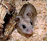 Cricetulus griseus (Chinese hamster) Cricetulus griseus (Chinese hamster) |
| Sequence | String: EVQLVESGGG LVKPGGSLKL SCAASGFTFS DYYMYWIRQT PEKRLEWVAT ISDGGFSTYY PDSVKGRFTI SRDNAKNNLF LQMSSLKSE DTAIYYCARF GAYSTFWGQQ GTLVTVSSAS TKGPSVFPLA PSSKSTSGGT AALGCLVKDY FPEPVTVSWN S GALTSGVH ...String: EVQLVESGGG LVKPGGSLKL SCAASGFTFS DYYMYWIRQT PEKRLEWVAT ISDGGFSTYY PDSVKGRFTI SRDNAKNNLF LQMSSLKSE DTAIYYCARF GAYSTFWGQQ GTLVTVSSAS TKGPSVFPLA PSSKSTSGGT AALGCLVKDY FPEPVTVSWN S GALTSGVH TFPAVLQSSG LYSLSSVVTV PSSSLGTQTY ICNVNHKPSN TKVDKKVEPK S |
-Macromolecule #3: 7C8 light chain
| Macromolecule | Name: 7C8 light chain / type: protein_or_peptide / ID: 3 / Number of copies: 2 / Enantiomer: LEVO |
|---|---|
| Source (natural) | Organism: Mus musculus (house mouse) |
| Molecular weight | Theoretical: 23.466078 KDa |
| Recombinant expression | Organism: Cricetulus griseus (Chinese hamster) |
| Sequence | String: QIVLSQSPAI LSASPGEKVT MTCRASSSVS YMYWYQQQKP GSPKPWIYAT SNLASGVPAR FSGSGSGTSY SLTIISRVEA EDATYYCQQ WSSDPPMTFG GGTTKLEIKR RTVAAPSVFI FPPSDEQLKS GTASVVCLLN NFYPREAKVQ WKVDNALQSG N SQESVTEQ ...String: QIVLSQSPAI LSASPGEKVT MTCRASSSVS YMYWYQQQKP GSPKPWIYAT SNLASGVPAR FSGSGSGTSY SLTIISRVEA EDATYYCQQ WSSDPPMTFG GGTTKLEIKR RTVAAPSVFI FPPSDEQLKS GTASVVCLLN NFYPREAKVQ WKVDNALQSG N SQESVTEQ DSKDSTYSLS STLTLSKADY EKHKVYACEV THQGLSSPVT KSFNRGE |
-Experimental details
-Structure determination
| Method | cryo EM |
|---|---|
 Processing Processing | single particle reconstruction |
| Aggregation state | particle |
-Sample preparation
| Concentration | 1 mg/mL |
|---|---|
| Buffer | pH: 8 |
| Grid | Model: UltrAuFoil R1.2/1.3 / Material: GOLD / Mesh: 300 / Pretreatment - Type: GLOW DISCHARGE / Pretreatment - Time: 15 sec. |
| Vitrification | Cryogen name: ETHANE / Chamber humidity: 100 % / Chamber temperature: 298 K / Instrument: FEI VITROBOT MARK IV |
- Electron microscopy
| Microscope | TFS KRIOS |
|---|---|
| Specialist optics | Energy filter - Name: GIF Bioquantum / Energy filter - Slit width: 10 eV |
| Image recording | Film or detector model: GATAN K3 BIOCONTINUUM (6k x 4k) / Number grids imaged: 1 / Number real images: 7270 / Average electron dose: 34.14 e/Å2 |
| Electron beam | Acceleration voltage: 300 kV / Electron source:  FIELD EMISSION GUN FIELD EMISSION GUN |
| Electron optics | Illumination mode: FLOOD BEAM / Imaging mode: BRIGHT FIELD / Nominal defocus max: 2.8000000000000003 µm / Nominal defocus min: 0.6 µm / Nominal magnification: 135000 |
| Sample stage | Specimen holder model: FEI TITAN KRIOS AUTOGRID HOLDER / Cooling holder cryogen: NITROGEN |
| Experimental equipment |  Model: Titan Krios / Image courtesy: FEI Company |
+Image processing

-Atomic model buiding 1
| Initial model |
| ||||||||||||||
|---|---|---|---|---|---|---|---|---|---|---|---|---|---|---|---|
| Refinement | Space: REAL / Protocol: RIGID BODY FIT | ||||||||||||||
| Output model | PDB-9oef: |