+ Open data
- Basic information
| Entry |  | |||||||||
|---|---|---|---|---|---|---|---|---|---|---|
| Title | Cryo-EM structure of AMT1-AMT7-AMTP1-AMTP2 complex | |||||||||
 Map data Map data | full map | |||||||||
 Sample Sample |
| |||||||||
 Keywords Keywords | 6mA methyltransferase / TRANSFERASE | |||||||||
| Function / homology |  Function and homology information Function and homology informationmRNA m6A methyltransferase / mRNA m(6)A methyltransferase activity / RNA N6-methyladenosine methyltransferase complex / methylation / membrane / nucleus Similarity search - Function | |||||||||
| Biological species |  Tetrahymena thermophila SB210 (eukaryote) Tetrahymena thermophila SB210 (eukaryote) | |||||||||
| Method | single particle reconstruction / cryo EM / Resolution: 3.59 Å | |||||||||
 Authors Authors | Song J / Shao Z | |||||||||
| Funding support |  United States, 1 items United States, 1 items
| |||||||||
 Citation Citation |  Journal: Protein Sci / Year: 2025 Journal: Protein Sci / Year: 2025Title: Structural insight into the substrate binding of the AMT complex via an inhibitor-trapped state. Authors: Zengyu Shao / Sol Yoon / Jiuwei Lu / Pranav Athavale / Yifan Liu / Jikui Song /  Abstract: N6-adenine (6mA) DNA methylation plays an important role in gene regulation and genome stability. The 6mA methylation in Tetrahymena thermophila is mainly mediated by the AMT complex, comprised of ...N6-adenine (6mA) DNA methylation plays an important role in gene regulation and genome stability. The 6mA methylation in Tetrahymena thermophila is mainly mediated by the AMT complex, comprised of the AMT1, AMT7, AMTP1, and AMTP2 subunits. To date, how this complex assembles on the DNA substrate remains elusive. Here we report the structure of the AMT complex bound to the OCR protein from bacteriophage T7, mimicking the AMT-DNA encounter complex. The AMT1-AMT7 heterodimer approaches OCR from one side, while the AMTP1 N-terminal domain, assuming a homeodomain fold, binds to OCR from the other side, resulting in a saddle-shaped architecture reminiscent of what was observed for prokaryotic 6mA writers. Mutation of the AMT1, AMT7, and AMTP1 residues on the OCR-contact points led to impaired DNA methylation activity to various extents, supporting a role for these residues in DNA binding. Furthermore, structural comparison of the AMT1-AMT7 subunits with the evolutionarily related METTL3-METTL14 and AMT1-AMT6 complexes reveals sequence conservation and divergence in the region corresponding to the OCR-binding site, shedding light on the substrate binding of the latter two complexes. Together, this study supports a model in which the AMT complex undergoes a substrate binding-induced open-to-closed conformational transition, with implications in its substrate binding and processive 6mA methylation. | |||||||||
| History |
|
- Structure visualization
| Supplemental images |
|---|
- Downloads & links
-EMDB archive
| Map data |  emd_70174.map.gz emd_70174.map.gz | 15.9 MB |  EMDB map data format EMDB map data format | |
|---|---|---|---|---|
| Header (meta data) |  emd-70174-v30.xml emd-70174-v30.xml emd-70174.xml emd-70174.xml | 22.4 KB 22.4 KB | Display Display |  EMDB header EMDB header |
| FSC (resolution estimation) |  emd_70174_fsc.xml emd_70174_fsc.xml | 6.6 KB | Display |  FSC data file FSC data file |
| Images |  emd_70174.png emd_70174.png | 38 KB | ||
| Filedesc metadata |  emd-70174.cif.gz emd-70174.cif.gz | 6.9 KB | ||
| Others |  emd_70174_half_map_1.map.gz emd_70174_half_map_1.map.gz emd_70174_half_map_2.map.gz emd_70174_half_map_2.map.gz | 28.3 MB 28.3 MB | ||
| Archive directory |  http://ftp.pdbj.org/pub/emdb/structures/EMD-70174 http://ftp.pdbj.org/pub/emdb/structures/EMD-70174 ftp://ftp.pdbj.org/pub/emdb/structures/EMD-70174 ftp://ftp.pdbj.org/pub/emdb/structures/EMD-70174 | HTTPS FTP |
-Related structure data
| Related structure data |  9o6kMC M: atomic model generated by this map C: citing same article ( |
|---|---|
| Similar structure data | Similarity search - Function & homology  F&H Search F&H Search |
- Links
Links
| EMDB pages |  EMDB (EBI/PDBe) / EMDB (EBI/PDBe) /  EMDataResource EMDataResource |
|---|---|
| Related items in Molecule of the Month |
- Map
Map
| File |  Download / File: emd_70174.map.gz / Format: CCP4 / Size: 30.5 MB / Type: IMAGE STORED AS FLOATING POINT NUMBER (4 BYTES) Download / File: emd_70174.map.gz / Format: CCP4 / Size: 30.5 MB / Type: IMAGE STORED AS FLOATING POINT NUMBER (4 BYTES) | ||||||||||||||||||||||||||||||||||||
|---|---|---|---|---|---|---|---|---|---|---|---|---|---|---|---|---|---|---|---|---|---|---|---|---|---|---|---|---|---|---|---|---|---|---|---|---|---|
| Annotation | full map | ||||||||||||||||||||||||||||||||||||
| Projections & slices | Image control
Images are generated by Spider. | ||||||||||||||||||||||||||||||||||||
| Voxel size | X=Y=Z: 1.3568 Å | ||||||||||||||||||||||||||||||||||||
| Density |
| ||||||||||||||||||||||||||||||||||||
| Symmetry | Space group: 1 | ||||||||||||||||||||||||||||||||||||
| Details | EMDB XML:
|
-Supplemental data
-Half map: half map
| File | emd_70174_half_map_1.map | ||||||||||||
|---|---|---|---|---|---|---|---|---|---|---|---|---|---|
| Annotation | half map | ||||||||||||
| Projections & Slices |
| ||||||||||||
| Density Histograms |
-Half map: half map
| File | emd_70174_half_map_2.map | ||||||||||||
|---|---|---|---|---|---|---|---|---|---|---|---|---|---|
| Annotation | half map | ||||||||||||
| Projections & Slices |
| ||||||||||||
| Density Histograms |
- Sample components
-Entire : AMT1-AMT7-AMTP1-AMTP2 complex
| Entire | Name: AMT1-AMT7-AMTP1-AMTP2 complex |
|---|---|
| Components |
|
-Supramolecule #1: AMT1-AMT7-AMTP1-AMTP2 complex
| Supramolecule | Name: AMT1-AMT7-AMTP1-AMTP2 complex / type: complex / ID: 1 / Parent: 0 / Macromolecule list: #1-#4 |
|---|---|
| Source (natural) | Organism:  Tetrahymena thermophila SB210 (eukaryote) Tetrahymena thermophila SB210 (eukaryote) |
-Macromolecule #1: AMT7
| Macromolecule | Name: AMT7 / type: protein_or_peptide / ID: 1 / Number of copies: 1 / Enantiomer: LEVO |
|---|---|
| Source (natural) | Organism:  Tetrahymena thermophila SB210 (eukaryote) Tetrahymena thermophila SB210 (eukaryote) |
| Molecular weight | Theoretical: 52.08343 KDa |
| Recombinant expression | Organism:  |
| Sequence | String: MGAPKKQEQE PIRLSTRTAS KKVDYLQLSN GKLEDFFDDL EEDNKPARNR SRSKKRGRKP LKKADSRSKT PSRVSNARGR SKSLGPRKT YPRKKNLSPD NQLSLLLKWR NDKIPLKSAS ETDNKCKVVN VKNIFKSDLS KYGANLQALF INALWKVKSR K EKEGLNIN ...String: MGAPKKQEQE PIRLSTRTAS KKVDYLQLSN GKLEDFFDDL EEDNKPARNR SRSKKRGRKP LKKADSRSKT PSRVSNARGR SKSLGPRKT YPRKKNLSPD NQLSLLLKWR NDKIPLKSAS ETDNKCKVVN VKNIFKSDLS KYGANLQALF INALWKVKSR K EKEGLNIN DLSNLKIPLS LMKNGILFIW SEKEILGQIV EIMEQKGFTY IENFSIMFLG LNKCLQSINH KDEDSQNSTA ST NNTNNEA ITSDLTLKDT SKFSDQIQDN HSEDSDQARK QQTPDDITQK KNKLLKKSSV PSIQKLFEED PVQTPSVNKP IEK SIEQVT QEKKFVMNNL DILKSTDINN LFLRNNYPYF KKTRHTLLMF RRIGDKNQKL ELRHQRTSDV VFEVTDEQDP SKVD TMMKE YVYQMIETLL PKAQFIPGVD KHLKMMELFA STDNYRPGWI SVIEK UniProtKB: Uncharacterized protein |
-Macromolecule #2: mRNA m(6)A methyltransferase
| Macromolecule | Name: mRNA m(6)A methyltransferase / type: protein_or_peptide / ID: 2 / Number of copies: 1 / Enantiomer: LEVO / EC number: mRNA m6A methyltransferase |
|---|---|
| Source (natural) | Organism:  Tetrahymena thermophila SB210 (eukaryote) Tetrahymena thermophila SB210 (eukaryote) |
| Molecular weight | Theoretical: 42.696059 KDa |
| Recombinant expression | Organism:  |
| Sequence | String: MSKAVNKKGL RPRKSDSILD HIKNKLDQEF LEDNENGEQS DEDYDQKSLN KAKKPYKKRQ TQNGSELVIS QQKTKAKASA NNKKSAKNS QKLDEEEKIV EEEDLSPQKN GAVSEDDQQQ EASTQEDDYL DRLPKSKKGL QGLLQDIEKR ILHYKQLFFK E QNEIANGK ...String: MSKAVNKKGL RPRKSDSILD HIKNKLDQEF LEDNENGEQS DEDYDQKSLN KAKKPYKKRQ TQNGSELVIS QQKTKAKASA NNKKSAKNS QKLDEEEKIV EEEDLSPQKN GAVSEDDQQQ EASTQEDDYL DRLPKSKKGL QGLLQDIEKR ILHYKQLFFK E QNEIANGK RSMVPDNSIP ICSDVTKLNF QALIDAQMRH AGKMFDVIMM DPPWQLSSSQ PSRGVAIAYD SLSDEKIQNM PI QSLQQDG FIFVWAINAK YRVTIKMIEN WGYKLVDEIT WVKKTVNGKI AKGHGFYLQH AKESCLIGVK GDVDNGRFKK NIA SDVIFS ERRGQSQKPE EIYQYINQLC PNGNYLEIFA RRNNLHDNWV SIGNEL UniProtKB: mRNA m(6)A methyltransferase |
-Macromolecule #3: Myb-like domain-containing protein
| Macromolecule | Name: Myb-like domain-containing protein / type: protein_or_peptide / ID: 3 / Number of copies: 1 / Enantiomer: LEVO |
|---|---|
| Source (natural) | Organism:  Tetrahymena thermophila SB210 (eukaryote) Tetrahymena thermophila SB210 (eukaryote) |
| Molecular weight | Theoretical: 41.602758 KDa |
| Recombinant expression | Organism:  |
| Sequence | String: MSLKKGKFQH NQSKSLWNYT LSPGWREEEV KILKSALQLF GIGKWKKIME SGCLPGKSIG QIYMQTQRLL GQQSLGDFMG LQIDLEAVF NQNMKKQDVL RKNNCIINTG DNPTKEERKR RIEQNRKIYG LSAKQIAEIK LPKVKKHAPQ YMTLEDIENE K FTNLEILT ...String: MSLKKGKFQH NQSKSLWNYT LSPGWREEEV KILKSALQLF GIGKWKKIME SGCLPGKSIG QIYMQTQRLL GQQSLGDFMG LQIDLEAVF NQNMKKQDVL RKNNCIINTG DNPTKEERKR RIEQNRKIYG LSAKQIAEIK LPKVKKHAPQ YMTLEDIENE K FTNLEILT HLYNLKAEIV RRLAEQGETI AQPSIIKSLN NLNHNLEQNQ NSNSSTETKV TLEQSGKKKY KVLAIEETEL QN GPIATNS QKKSINGKRK NNRKINSDSE GNEEDISLED IDSQESEINS EEIVEDDEED EQIEEPSKIK KRKKNPEQES EED DIEEDQ EEDELVVNEE EIFEDDDDDE DNQDSSEDDD DDED UniProtKB: Myb-like domain-containing protein |
-Macromolecule #4: AMTP2
| Macromolecule | Name: AMTP2 / type: protein_or_peptide / ID: 4 / Number of copies: 1 / Enantiomer: LEVO |
|---|---|
| Source (natural) | Organism:  Tetrahymena thermophila SB210 (eukaryote) Tetrahymena thermophila SB210 (eukaryote) |
| Molecular weight | Theoretical: 16.612893 KDa |
| Recombinant expression | Organism:  |
| Sequence | String: MKKNGKSQNQ PLDFTQYAKN MRKDLSNQDI CLEDGALNHS YFLTKKGQYW TPLNQKALQR GIELFGVGNW KEINYDEFSG KANIVELEL RTCMILGIND ITEYYGKKIS EEEQEEIKKS NIAKGKKENK LKDNIYQKLQ QMQ UniProtKB: Transmembrane protein, putative |
-Macromolecule #5: S-ADENOSYL-L-HOMOCYSTEINE
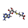
| Macromolecule | Name: S-ADENOSYL-L-HOMOCYSTEINE / type: ligand / ID: 5 / Number of copies: 1 / Formula: SAH |
|---|---|
| Molecular weight | Theoretical: 384.411 Da |
| Chemical component information |  ChemComp-SAH: |
-Experimental details
-Structure determination
| Method | cryo EM |
|---|---|
 Processing Processing | single particle reconstruction |
| Aggregation state | particle |
- Sample preparation
| Buffer | pH: 7.5 |
|---|---|
| Vitrification | Cryogen name: ETHANE |
- Electron microscopy
| Microscope | TFS KRIOS |
|---|---|
| Image recording | Film or detector model: GATAN K3 (6k x 4k) / Average electron dose: 53.55 e/Å2 |
| Electron beam | Acceleration voltage: 300 kV / Electron source:  FIELD EMISSION GUN FIELD EMISSION GUN |
| Electron optics | Illumination mode: FLOOD BEAM / Imaging mode: BRIGHT FIELD / Nominal defocus max: 3.1 µm / Nominal defocus min: 0.4 µm |
| Experimental equipment |  Model: Titan Krios / Image courtesy: FEI Company |
 Movie
Movie Controller


 Z (Sec.)
 Y (Row.)
 X (Col.)


