[English] 日本語
Yorodumi
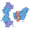
- EMDB-69118: Structure of mouse DNMT3A-TCL1A complex -

+
Open data


ID or keywords:

Loading...

-
Basic information

Entry
Database: EMDB / ID: EMD-69118
TitleStructure of mouse DNMT3A-TCL1A complex
Map data
Sample
  • Complex: DNMT3A-TCL1A complex
    • Protein or peptide: T-cell leukemia/lymphoma protein 1A
    • Protein or peptide: DNA (cytosine-5)-methyltransferase 3A
KeywordsDNA methyltransferase / complex / TRANSFERASE
Function / homology
Function and homology information


DNA (cytosine-5-)-methyltransferase activity, acting on CpN substrates / autosome genomic imprinting / cellular response to bisphenol A / epigenetic programming of gene expression / PRC2 methylates histones and DNA / genomic imprinting / transposable element silencing by piRNA-mediated DNA methylation / RMTs methylate histone arginines / protein-cysteine methyltransferase activity / positive regulation of cellular response to hypoxia ...DNA (cytosine-5-)-methyltransferase activity, acting on CpN substrates / autosome genomic imprinting / cellular response to bisphenol A / epigenetic programming of gene expression / PRC2 methylates histones and DNA / genomic imprinting / transposable element silencing by piRNA-mediated DNA methylation / RMTs methylate histone arginines / protein-cysteine methyltransferase activity / positive regulation of cellular response to hypoxia / regulatory ncRNA-mediated heterochromatin formation / unmethylated CpG binding / DNA (cytosine-5-)-methyltransferase / DNA (cytosine-5-)-methyltransferase activity / hepatocyte apoptotic process / response to vitamin A / post-embryonic development / XY body / DNA methylation-dependent constitutive heterochromatin formation / activation of protein kinase B activity / negative regulation of gene expression via chromosomal CpG island methylation / response to ionizing radiation / lncRNA binding / catalytic complex / cellular response to ethanol / chromosome, centromeric region / heterochromatin / protein serine/threonine kinase activator activity / Transferases; Transferring one-carbon groups; Methyltransferases / cellular response to amino acid stimulus / response to cocaine / euchromatin / response to toxic substance / response to lead ion / nuclear matrix / response to estradiol / neuron differentiation / methylation / heterochromatin formation / spermatogenesis / cellular response to hypoxia / RNA polymerase II-specific DNA-binding transcription factor binding / intracellular signal transduction / response to xenobiotic stimulus / RNA polymerase II cis-regulatory region sequence-specific DNA binding / negative regulation of DNA-templated transcription / chromatin binding / negative regulation of transcription by RNA polymerase II / endoplasmic reticulum / DNA binding / DNA-templated transcription / nucleoplasm / zinc ion binding / identical protein binding / nucleus / cytoplasm
Similarity search - Function
TCL1/MTCP1 / TCL1/MTCP1 superfamily / TCL1/MTCP1 family / DNA (cytosine-5)-methyltransferase 3A, ADD domain / : / DNA (cytosine-5-)-methyltransferase, N-terminal / DNMT3, cysteine rich ADD domain / : / DNMT3, cysteine rich ADD domain, GATA1-like zinc finger / DNMT3, ADD PHD zinc finger ...TCL1/MTCP1 / TCL1/MTCP1 superfamily / TCL1/MTCP1 family / DNA (cytosine-5)-methyltransferase 3A, ADD domain / : / DNA (cytosine-5-)-methyltransferase, N-terminal / DNMT3, cysteine rich ADD domain / : / DNMT3, cysteine rich ADD domain, GATA1-like zinc finger / DNMT3, ADD PHD zinc finger / ADD domain / ADD domain profile. / : / DNA methylase, C-5 cytosine-specific, active site / C-5 cytosine-specific DNA methylases active site. / C-5 cytosine-specific DNA methylase (Dnmt) domain profile. / C-5 cytosine methyltransferase / C-5 cytosine-specific DNA methylase / domain with conserved PWWP motif / PWWP domain / PWWP domain profile. / PWWP domain / S-adenosyl-L-methionine-dependent methyltransferase superfamily
Similarity search - Domain/homology
DNA (cytosine-5)-methyltransferase 3A / T-cell leukemia/lymphoma protein 1A
Similarity search - Component
Biological speciesMus musculus (house mouse)
Methodsingle particle reconstruction / cryo EM / Resolution: 3.32 Å
AuthorsLi W / Liu Q / Li J / Wang X / Guo L / He G
Funding support China, 1 items
OrganizationGrant numberCountry
National Natural Science Foundation of China (NSFC)2021YFC2701501 China
CitationJournal: J Struct Biol / Year: 2026
Title: Cryo-EM structure of the murine DNMT3A-TCL1A complex.
Authors: Wei Li / Qingting Liu / Jinhong Li / Xiang Wang / Guolin He / Li Guo /
Abstract: DNA methyltransferase DNMT3A is a key enzyme responsible for establishing DNA methylation patterns during mammalian development. T-cell leukemia/lymphoma 1 A (TCL1A) is a proto-oncogene expressed ...DNA methyltransferase DNMT3A is a key enzyme responsible for establishing DNA methylation patterns during mammalian development. T-cell leukemia/lymphoma 1 A (TCL1A) is a proto-oncogene expressed mainly in embryonic and fetal tissues, as well as in specific lymphocyte populations. In this study, we determined the structure of the murine DNMT3A-TCL1A complex using single-particle cryo-electron microscopy. The complex adopts a linear conformation, with two TCL1A dimers bound to the catalytic domain of DNMT3A to form a heterohexamer. TCL1A competitively binds to the same structural interface on DNMT3A as DNMT3L, but produces an inhibitory-rather than an activating-effect on the catalytic activity of DNMT3A. Furthermore, comparative analysis with previously reported assembly modes of murine TCL1A revealed that the TCL1A dimer complex we resolved adopts distinct molecular conformations and interaction mechanisms. Our findings elucidate the allosteric mechanism by which murine TCL1A inhibits DNMT3A activity, providing a structural basis for understanding mammalian epigenetic reprogramming.
History
DepositionFeb 11, 2026-
Header (metadata) releaseAug 19, 2026-
Map releaseAug 19, 2026-
UpdateAug 19, 2026-
Current statusAug 19, 2026Processing site: PDBc / Status: Released

-
Structure visualization

Supplemental images

Downloads & links

-
Map

FileDownload / File: emd_69118.map.gz / Format: CCP4 / Size: 64 MB / Type: IMAGE STORED AS FLOATING POINT NUMBER (4 BYTES)
Projections & slices

Image control

Size
Brightness
Contrast
Others
AxesZ (Sec.)Y (Row.)X (Col.)
1.1 Å/pix.
x 256 pix.
= 281.6 Å
1.1 Å/pix.
x 256 pix.
= 281.6 Å
1.1 Å/pix.
x 256 pix.
= 281.6 Å

Surface

Projections

Slices (1/3)

Slices (1/2)

Slices (2/3)

Images are generated by Spider.

Voxel sizeX=Y=Z: 1.1 Å
Density
Contour LevelBy AUTHOR: 0.068
Minimum - Maximum-0.2492243 - 0.6887947
Average (Standard dev.)0.00093856594 (±0.0162285)
SymmetrySpace group: 1
Details

EMDB XML:

Map geometry
Axis orderXYZ
Origin000
Dimensions256256256
Spacing256256256
CellA=B=C: 281.6 Å
α=β=γ: 90.0 °

-
Supplemental data

-
Mask #1

Fileemd_69118_msk_1.map
Projections & Slices
AxesZYX

Projections

Slices (1/2)
Density Histograms

-
Half map: #2

Fileemd_69118_half_map_1.map
Projections & Slices
AxesZYX

Projections

Slices (1/2)
Density Histograms

-
Half map: #1

Fileemd_69118_half_map_2.map
Projections & Slices
AxesZYX

Projections

Slices (1/2)
Density Histograms

-
Sample components

-
Entire : DNMT3A-TCL1A complex

EntireName: DNMT3A-TCL1A complex
Components
  • Complex: DNMT3A-TCL1A complex
    • Protein or peptide: T-cell leukemia/lymphoma protein 1A
    • Protein or peptide: DNA (cytosine-5)-methyltransferase 3A

-
Supramolecule #1: DNMT3A-TCL1A complex

SupramoleculeName: DNMT3A-TCL1A complex / type: complex / ID: 1 / Parent: 0 / Macromolecule list: all
Source (natural)Organism: Mus musculus (house mouse)

-
Macromolecule #1: T-cell leukemia/lymphoma protein 1A

MacromoleculeName: T-cell leukemia/lymphoma protein 1A / type: protein_or_peptide / ID: 1 / Number of copies: 4 / Enantiomer: LEVO
Source (natural)Organism: Mus musculus (house mouse)
Molecular weightTheoretical: 14.130103 KDa
Recombinant expressionOrganism: Spodoptera frugiperda (fall armyworm)
SequenceString:
MATQRAHRAE TPAHPNRLWI WEKHVYLDEF RRSWLPVVIK SNEKFQVILR QEDVTLGEAM SPSQLVPYEL PLMWQLYPKD RYRSCDSMY WQILYHIKFR DVEDMLLELI DSESNDE

UniProtKB: T-cell leukemia/lymphoma protein 1A

-
Macromolecule #2: DNA (cytosine-5)-methyltransferase 3A

MacromoleculeName: DNA (cytosine-5)-methyltransferase 3A / type: protein_or_peptide / ID: 2 / Number of copies: 2 / Enantiomer: LEVO / EC number: DNA (cytosine-5-)-methyltransferase
Source (natural)Organism: Mus musculus (house mouse)
Molecular weightTheoretical: 93.76643 KDa
Recombinant expressionOrganism: Spodoptera frugiperda (fall armyworm)
SequenceString: PMAQDSGPSD LLPNGDLEKR SEPQPEEGSP AAGQKGGAPA EGEGTETPPE ASRAVENGCC VTKEGRGASA GEGKEQKQTN IESMKMEGS RGRLRGGLGW ESSLRQRPMP RLTFQAGDPY YISKRKRDEW LARWKREAEK KAKVIAVMNA VEENQASGES Q KVEEASPP ...String:
PMAQDSGPSD LLPNGDLEKR SEPQPEEGSP AAGQKGGAPA EGEGTETPPE ASRAVENGCC VTKEGRGASA GEGKEQKQTN IESMKMEGS RGRLRGGLGW ESSLRQRPMP RLTFQAGDPY YISKRKRDEW LARWKREAEK KAKVIAVMNA VEENQASGES Q KVEEASPP AVQQPTDPAS PTVATTPEPV GGDAGDKNAT KAADDEPEYE DGRGFGIGEL VWGKLRGFSW WPGRIVSWWM TG RSRAAEG TRWVMWFGDG KFSVVCVEKL MPLSSFCSAF HQATYNKQPM YRKAIYEVLQ VASSRAGKLF PACHDSDESD SGK AVEVQN KQMIEWALGG FQPSGPKGLE PPEEEKNPYK EVYTDMWVEP EAAAYAPPPP AKKPRKSTTE KPKVKEIIDE RTRE RLVYE VRQKCRNIED ICISCGSLNV TLEHPLFIGG MCQNCKNCFL ECAYQYDDDG YQSYCTICCG GREVLMCGNN NCCRC FCVE CVDLLVGPGA AQAAIKEDPW NCYMCGHKGT YGLLRRREDW PSRLQMFFAN NHDQEFDPPK VYPPVPAEKR KPIRVL SLF DGIATGLLVL KDLGIQVDRY IASEVCEDSI TVGMVRHQGK IMYVGDVRSV TQKHIQEWGP FDLVIGGSPC NDLSIVN PA RKGLYEGTGR LFFEFYRLLH DARPKEGDDR PFFWLFENVV AMGVSDKRDI SRFLESNPVM IDAKEVSAAH RARYFWGN L PGMNRPLAST VNDKLELQEC LEHGRIAKFS KVRTITTRSN SIKQGKDQHF PVFMNEKEDI LWCTEMERVF GFPVHYTDV SNMSRLARQR LLGRSWSVPV IRHLFAPLKE YFACV

UniProtKB: DNA (cytosine-5)-methyltransferase 3A

-
Experimental details

-
Structure determination

Methodcryo EM
Processingsingle particle reconstruction
Aggregation stateparticle

-
Sample preparation

BufferpH: 7.5
VitrificationCryogen name: ETHANE

-
Electron microscopy

MicroscopeTFS KRIOS
Image recordingFilm or detector model: GATAN K2 SUMMIT (4k x 4k) / Average electron dose: 55.13 e/Å2
Electron beamAcceleration voltage: 300 kV / Electron source: FIELD EMISSION GUN
Electron opticsIllumination mode: FLOOD BEAM / Imaging mode: BRIGHT FIELD / Nominal defocus max: 1.8 µm / Nominal defocus min: 1.1 µm
Experimental equipment
Model: Titan Krios / Image courtesy: FEI Company

+
Image processing

CTF correctionType: NONE
Startup modelType of model: NONE
Final reconstructionResolution.type: BY AUTHOR / Resolution: 3.32 Å / Resolution method: FSC 0.143 CUT-OFF / Software - Name: cryoSPARC / Number images used: 76682
Initial angle assignmentType: ANGULAR RECONSTITUTION
Final angle assignmentType: ANGULAR RECONSTITUTION
FSC plot (resolution estimation)

+
About Yorodumi

-
News

-
Feb 9, 2022. New format data for meta-information of EMDB entries

New format data for meta-information of EMDB entries

  • Version 3 of the EMDB header file is now the official format.
  • The previous official version 1.9 will be removed from the archive.

Related info.:EMDB header

External links:wwPDB to switch to version 3 of the EMDB data model

-
Aug 12, 2020. Covid-19 info

Covid-19 info

URL: https://pdbj.org/emnavi/covid19.php

New page: Covid-19 featured information page in EM Navigator.

Related info.:Covid-19 info / Mar 5, 2020. Novel coronavirus structure data

+
Mar 5, 2020. Novel coronavirus structure data

Novel coronavirus structure data

Related info.:Yorodumi Speices / Aug 12, 2020. Covid-19 info

External links:COVID-19 featured content - PDBj / Molecule of the Month (242):Coronavirus Proteases

+
Jan 31, 2019. EMDB accession codes are about to change! (news from PDBe EMDB page)

EMDB accession codes are about to change! (news from PDBe EMDB page)

  • The allocation of 4 digits for EMDB accession codes will soon come to an end. Whilst these codes will remain in use, new EMDB accession codes will include an additional digit and will expand incrementally as the available range of codes is exhausted. The current 4-digit format prefixed with “EMD-” (i.e. EMD-XXXX) will advance to a 5-digit format (i.e. EMD-XXXXX), and so on. It is currently estimated that the 4-digit codes will be depleted around Spring 2019, at which point the 5-digit format will come into force.
  • The EM Navigator/Yorodumi systems omit the EMD- prefix.

Related info.:Q: What is EMD? / ID/Accession-code notation in Yorodumi/EM Navigator

External links:EMDB Accession Codes are Changing Soon! / Contact to PDBj

+
Jul 12, 2017. Major update of PDB

Major update of PDB

  • wwPDB released updated PDB data conforming to the new PDBx/mmCIF dictionary.
  • This is a major update changing the version number from 4 to 5, and with Remediation, in which all the entries are updated.
  • In this update, many items about electron microscopy experimental information are reorganized (e.g. em_software).
  • Now, EM Navigator and Yorodumi are based on the updated data.

External links:wwPDB Remediation / Enriched Model Files Conforming to OneDep Data Standards Now Available in the PDB FTP Archive

-
Yorodumi

Thousand views of thousand structures

  • Yorodumi is a browser for structure data from EMDB, PDB, SASBDB, etc.
  • This page is also the successor to EM Navigator detail page, and also detail information page/front-end page for Omokage search.
  • The word "yorodu" (or yorozu) is an old Japanese word meaning "ten thousand". "mi" (miru) is to see.

Related info.:EMDB / PDB / SASBDB / Comparison of 3 databanks / Yorodumi Search / Aug 31, 2016. New EM Navigator & Yorodumi / Yorodumi Papers / Jmol/JSmol / Function and homology information / Changes in new EM Navigator and Yorodumi

Read more