 Movie
Movie Controller

[English] 日本語
 Yorodumi
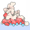
- EMDB-68645: Client peptide-bound structure of a MucD trimer within a 24mer cage -
+ Open data

- Basic information
| Entry |  | |||||||||
|---|---|---|---|---|---|---|---|---|---|---|
| Title | Client peptide-bound structure of a MucD trimer within a 24mer cage | |||||||||
 Map data Map data | ||||||||||
 Sample Sample |
| |||||||||
 Keywords Keywords | Complex / Protease / Cryo-EM / HYDROLASE | |||||||||
| Function / homology |  Function and homology information Function and homology informationalginic acid biosynthetic process / peptidase Do / cell outer membrane / periplasmic space / serine-type endopeptidase activity / signal transduction / proteolysis Similarity search - Function | |||||||||
| Biological species |  Pseudomonas aeruginosa PAO1 (bacteria) Pseudomonas aeruginosa PAO1 (bacteria) | |||||||||
| Method | single particle reconstruction / cryo EM / Resolution: 3.0 Å | |||||||||
 Authors Authors | Jiang YJ / Gao YG | |||||||||
| Funding support |  Singapore, 1 items Singapore, 1 items
| |||||||||
 Citation Citation | Journal: PLoS Biol / Year: 2026 Title: MucD regulates alginate biosynthesis through the proteolytic control of AlgX and AlgK in Pseudomonas aeruginosa. Authors: Yujun Jiang / Xin-Fu Yan / Rya Ero / Chao Wang / Kanaga Sabapathy / Yong-Gui Gao / Abstract: Pseudomonas aeruginosa is an opportunistic human pathogen capable of infecting a wide range of tissues and organs. Its persistence during chronic infection is strongly associated with biofilm ...Pseudomonas aeruginosa is an opportunistic human pathogen capable of infecting a wide range of tissues and organs. Its persistence during chronic infection is strongly associated with biofilm formation, which depends on extracellular polysaccharides such as alginate. The HtrA-like periplasmic serine protease MucD is a key regulator of bacterial virulence, stress response, and alginate production, yet its molecular mechanism has remained largely unclear. Here, we discovered the alginate acetylation and export proteins AlgX and AlgK as MucD substrates, and characterized their degradation by mass spectrometry and bioinformatic analysis. We further determined the cryo-EM structure of MucD bound to an AlgK-derived substrate peptide, offering atomic insights into MucD oligomerization assembly, substrate recognition, and specificity. Together with structure-guided mutagenesis and biochemical assays, our results revealed that MucD proteolytic activity is governed by an equilibrium between a resting 12-mer and an active trimer. Crucially, we demonstrate that MucD represses alginate biosynthesis post-translationally, in addition to its previously implicated role in transcriptional regulation. These findings define a distinct activation mechanism and regulatory function for MucD and provide new insight into bacterial HtrA-like serine proteases. | |||||||||
| History |
|
- Structure visualization
| Supplemental images |
|---|

- Downloads & links
-EMDB archive
| Map data | emd_68645.map.gz | 122.6 MB | EMDB map data format | |
|---|---|---|---|---|
| Header (meta data) | emd-68645-v30.xmlemd-68645.xml | 18.9 KB 18.9 KB | Display Display | EMDB header |
| FSC (resolution estimation) | emd_68645_fsc.xml | 13.2 KB | Display | FSC data file |
| Images |  emd_68645.png emd_68645.png | 46.3 KB | ||
| Filedesc metadata | emd-68645.cif.gz | 6.1 KB | ||
| Others | emd_68645_half_map_1.map.gzemd_68645_half_map_2.map.gz | 226.7 MB 226.7 MB | ||
| Archive directory |  https://data.pdbj.org/pub/emdb/structures/EMD-68645ftp://data.pdbj.org/pub/emdb/structures/EMD-68645 https://data.pdbj.org/pub/emdb/structures/EMD-68645ftp://data.pdbj.org/pub/emdb/structures/EMD-68645 | HTTPS FTP |
-Related structure data
| Related structure data |  22skMC  22kgC  24xxC  24yeC M: atomic model generated by this map C: citing same article ( |
|---|---|
| Similar structure data |


-Links
| EMDB pages | EMDB (EBI/PDBe) / EMDataResource |
|---|---|
| Related items in Molecule of the Month |
-Map
| File | Download / File: emd_68645.map.gz / Format: CCP4 / Size: 244.1 MB / Type: IMAGE STORED AS FLOATING POINT NUMBER (4 BYTES) | ||||||||||||||||||||||||||||||||||||
|---|---|---|---|---|---|---|---|---|---|---|---|---|---|---|---|---|---|---|---|---|---|---|---|---|---|---|---|---|---|---|---|---|---|---|---|---|---|
| Projections & slices | Image control
Images are generated by Spider. | ||||||||||||||||||||||||||||||||||||
| Voxel size | X=Y=Z: 0.97 Å | ||||||||||||||||||||||||||||||||||||
| Density |
| ||||||||||||||||||||||||||||||||||||
| Symmetry | Space group: 1 | ||||||||||||||||||||||||||||||||||||
| Details | EMDB XML:
|
 Z (Sec.)
 Y (Row.)
 X (Col.)


-Supplemental data
-Half map: #2
| File | emd_68645_half_map_1.map | ||||||||||||
|---|---|---|---|---|---|---|---|---|---|---|---|---|---|
| Projections & Slices |
| ||||||||||||
| Density Histograms |


-Half map: #1
| File | emd_68645_half_map_2.map | ||||||||||||
|---|---|---|---|---|---|---|---|---|---|---|---|---|---|
| Projections & Slices |
| ||||||||||||
| Density Histograms |


- Sample components
-Entire : Client peptide-bound structure of a MucD trimer within a 24mer cage
| Entire | Name: Client peptide-bound structure of a MucD trimer within a 24mer cage |
|---|---|
| Components |
|
-Supramolecule #1: Client peptide-bound structure of a MucD trimer within a 24mer cage
| Supramolecule | Name: Client peptide-bound structure of a MucD trimer within a 24mer cage type: complex / ID: 1 / Parent: 0 / Macromolecule list: all |
|---|---|
| Source (natural) | Organism: Pseudomonas aeruginosa PAO1 (bacteria) |
-Macromolecule #1: Probable periplasmic serine endoprotease DegP-like
| Macromolecule | Name: Probable periplasmic serine endoprotease DegP-like / type: protein_or_peptide / ID: 1 Details: All chains contain S217A mutation and 6*His tag at C-terminus. Number of copies: 6 / Enantiomer: LEVO / EC number: peptidase Do |
|---|---|
| Source (natural) | Organism: Pseudomonas aeruginosa PAO1 (bacteria) |
| Molecular weight | Theoretical: 51.192441 KDa |
| Recombinant expression | Organism: Pseudomonas aeruginosa PAO1 (bacteria) |
| Sequence | String: MHTLKRCMAA MVALLALSLA MTARAELPDF TPLVEQASPA VVNISTRQKL PDRAMARGQL SIPDLEGLPP MFRDFLERSI PQVPRNPRG QQREAQSLGS GFIISNDGYI LTNNHVVADA DEILVRLSDR SEHKAKLIGA DPRSDVAVLK IEAKNLPTLK L GDSNKLKV ...String: MHTLKRCMAA MVALLALSLA MTARAELPDF TPLVEQASPA VVNISTRQKL PDRAMARGQL SIPDLEGLPP MFRDFLERSI PQVPRNPRG QQREAQSLGS GFIISNDGYI LTNNHVVADA DEILVRLSDR SEHKAKLIGA DPRSDVAVLK IEAKNLPTLK L GDSNKLKV GEWVLAIGSP FGFDHSVTAG IVSAKGRSLP NESYVPFIQT DVAINPGNAG GPLLNLQGEV VGINSQIFTR SG GFMGLSF AIPIDVALNV ADQLKKAGKV SRGWLGVVIQ EVNKDLAESF GLDKPSGALV AQLVEDGPAA KGGLQVGDVI LSL NGQSIN ESADLPHLVG NMKPGDKINL DVIRNGQRKS LSMAVGSLPD DDEEIASMGA PGAERSSNRL GVTVADLTAE QRKS LDIQG GVVIKEVQDG PAAVIGLRPG DVITHLDNKA VTSTKVFADV AKALPKNRSV SMRVLRQGRA SFITFKLAEH HHHHH UniProtKB: Probable periplasmic serine endoprotease DegP-like |
-Macromolecule #2: Alginate biosynthesis protein AlgK
| Macromolecule | Name: Alginate biosynthesis protein AlgK / type: protein_or_peptide / ID: 2 / Number of copies: 3 / Enantiomer: LEVO |
|---|---|
| Source (natural) | Organism: Pseudomonas aeruginosa PAO1 (bacteria) |
| Molecular weight | Theoretical: 1.996292 KDa |
| Sequence | String: VDHLILAARA GQASADMALA UniProtKB: Alginate biosynthesis protein AlgK |
-Experimental details
-Structure determination
| Method | cryo EM |
|---|---|
 Processing Processing | single particle reconstruction |
| Aggregation state | particle |
-Sample preparation
| Buffer | pH: 8 |
|---|---|
| Vitrification | Cryogen name: ETHANE |
- Electron microscopy
| Microscope | TFS KRIOS |
|---|---|
| Image recording | Film or detector model: GATAN K3 (6k x 4k) / Average electron dose: 40.0 e/Å2 |
| Electron beam | Acceleration voltage: 300 kV / Electron source:  FIELD EMISSION GUN FIELD EMISSION GUN |
| Electron optics | Illumination mode: OTHER / Imaging mode: BRIGHT FIELD / Nominal defocus max: 1.2 µm / Nominal defocus min: 0.6 µm |
| Experimental equipment |  Model: Titan Krios / Image courtesy: FEI Company |

