Journal: Proc Natl Acad Sci U S A / Year: 2018 Title: Flagellum couples cell shape to motility in . Authors: Stella Y Sun / Jason T Kaelber / Muyuan Chen / Xiaoduo Dong / Yasaman Nematbakhsh / Jian Shi / Matthew Dougherty / Chwee Teck Lim / Michael F Schmid / Wah Chiu / Cynthia Y He / Abstract: In the unicellular parasite the causative agent of human African sleeping sickness, complex swimming behavior is driven by a flagellum laterally attached to the long and slender cell body. Using ...In the unicellular parasite the causative agent of human African sleeping sickness, complex swimming behavior is driven by a flagellum laterally attached to the long and slender cell body. Using microfluidic assays, we demonstrated that can penetrate through an orifice smaller than its maximum diameter. Efficient motility and penetration depend on active flagellar beating. To understand how active beating of the flagellum affects the cell body, we genetically engineered to produce anucleate cytoplasts (zoids and minis) with different flagellar attachment configurations and different swimming behaviors. We used cryo-electron tomography (cryo-ET) to visualize zoids and minis vitrified in different motility states. We showed that flagellar wave patterns reflective of their motility states are coupled to cytoskeleton deformation. Based on these observations, we propose a mechanism for how flagellum beating can deform the cell body via a flexible connection between the flagellar axoneme and the cell body. This mechanism may be critical for to disseminate in its host through size-limiting barriers.
In the structure databanks used in Yorodumi, some data are registered as the other names, "COVID-19 virus" and "2019-nCoV". Here are the details of the virus and the list of structure data.
Jan 31, 2019. EMDB accession codes are about to change! (news from PDBe EMDB page)
EMDB accession codes are about to change! (news from PDBe EMDB page)
The allocation of 4 digits for EMDB accession codes will soon come to an end. Whilst these codes will remain in use, new EMDB accession codes will include an additional digit and will expand incrementally as the available range of codes is exhausted. The current 4-digit format prefixed with “EMD-” (i.e. EMD-XXXX) will advance to a 5-digit format (i.e. EMD-XXXXX), and so on. It is currently estimated that the 4-digit codes will be depleted around Spring 2019, at which point the 5-digit format will come into force.
The EM Navigator/Yorodumi systems omit the EMD- prefix.
Related info.:Q: What is EMD? / ID/Accession-code notation in Yorodumi/EM Navigator
Yorodumi is a browser for structure data from EMDB, PDB, SASBDB, etc.
This page is also the successor to EM Navigator detail page, and also detail information page/front-end page for Omokage search.
The word "yorodu" (or yorozu) is an old Japanese word meaning "ten thousand". "mi" (miru) is to see.
Related info.:EMDB / PDB / SASBDB / Comparison of 3 databanks / Yorodumi Search / Aug 31, 2016. New EM Navigator & Yorodumi / Yorodumi Papers / Jmol/JSmol / Function and homology information / Changes in new EM Navigator and Yorodumi
 Movie
Movie Controller


 Open data
 Basic information
 Map data
 Sample
 Authors
 Citation

 Structure visualization
 Movie viewer
 Downloads & links
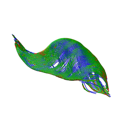 emd_6748.png
 http://ftp.pdbj.org/pub/emdb/structures/EMD-6748
 Sample components
 Processing
 Electron microscopy
 FIELD EMISSION GUN
