 Movie
Movie Controller

[English] 日本語
 Yorodumi
- EMDB-66913: Structure of dodecameric TpkB bound to AMP-PNP from Thermus therm... -
+ Open data

- Basic information
| Entry |  | |||||||||
|---|---|---|---|---|---|---|---|---|---|---|
| Title | Structure of dodecameric TpkB bound to AMP-PNP from Thermus thermophilus | |||||||||
 Map data Map data | full map | |||||||||
 Sample Sample |
| |||||||||
 Keywords Keywords | HYPOTHETICAL PROTEIN / ATP-BINDING / PRK/YEAG FAMILY / STRUCTURAL GENOMICS | |||||||||
| Function / homology | PrkA C-terminal domain / PrkA AAA domain / Serine-protein kinase, PrkA / PrkA serine protein kinase C-terminal domain / PrkA AAA domain / PrkA AAA domain / protein kinase activity / P-loop containing nucleoside triphosphate hydrolase / Serine protein kinase Function and homology information Function and homology information | |||||||||
| Biological species |   Thermus thermophilus HB8 (bacteria) Thermus thermophilus HB8 (bacteria) | |||||||||
| Method | single particle reconstruction / cryo EM / Resolution: 3.2 Å | |||||||||
 Authors Authors | Torri M / Kanno R / Mizoguchi A / Humbel B / Tani K / Masui R | |||||||||
| Funding support |  Japan, 2 items Japan, 2 items
| |||||||||
 Citation Citation | Journal: J Biochem / Year: 2026 Title: Structure and biochemical analyses suggest that PrkA/YeaG protein of Thermus thermophilus functions as a putative molecular chaperone. Authors: Masayuki Torii / Ryo Kanno / Akira Mizoguchi / Bruno M Humbel / Kazutoshi Tani / Ryoji Masui / Abstract: Protein phosphorylation, a key post-translational modification, is mediated by various protein kinases. The bacterial PrkA/YeaG is recognized as an atypical protein kinase, but its activity remains ...Protein phosphorylation, a key post-translational modification, is mediated by various protein kinases. The bacterial PrkA/YeaG is recognized as an atypical protein kinase, but its activity remains elusive. This study investigated the structural and functional characteristics of a PrkA/YeaG homologue, TpkB, from Thermus thermophilus HB8. Using cryo-electron microscopy, the structures of TpkB in apo and AMPPNP-bound forms were determined, revealing a hexameric ring architecture characteristic of AAA + superfamily proteins. The TpkB protomer is comprised of an N-terminal domain, an ATPase domain, and a LID domain. The ATPase domain contains conserved sequence motifs associated with ATPase activity, whereas the other domains present a novel fold. TpkB exhibits structural similarity to MoxR family proteins, which possess chaperone-like functions in conjunction with von Willebrand factor A (vWA) domain proteins. Structural and gene neighborhood analyses suggested a functional link between PrkA/YeaG proteins and vWA domain proteins. Biochemical analyses demonstrated that TpkB exhibited ATPase activity and chaperone-like activity, but lacked detectable protein kinase activity. These findings establish TpkB as a novel member of the AAA+ superfamily with potential chaperone functions, providing new insights into the PrkA/YeaG family. | |||||||||
| History |
|
- Structure visualization
| Supplemental images |
|---|

- Downloads & links
-EMDB archive
| Map data | emd_66913.map.gz | 445.9 MB | EMDB map data format | |
|---|---|---|---|---|
| Header (meta data) | emd-66913-v30.xmlemd-66913.xml | 19.3 KB 19.3 KB | Display Display | EMDB header |
| FSC (resolution estimation) | emd_66913_fsc.xml | 18 KB | Display | FSC data file |
| Images |  emd_66913.png emd_66913.png | 108.8 KB | ||
| Filedesc metadata | emd-66913.cif.gz | 6.4 KB | ||
| Others | emd_66913_half_map_1.map.gzemd_66913_half_map_2.map.gz | 380 MB 380.1 MB | ||
| Archive directory |  http://ftp.pdbj.org/pub/emdb/structures/EMD-66913ftp://ftp.pdbj.org/pub/emdb/structures/EMD-66913 http://ftp.pdbj.org/pub/emdb/structures/EMD-66913ftp://ftp.pdbj.org/pub/emdb/structures/EMD-66913 | HTTPS FTP |
-Related structure data
| Related structure data |  9xirMC  9xipC  9xiqC M: atomic model generated by this map C: citing same article ( |
|---|---|
| Similar structure data |


-Links
| EMDB pages | EMDB (EBI/PDBe) / EMDataResource |
|---|
-Map
| File | Download / File: emd_66913.map.gz / Format: CCP4 / Size: 476.8 MB / Type: IMAGE STORED AS FLOATING POINT NUMBER (4 BYTES) | ||||||||||||||||||||||||||||||||||||
|---|---|---|---|---|---|---|---|---|---|---|---|---|---|---|---|---|---|---|---|---|---|---|---|---|---|---|---|---|---|---|---|---|---|---|---|---|---|
| Annotation | full map | ||||||||||||||||||||||||||||||||||||
| Projections & slices | Image control
Images are generated by Spider. | ||||||||||||||||||||||||||||||||||||
| Voxel size | X=Y=Z: 0.82 Å | ||||||||||||||||||||||||||||||||||||
| Density |
| ||||||||||||||||||||||||||||||||||||
| Symmetry | Space group: 1 | ||||||||||||||||||||||||||||||||||||
| Details | EMDB XML:
|
 Z (Sec.)
 Y (Row.)
 X (Col.)


-Supplemental data
-Half map: odd half map
| File | emd_66913_half_map_1.map | ||||||||||||
|---|---|---|---|---|---|---|---|---|---|---|---|---|---|
| Annotation | odd half map | ||||||||||||
| Projections & Slices |
| ||||||||||||
| Density Histograms |


-Half map: even half map
| File | emd_66913_half_map_2.map | ||||||||||||
|---|---|---|---|---|---|---|---|---|---|---|---|---|---|
| Annotation | even half map | ||||||||||||
| Projections & Slices |
| ||||||||||||
| Density Histograms |


- Sample components
-Entire : Dodecameric TpkB
| Entire | Name: Dodecameric TpkB |
|---|---|
| Components |
|
-Supramolecule #1: Dodecameric TpkB
| Supramolecule | Name: Dodecameric TpkB / type: complex / ID: 1 / Parent: 0 / Macromolecule list: #1 |
|---|---|
| Source (natural) | Organism: Thermus thermophilus HB8 (bacteria) |
-Macromolecule #1: Serine protein kinase
| Macromolecule | Name: Serine protein kinase / type: protein_or_peptide / ID: 1 / Number of copies: 12 / Enantiomer: LEVO |
|---|---|
| Source (natural) | Organism: Thermus thermophilus HB8 (bacteria) |
| Molecular weight | Theoretical: 79.879211 KDa |
| Recombinant expression | Organism:  Escherichia coli (E. coli) Escherichia coli (E. coli) |
| Sequence | String: MGSSHHHHHH SSGENLYFQG GLNHMSELDF VRRGQDLKAY RALNWEGSFA DYLRLLKEDP RPLRTSFQRV HDMILAHGVE EYTRFKEKL LHYRFFDDPF EGGKDAVFGL DKPLMRLVAT LKAAAHRLGP ERRILLLHGP VGSAKSTIAR LLKKGLEAYS R TEEGKLFT ...String: MGSSHHHHHH SSGENLYFQG GLNHMSELDF VRRGQDLKAY RALNWEGSFA DYLRLLKEDP RPLRTSFQRV HDMILAHGVE EYTRFKEKL LHYRFFDDPF EGGKDAVFGL DKPLMRLVAT LKAAAHRLGP ERRILLLHGP VGSAKSTIAR LLKKGLEAYS R TEEGKLFT FYWKTEEGPL PCPMHEEPLH LLPQDLREAF LEELRALHPD YPYPLEVEGD LCPVCRFQMR EGLRKYEGDL AA LLEHEVV VKRLVLSEKD RVGIGTFQPK DEKNQDSTEL TGDINYRKVA LYGSDSDPRA FNFDGELNIA NRGIVEFIEI LKL DVAFLY DLLTASQEHK IKSKKFAQTD IDEIVLGHTN EPEYRKLQAN EYMEALRDRT IKIDVPYILR VSDEVRIYQR DFAK VRGKH IAPHTLEMAA TWAVLTRLEP PKRAGLTLMQ KLKLYDGKLL PGWTEEAVRE LMGEAKREGL EGISPRYIQD KISNV LVTS EEPCINPFMV MNELEEGLKH HSLISDERTR ERYRALLQEV KAEYAEIVKN EVQRAIAADE EALNRLFHNY IDHVKA YVL GEKVKNPYTG APEPPNERLM RSIEERIDIP ESRKDDFRRE IMNYIGALAL EGKPFTYKDN DRLRRALELK LFDDQKD TI RLSALVSGVV DPETQAKIDV VKARLIRDYG YCEHCASGVL EFAASLFARS UniProtKB: Serine protein kinase |
-Macromolecule #2: PHOSPHOAMINOPHOSPHONIC ACID-ADENYLATE ESTER
| Macromolecule | Name: PHOSPHOAMINOPHOSPHONIC ACID-ADENYLATE ESTER / type: ligand / ID: 2 / Number of copies: 12 / Formula: ANP |
|---|---|
| Molecular weight | Theoretical: 506.196 Da |
| Chemical component information | 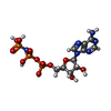 ChemComp-ANP: |
-Experimental details
-Structure determination
| Method | cryo EM |
|---|---|
 Processing Processing | single particle reconstruction |
| Aggregation state | particle |
-Sample preparation
| Concentration | 5.6 mg/mL | ||||||||||||
|---|---|---|---|---|---|---|---|---|---|---|---|---|---|
| Buffer | pH: 7.5 Component:
| ||||||||||||
| Vitrification | Cryogen name: ETHANE / Chamber humidity: 80 % / Chamber temperature: 277 K / Instrument: LEICA EM GP |
- Electron microscopy
| Microscope | TFS KRIOS |
|---|---|
| Image recording | Film or detector model: FEI FALCON III (4k x 4k) / Detector mode: COUNTING / Average electron dose: 1.0 e/Å2 |
| Electron beam | Acceleration voltage: 300 kV / Electron source:  FIELD EMISSION GUN FIELD EMISSION GUN |
| Electron optics | Illumination mode: FLOOD BEAM / Imaging mode: BRIGHT FIELD / Nominal defocus max: 3.3000000000000003 µm / Nominal defocus min: 1.0 µm |
| Sample stage | Specimen holder model: FEI TITAN KRIOS AUTOGRID HOLDER |
| Experimental equipment |  Model: Titan Krios / Image courtesy: FEI Company |
+Image processing

-Atomic model buiding 1
| Refinement | Protocol: AB INITIO MODEL |
|---|---|
| Output model | PDB-9xir: |
