 Movie
Movie Controller


+ Open data

- Basic information
| Entry |  | |||||||||
|---|---|---|---|---|---|---|---|---|---|---|
| Title | Cryo-EM structure of Ceg14-AnkJ-Actin complex | |||||||||
 Map data Map data | ||||||||||
 Sample Sample |
| |||||||||
 Keywords Keywords | Legionella / Ceg14 / AnkJ / Actin / CELL INVASION | |||||||||
| Biological species |  Legionella (bacteria) / Legionella (bacteria) /   Lepus (hares) Lepus (hares) | |||||||||
| Method | single particle reconstruction / cryo EM / Resolution: 2.52 Å | |||||||||
 Authors Authors | Li Y / Li S / Zheng Q / Wu Y | |||||||||
| Funding support | 1 items
| |||||||||
 Citation Citation | Journal: Nat Commun / Year: 2026 Title: Molecular basis of host ATP level modulation by actin-dependent secreted bacterial ATPase and its metaeffector. Authors: Hongxin Guan / Yu Li / Luhao Zhang / Song Xie / Yuchen Jiang / Chunlin He / Fan Li / Yin-di Jiao / Mengrou Gan / Yiran Sha / Fengjie Zheng / Kaiqiong Zhang / Zhao-Qing Luo / Qi Lai / Jinfu ...Authors: Hongxin Guan / Yu Li / Luhao Zhang / Song Xie / Yuchen Jiang / Chunlin He / Fan Li / Yin-di Jiao / Mengrou Gan / Yiran Sha / Fengjie Zheng / Kaiqiong Zhang / Zhao-Qing Luo / Qi Lai / Jinfu Su / Ningshao Xia / Qingbing Zheng / Jinyu Li / Shaowei Li / Songying Ouyang /  Abstract: Legionella pneumophila employs effectors including kinases, phosphoryl-AMPylases, ATPases, etc. to exploit host ATP for infection. Ceg14, a member of the S-HxxxE family, modulates host-cell energy ...Legionella pneumophila employs effectors including kinases, phosphoryl-AMPylases, ATPases, etc. to exploit host ATP for infection. Ceg14, a member of the S-HxxxE family, modulates host-cell energy levels in concert with host actin and the metaeffector AnkJ: actin activates while AnkJ inhibits Ceg14 ATPase activity. However, the molecular basis of this regulation remains unclear. Here we present Cryo-EM structures of Ceg14-actin, Ceg14-AnkJ, and Ceg14-actin-AnkJ complexes at 2.89 Å, 2.93 Å, and 2.52 Å, respectively. Actin binds to the C-terminal α-helix of the Ceg14 catalytic domain (CD), inducing rearrangement of its N-terminal domain (NTD) and reconfiguration of the flexible Lid domain. Surprisingly, AnkJ binds to the surface opposite the catalytic pocket rather than occupying the pocket. Using integrated in silico, in vitro, and in cellulo approaches, we propose a mechanism for Ceg14-mediated ATP hydrolysis. Actin binding triggers NTD rotation, driving the catalytic pocket through open, intermediate, and closed states. In the open state, H571 catalyzes ATP conversion to AMP and PPi. AnkJ binding to an allosteric site locks the intermediate state, thereby inhibiting hydrolysis. Together, our study reveals the molecular mechanism by which actin and AnkJ reciprocally regulate Ceg14 activity. | |||||||||
| History |
|
- Structure visualization
| Supplemental images |
|---|

- Downloads & links
-EMDB archive
| Map data | emd_66735.map.gz | 113.5 MB |  EMDB map data format EMDB map data format | |
|---|---|---|---|---|
| Header (meta data) | emd-66735-v30.xmlemd-66735.xml | 18 KB 18 KB | Display Display | EMDB header |
| Images |  emd_66735.png emd_66735.png | 57.3 KB | ||
| Filedesc metadata | emd-66735.cif.gz | 6.3 KB | ||
| Others | emd_66735_half_map_1.map.gzemd_66735_half_map_2.map.gz | 120.5 MB 120.5 MB | ||
| Archive directory |  https://data.pdbj.org/pub/emdb/structures/EMD-66735ftp://data.pdbj.org/pub/emdb/structures/EMD-66735 https://data.pdbj.org/pub/emdb/structures/EMD-66735ftp://data.pdbj.org/pub/emdb/structures/EMD-66735 | HTTPS FTP |
-Related structure data
| Related structure data |  9xcmMC  9xchC  9xfhC M: atomic model generated by this map C: citing same article ( |
|---|


-Links
| EMDB pages | EMDB (EBI/PDBe) / EMDataResource |
|---|
-Map
| File | Download / File: emd_66735.map.gz / Format: CCP4 / Size: 129.7 MB / Type: IMAGE STORED AS FLOATING POINT NUMBER (4 BYTES) | ||||||||||||||||||||||||||||||||||||
|---|---|---|---|---|---|---|---|---|---|---|---|---|---|---|---|---|---|---|---|---|---|---|---|---|---|---|---|---|---|---|---|---|---|---|---|---|---|
| Projections & slices | Image control
Images are generated by Spider. | ||||||||||||||||||||||||||||||||||||
| Voxel size | X=Y=Z: 0.65 Å | ||||||||||||||||||||||||||||||||||||
| Density |
| ||||||||||||||||||||||||||||||||||||
| Symmetry | Space group: 1 | ||||||||||||||||||||||||||||||||||||
| Details | EMDB XML:
|
 Z (Sec.)
 Y (Row.)
 X (Col.)


-Supplemental data
-Half map: #1
| File | emd_66735_half_map_1.map | ||||||||||||
|---|---|---|---|---|---|---|---|---|---|---|---|---|---|
| Projections & Slices |
| ||||||||||||
| Density Histograms |


-Half map: #2
| File | emd_66735_half_map_2.map | ||||||||||||
|---|---|---|---|---|---|---|---|---|---|---|---|---|---|
| Projections & Slices |
| ||||||||||||
| Density Histograms |


- Sample components
-Entire : Ceg14-AnkJ-Actin complex
| Entire | Name: Ceg14-AnkJ-Actin complex |
|---|---|
| Components |
|
-Supramolecule #1: Ceg14-AnkJ-Actin complex
| Supramolecule | Name: Ceg14-AnkJ-Actin complex / type: complex / ID: 1 / Parent: 0 / Macromolecule list: #1-#3 |
|---|---|
| Source (natural) | Organism: Legionella (bacteria) |
-Macromolecule #1: Actin
| Macromolecule | Name: Actin / type: protein_or_peptide / ID: 1 / Number of copies: 1 / Enantiomer: LEVO |
|---|---|
| Source (natural) | Organism: Lepus (hares) |
| Molecular weight | Theoretical: 42.096953 KDa |
| Recombinant expression | Organism: Lepus (hares) |
| Sequence | String: MCDEDETTAL VCDNGSGLVK AGFAGDDAPR AVFPSIVGRP RHQGVMVGMG QKDSYVGDEA QSKRGILTLK YPIEHGIITN WDDMEKIWH HTFYNELRVA PEEHPTLLTE APLNPKANRE KMTQIMFETF NVPAMYVAIQ AVLSLYASGR TTGIVLDSGD G VTHNVPIY ...String: MCDEDETTAL VCDNGSGLVK AGFAGDDAPR AVFPSIVGRP RHQGVMVGMG QKDSYVGDEA QSKRGILTLK YPIEHGIITN WDDMEKIWH HTFYNELRVA PEEHPTLLTE APLNPKANRE KMTQIMFETF NVPAMYVAIQ AVLSLYASGR TTGIVLDSGD G VTHNVPIY EGYALPHAIM RLDLAGRDLT DYLMKILTER GYSFVTTAER EIVRDIKEKL CYVALDFENE MATAASSSSL EK SYELPDG QVITIGNERF RCPETLFQPS FIGMESAGIH ETTYNSIMKC DIDIRKDLYA NNVMSGGTTM YPGIADRMQK EIT ALAPST MKIKIIAPPE RKYSVWIGGS ILASLSTFQQ MWITKQEYDE AGPSIVHRKC F |
-Macromolecule #2: Ceg14
| Macromolecule | Name: Ceg14 / type: protein_or_peptide / ID: 2 / Number of copies: 1 / Enantiomer: LEVO |
|---|---|
| Source (natural) | Organism: Legionella (bacteria) |
| Molecular weight | Theoretical: 75.982969 KDa |
| Recombinant expression | Organism:  Escherichia coli (E. coli) Escherichia coli (E. coli) |
| Sequence | String: MQNLDEILKK LRKDLKKTSK ETTEKLSVLS KKVAEDVVKV SNQTVQKINE VTKKFTVDTT VSTIDTQKIL DDFMKNIRFS RFKQIWQAI EKTPKSKHIL LKSLFLKGLL THNKPLLEEI IRQIELIPYS SDLEMLGAFS QDKAHPLSPE LLEFVQEMLA N ESEKVLLT ...String: MQNLDEILKK LRKDLKKTSK ETTEKLSVLS KKVAEDVVKV SNQTVQKINE VTKKFTVDTT VSTIDTQKIL DDFMKNIRFS RFKQIWQAI EKTPKSKHIL LKSLFLKGLL THNKPLLEEI IRQIELIPYS SDLEMLGAFS QDKAHPLSPE LLEFVQEMLA N ESEKVLLT ILMNAFQSIP ELSLDSKTGT LSMKTKKALL PLLIEKVDTS IAKSIMSQLT DISFDSVLPA FRPSIGDPSY QK TNINLNK YLSLVGNKTD ISEFLILTIG LFTSLKIGDK GFLQELSADY LFVRYDDCLH KIIEKLKEKE VIEQLGEKEV QKI LEASGN LKRFKPTSNN PRRFFETRLA QYINGLSHSE KTIEIDSIGE DKEPEDEELR DNTLKACNKI LQFFLGDCGR VDTP REMEQ KICIFANGYK DTSKFTKKEV EFITGVLHHA GAHKGRDRVE RDKLTGIKER KFETDSEKVG SDVWNCGGGV MKGCS PVFY DAQRDPMRNM LVDSSTVAPD NKEGKQWFWN NNRQYSSYVG SISGHTCNIV GMLAKYMTEY KEDLDLQNDI NLFLIQ VIG VYAKRGFHAM LEVIDVLHDP YVQDIFKGYG VQVNLYSYFK ENPELAGFLQ HAMNDATTYT QALVNKKHVK EALKSNS FF SKKDEESGDK KNDHTPKDGT VYGC |
-Macromolecule #3: AnkJ
| Macromolecule | Name: AnkJ / type: protein_or_peptide / ID: 3 / Number of copies: 1 / Enantiomer: LEVO |
|---|---|
| Source (natural) | Organism: Legionella (bacteria) |
| Molecular weight | Theoretical: 30.693082 KDa |
| Recombinant expression | Organism: Escherichia coli (E. coli) |
| Sequence | String: MIKMGRSEMK IASAELRELM KAVSEGHYET VNTILDKDPE LVNQYAPPTY DSPLARVLNK KHIDYKMLDI LVKHHVDFDY PINYHKETP IELACKNQDL QLFKYLVQHN APISEQAPHF LLVNSTNIKY LTEDKIKNTC EIIKLMGGLE AVSSKCDAEG N RFGEQARK ...String: MIKMGRSEMK IASAELRELM KAVSEGHYET VNTILDKDPE LVNQYAPPTY DSPLARVLNK KHIDYKMLDI LVKHHVDFDY PINYHKETP IELACKNQDL QLFKYLVQHN APISEQAPHF LLVNSTNIKY LTEDKIKNTC EIIKLMGGLE AVSSKCDAEG N RFGEQARK SQLINRFGGI VKYDYMQLLQ SVYPIVDREV DAPTIHGSTE VLTNLLNKIR GQFSSKETYD QQNLKDSISL FF MTGGEIP PSRKVPESRF EEAGIDTPKN AL |
-Macromolecule #4: ADENOSINE-5'-TRIPHOSPHATE
| Macromolecule | Name: ADENOSINE-5'-TRIPHOSPHATE / type: ligand / ID: 4 / Number of copies: 1 / Formula: ATP |
|---|---|
| Molecular weight | Theoretical: 507.181 Da |
| Chemical component information | 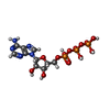 ChemComp-ATP: |
-Experimental details
-Structure determination
| Method | cryo EM |
|---|---|
 Processing Processing | single particle reconstruction |
| Aggregation state | particle |
-Sample preparation
| Buffer | pH: 7.4 |
|---|---|
| Vitrification | Cryogen name: ETHANE |
- Electron microscopy
| Microscope | TFS KRIOS |
|---|---|
| Image recording | Film or detector model: GATAN K3 (6k x 4k) / Average electron dose: 48.0 e/Å2 |
| Electron beam | Acceleration voltage: 300 kV / Electron source:  FIELD EMISSION GUN FIELD EMISSION GUN |
| Electron optics | Illumination mode: FLOOD BEAM / Imaging mode: BRIGHT FIELD / Nominal defocus max: 1.8 µm / Nominal defocus min: 0.8 µm |
| Experimental equipment |  Model: Titan Krios / Image courtesy: FEI Company |
