 Movie
Movie Controller

[English] 日本語
 Yorodumi
- EMDB-66440: Cryo-EM Structure of Alcohol Dehydrogenase Variant from Gluconoba... -
+ Open data

- Basic information
| Entry |  | ||||||||||||
|---|---|---|---|---|---|---|---|---|---|---|---|---|---|
| Title | Cryo-EM Structure of Alcohol Dehydrogenase Variant from Gluconobacter oxydans Truncating Membrane-Binding Regions (Form 2) | ||||||||||||
 Map data Map data | |||||||||||||
 Sample Sample |
| ||||||||||||
 Keywords Keywords | Comples / Oxidoreductase / Membrane-bound protein | ||||||||||||
| Function / homology |  Function and homology information Function and homology informationalcohol dehydrogenase (quinone) / oxidoreductase activity, acting on CH-OH group of donors / respiratory electron transport chain / outer membrane-bounded periplasmic space / electron transfer activity / iron ion binding / heme binding / calcium ion binding / plasma membrane Similarity search - Function | ||||||||||||
| Biological species |  Gluconobacter oxydans (bacteria) Gluconobacter oxydans (bacteria) | ||||||||||||
| Method | single particle reconstruction / cryo EM / Resolution: 2.69 Å | ||||||||||||
 Authors Authors | Ichikawa K / Adachi T / Miyata T / Makino F / Namba K / Kitazumi Y / Shirai O / Sowa K | ||||||||||||
| Funding support |  Japan, 3 items Japan, 3 items
| ||||||||||||
 Citation Citation | Journal: Chem Commun (Camb) / Year: 2026 Title: Structure-guided engineering of membrane-binding regions for surfactant-free solubilization of direct electron transfer-type alcohol dehydrogenase. Authors: Konatsu Ichikawa / Taiki Adachi / Tomoko Miyata / Fumiaki Makino / Keiichi Namba / Yuki Kitazumi / Osamu Shirai / Keisei Sowa / Abstract: Membrane-bound alcohol dehydrogenase (ADH) from is a direct electron transfer-type biocatalyst for ethanol oxidation. To improve its bioelectrocatalysis, membrane-binding regions of ADH were ...Membrane-bound alcohol dehydrogenase (ADH) from is a direct electron transfer-type biocatalyst for ethanol oxidation. To improve its bioelectrocatalysis, membrane-binding regions of ADH were predicted, resulting in the construction of a soluble ADH variant by enzyme engineering. The variant was purified and characterized using structural and bioelectrochemical approaches. | ||||||||||||
| History |
|
- Structure visualization
| Supplemental images |
|---|

- Downloads & links
-EMDB archive
| Map data | emd_66440.map.gz | 62.9 MB | EMDB map data format | |
|---|---|---|---|---|
| Header (meta data) | emd-66440-v30.xmlemd-66440.xml | 26.4 KB 26.4 KB | Display Display | EMDB header |
| FSC (resolution estimation) | emd_66440_fsc.xml | 10.5 KB | Display | FSC data file |
| Images |  emd_66440.png emd_66440.png | 86.4 KB | ||
| Masks | emd_66440_msk_1.map | 125 MB | Mask map | |
| Filedesc metadata | emd-66440.cif.gz | 7.4 KB | ||
| Others | emd_66440_half_map_1.map.gzemd_66440_half_map_2.map.gz | 116 MB 116 MB | ||
| Archive directory |  http://ftp.pdbj.org/pub/emdb/structures/EMD-66440ftp://ftp.pdbj.org/pub/emdb/structures/EMD-66440 http://ftp.pdbj.org/pub/emdb/structures/EMD-66440ftp://ftp.pdbj.org/pub/emdb/structures/EMD-66440 | HTTPS FTP |
-Related structure data
| Related structure data |  9x0rMC  9x0qC M: atomic model generated by this map C: citing same article ( |
|---|---|
| Similar structure data |

-Links
| EMDB pages | EMDB (EBI/PDBe) / EMDataResource |
|---|---|
| Related items in Molecule of the Month |
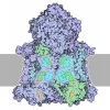
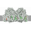
-Map
| File | Download / File: emd_66440.map.gz / Format: CCP4 / Size: 125 MB / Type: IMAGE STORED AS FLOATING POINT NUMBER (4 BYTES) | ||||||||||||||||||||||||||||||||||||
|---|---|---|---|---|---|---|---|---|---|---|---|---|---|---|---|---|---|---|---|---|---|---|---|---|---|---|---|---|---|---|---|---|---|---|---|---|---|
| Projections & slices | Image control
Images are generated by Spider. | ||||||||||||||||||||||||||||||||||||
| Voxel size | X=Y=Z: 0.859 Å | ||||||||||||||||||||||||||||||||||||
| Density |
| ||||||||||||||||||||||||||||||||||||
| Symmetry | Space group: 1 | ||||||||||||||||||||||||||||||||||||
| Details | EMDB XML:
|
 Z (Sec.)
 Y (Row.)
 X (Col.)


-Supplemental data
-Mask #1
| File | emd_66440_msk_1.map | ||||||||||||
|---|---|---|---|---|---|---|---|---|---|---|---|---|---|
| Projections & Slices |
| ||||||||||||
| Density Histograms |


-Half map: #1
| File | emd_66440_half_map_1.map | ||||||||||||
|---|---|---|---|---|---|---|---|---|---|---|---|---|---|
| Projections & Slices |
| ||||||||||||
| Density Histograms |


-Half map: #2
| File | emd_66440_half_map_2.map | ||||||||||||
|---|---|---|---|---|---|---|---|---|---|---|---|---|---|
| Projections & Slices |
| ||||||||||||
| Density Histograms |


- Sample components
-Entire : Cryo-EM Structure of Alcohol Dehydrogenase Variant from Gluconoba...
| Entire | Name: Cryo-EM Structure of Alcohol Dehydrogenase Variant from Gluconobacter oxydans Truncating Membrane-Bound Regions (Form 2) |
|---|---|
| Components |
|
-Supramolecule #1: Cryo-EM Structure of Alcohol Dehydrogenase Variant from Gluconoba...
| Supramolecule | Name: Cryo-EM Structure of Alcohol Dehydrogenase Variant from Gluconobacter oxydans Truncating Membrane-Bound Regions (Form 2) type: complex / ID: 1 / Parent: 0 / Macromolecule list: #1-#3 |
|---|---|
| Source (natural) | Organism: Gluconobacter oxydans (bacteria) |
| Molecular weight | Theoretical: 140 KDa |
-Macromolecule #1: Alcohol dehydrogenase (quinone), dehydrogenase subunit
| Macromolecule | Name: Alcohol dehydrogenase (quinone), dehydrogenase subunit type: protein_or_peptide / ID: 1 / Number of copies: 1 / Enantiomer: LEVO / EC number: alcohol dehydrogenase (quinone) |
|---|---|
| Source (natural) | Organism: Gluconobacter oxydans (bacteria) |
| Molecular weight | Theoretical: 82.938906 KDa |
| Recombinant expression | Organism: Gluconobacter oxydans (bacteria) |
| Sequence | String: MTSGLLTPIK VTKKRLLSCA AALAFSAAVP VAFAQEDTGT AITSSDNGGH PGDWLSYGRS YSEQRYSPLD QINTENVGKL KLAWHYDLD TNRGQEGTPL IVNGVMYATT NWSKMKALDA ATGKLLWSYD PKVPGNIADR GCCDTVSRGA AYWNGKVYFG T FDGRLIAL ...String: MTSGLLTPIK VTKKRLLSCA AALAFSAAVP VAFAQEDTGT AITSSDNGGH PGDWLSYGRS YSEQRYSPLD QINTENVGKL KLAWHYDLD TNRGQEGTPL IVNGVMYATT NWSKMKALDA ATGKLLWSYD PKVPGNIADR GCCDTVSRGA AYWNGKVYFG T FDGRLIAL DAKTGKLVWS VYTIPKEAQL GHQRSYTVDG APRIAKGKVL IGNGGAEFGA RGFVSAFDAE TGKLDWRFFT VP NPENKPD GAASDDILMS KAYPTWGKNG AWKQQGGGGT VWDSLVYDPV TDLVYLGVGN GSPWNYKFRS EGKGDNLFLG SIV AINPDT GKYVWHFQET PMDEWDYTSV QQIMTLDMPV NGEMRHVIVH APKNGFFYII DAKTGKFITG KPYTYENWAN GLDP VTGRP NYVPDALWTL TGKPWLGIPG ELGGHNFAAM AYSPKTKLVY IPAQQIPLLY DGQKGGFKAY HDAWNLGLDM NKIGL FDDN DPEHVAAKKD FLKVLKGWTV AWDPEKMAPA FTINHKGPWN GGLLATAGNV IFQGLANGEF HAYDATNGND LYSFPA QSA IIAPPVTYTA NGKQYVAVEV GWGGIYPFLY GGVARTSGWT VNHSRVIAFS LDGKDSLPPK NELGFTPVKP VPTYDEA RQ KDGYFMYQTF CSACHGDNAI SGGVLPDLRW SGAPRGRESF YKLVGRGALT AYGMDRFDTS MTPEQIEDIR NFIVKRAN E SYDDEVKARE NSTGVPNDQF LNVPQSTADV PTADHP UniProtKB: Alcohol dehydrogenase (quinone), dehydrogenase subunit |
-Macromolecule #2: Alcohol dehydrogenase (quinone), cytochrome c subunit
| Macromolecule | Name: Alcohol dehydrogenase (quinone), cytochrome c subunit / type: protein_or_peptide / ID: 2 / Number of copies: 1 / Enantiomer: LEVO / EC number: alcohol dehydrogenase (quinone) |
|---|---|
| Source (natural) | Organism: Gluconobacter oxydans (bacteria) |
| Molecular weight | Theoretical: 48.939645 KDa |
| Recombinant expression | Organism: Gluconobacter oxydans (bacteria) |
| Sequence | String: MLNALTRDRL VSEMKQGWKL AAAIGLMAVS FGAAHAQDAD EALIKRGEYV ARLSDCIACH TALHGQPYAG GLEIKSGGGT IYSTNITPD PEHGIGNYTL EDFTKALRKG IRKDGATVYP AMPYPEFARL SDDDIRAMYA FFMHGVKPVA LQNKAPDISG G GGVPSMTP ...String: MLNALTRDRL VSEMKQGWKL AAAIGLMAVS FGAAHAQDAD EALIKRGEYV ARLSDCIACH TALHGQPYAG GLEIKSGGGT IYSTNITPD PEHGIGNYTL EDFTKALRKG IRKDGATVYP AMPYPEFARL SDDDIRAMYA FFMHGVKPVA LQNKAPDISG G GGVPSMTP GVDKSISDPE VARGEYLVNG PGHCGECHTP RQVKAYGTAG GNAYLAGGAP IDNWIAPSLR SNSDTGLGRW SE DDIVTFL KSGRIDHSAV FGGMADVVAY STQHWSDDDL RATAKYLKSM PAVPEGKNLG QDDGQTTALL NKGGQGNAGA EVY LHNCAI CHMNDGTGVN RMFPPLAGNP VVITDDPTSL ANVVAFGGIL PPTNSAPSAV AMPGFKNHLS DQEMADVVNF MRKG WGNNA PGTVSASDIQ KLRTTGAPVS TAGWNVSSKG WMAYMPQPYG EDWTFSPQTH TGVDDAQ UniProtKB: Alcohol dehydrogenase (quinone), cytochrome c subunit, Alcohol dehydrogenase (quinone), cytochrome c subunit |
-Macromolecule #3: Small subunit of alcohol dehydrogenase
| Macromolecule | Name: Small subunit of alcohol dehydrogenase / type: protein_or_peptide / ID: 3 / Number of copies: 1 / Enantiomer: LEVO / EC number: alcohol dehydrogenase (quinone) |
|---|---|
| Source (natural) | Organism: Gluconobacter oxydans (bacteria) |
| Molecular weight | Theoretical: 14.282063 KDa |
| Recombinant expression | Organism: Gluconobacter oxydans (bacteria) |
| Sequence | String: MFRRIVPVLG LALGLGLASQ AAMAQEQSPP PPPAVQGTPG KDFTGVSPAN LAGIMNYCVE QQYVSYDEGN PVLYGLSEKY KATEQTVGN FDYALGTAGY FDSNGKRFYL VAYTNEDDRR AACHAAVKAA QPML UniProtKB: Alcohol dehydrogenase, 15 kDa subunit |
-Macromolecule #4: HEME C
| Macromolecule | Name: HEME C / type: ligand / ID: 4 / Number of copies: 4 / Formula: HEC |
|---|---|
| Molecular weight | Theoretical: 620.519 Da |
| Chemical component information | 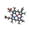 ChemComp-HEC: |
-Macromolecule #5: PYRROLOQUINOLINE QUINONE
| Macromolecule | Name: PYRROLOQUINOLINE QUINONE / type: ligand / ID: 5 / Number of copies: 1 / Formula: PQQ |
|---|---|
| Molecular weight | Theoretical: 330.206 Da |
| Chemical component information | 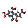 ChemComp-PQQ: |
-Macromolecule #6: CALCIUM ION
| Macromolecule | Name: CALCIUM ION / type: ligand / ID: 6 / Number of copies: 1 / Formula: CA |
|---|---|
| Molecular weight | Theoretical: 40.078 Da |
-Experimental details
-Structure determination
| Method | cryo EM |
|---|---|
 Processing Processing | single particle reconstruction |
| Aggregation state | particle |
-Sample preparation
| Concentration | 3 mg/mL | ||||||||||||
|---|---|---|---|---|---|---|---|---|---|---|---|---|---|
| Buffer | pH: 6 Component:
| ||||||||||||
| Grid | Model: Quantifoil R1.2/1.3 / Material: COPPER / Mesh: 200 / Support film - Material: CARBON / Support film - topology: HOLEY / Support film - Film thickness: 500 / Pretreatment - Type: GLOW DISCHARGE / Pretreatment - Time: 20 sec. | ||||||||||||
| Vitrification | Cryogen name: ETHANE / Chamber humidity: 100 % / Chamber temperature: 277 K / Instrument: FEI VITROBOT MARK IV |
- Electron microscopy
| Microscope | JEOL CRYO ARM 300 |
|---|---|
| Temperature | Min: 80.0 K / Max: 80.0 K |
| Alignment procedure | Coma free - Residual tilt: 0.01 mrad |
| Specialist optics | Energy filter - Name: In-column Omega Filter / Energy filter - Slit width: 20 eV |
| Image recording | Film or detector model: GATAN K3 (6k x 4k) / Number grids imaged: 1 / Number real images: 20066 / Average exposure time: 3.0 sec. / Average electron dose: 80.0 e/Å2 |
| Electron beam | Acceleration voltage: 300 kV / Electron source:  FIELD EMISSION GUN FIELD EMISSION GUN |
| Electron optics | C2 aperture diameter: 50.0 µm / Calibrated defocus max: 2.5 µm / Calibrated defocus min: 0.5 µm / Calibrated magnification: 56754 / Illumination mode: FLOOD BEAM / Imaging mode: BRIGHT FIELD / Cs: 2.7 mm / Nominal defocus max: 2.5 µm / Nominal defocus min: 0.5 µm / Nominal magnification: 60000 |
| Sample stage | Specimen holder model: JEOL CRYOSPECPORTER / Cooling holder cryogen: NITROGEN |
+Image processing

-Atomic model buiding 1
| Refinement | Space: REAL / Protocol: FLEXIBLE FIT |
|---|---|
| Output model | PDB-9x0r: |
