[English] 日本語
Yorodumi
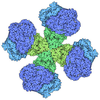
- EMDB-66321: Wild-type Escherichia coli transhydrogenase single dIII attached ... -

+
Open data


ID or keywords:

Loading...

-
Basic information

Entry
Database: EMDB / ID: EMD-66321
TitleWild-type Escherichia coli transhydrogenase single dIII attached to dII in the presence of both NADP+ and NAD+.
Map data
Sample
  • Complex: The E. coli transhydrogenase complex.
    • Protein or peptide: NAD(P) transhydrogenase subunit alpha
    • Protein or peptide: NAD(P) transhydrogenase subunit beta
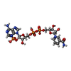
  • Ligand: NADP NICOTINAMIDE-ADENINE-DINUCLEOTIDE PHOSPHATE
Keywordsnicotinamide nucleotide transhydrogenase / hydride transfer / proton pump / conformational dynamics / NADPH production / MEMBRANE PROTEIN
Function / homology
Function and homology information


proton export across plasma membrane / proton-translocating NAD(P)+ transhydrogenase activity / proton-translocating NAD(P)+ transhydrogenase / NADPH regeneration / NADP binding / oxidoreductase activity / protein dimerization activity / plasma membrane
Similarity search - Function
NAD(P) transhydrogenase, alpha subunit / NADP transhydrogenase, beta subunit / NAD(P) transhydrogenase, alpha subunit, C-terminal / 4TM region of pyridine nucleotide transhydrogenase, mitoch / NADP transhydrogenase beta-like domain / NAD(P) transhydrogenase beta subunit / Alanine dehydrogenase/NAD(P) transhydrogenase, conserved site-1 / Alanine dehydrogenase & pyridine nucleotide transhydrogenase signature 1. / Alanine dehydrogenase/pyridine nucleotide transhydrogenase, conserved site-2 / Alanine dehydrogenase & pyridine nucleotide transhydrogenase signature 2. ...NAD(P) transhydrogenase, alpha subunit / NADP transhydrogenase, beta subunit / NAD(P) transhydrogenase, alpha subunit, C-terminal / 4TM region of pyridine nucleotide transhydrogenase, mitoch / NADP transhydrogenase beta-like domain / NAD(P) transhydrogenase beta subunit / Alanine dehydrogenase/NAD(P) transhydrogenase, conserved site-1 / Alanine dehydrogenase & pyridine nucleotide transhydrogenase signature 1. / Alanine dehydrogenase/pyridine nucleotide transhydrogenase, conserved site-2 / Alanine dehydrogenase & pyridine nucleotide transhydrogenase signature 2. / Alanine dehydrogenase/PNT, C-terminal domain / Alanine dehydrogenase/pyridine nucleotide transhydrogenase, N-terminal / Alanine dehydrogenase/PNT, N-terminal domain / Alanine dehydrogenase/PNT, C-terminal domain / Alanine dehydrogenase/PNT, N-terminal domain / Alanine dehydrogenase/pyridine nucleotide transhydrogenase, NAD(H)-binding domain / DHS-like NAD/FAD-binding domain superfamily / NAD(P)-binding domain superfamily
Similarity search - Domain/homology
NAD(P) transhydrogenase subunit alpha / NAD(P) transhydrogenase subunit beta
Similarity search - Component
Biological speciesEscherichia coli K-12 (bacteria)
Methodsingle particle reconstruction / cryo EM / Resolution: 3.42 Å
AuthorsZhu JP / Zhang K / Li J / Zheng W / Gu M
Funding support1 items
OrganizationGrant numberCountry
Not funded
CitationJournal: To Be Published
Title: Wild-type Escherichia coli transhydrogenase single dIII attached to dII in the presence of both NADP+ and NAD+.
Authors: Zhu JP / Zhang K / Li J
History
DepositionSep 24, 2025-
Header (metadata) releaseSep 30, 2026-
Map releaseSep 30, 2026-
UpdateSep 30, 2026-
Current statusSep 30, 2026Processing site: PDBc / Status: Released

-
Structure visualization

Supplemental images

Downloads & links

-
Map

FileDownload / File: emd_66321.map.gz / Format: CCP4 / Size: 178 MB / Type: IMAGE STORED AS FLOATING POINT NUMBER (4 BYTES)
Projections & slices

Image control

Size
Brightness
Contrast
Others
AxesZ (Sec.)Y (Row.)X (Col.)
0.83 Å/pix.
x 360 pix.
= 299.52 Å
0.83 Å/pix.
x 360 pix.
= 299.52 Å
0.83 Å/pix.
x 360 pix.
= 299.52 Å

Surface

Projections

Slices (1/3)

Slices (1/2)

Slices (2/3)

Images are generated by Spider.

Voxel sizeX=Y=Z: 0.832 Å
Density
Contour LevelBy AUTHOR: 0.109
Minimum - Maximum-0.4094992 - 0.6726513
Average (Standard dev.)-0.00022136277 (±0.014027352)
SymmetrySpace group: 1
Details

EMDB XML:

Map geometry
Axis orderXYZ
Origin000
Dimensions360360360
Spacing360360360
CellA=B=C: 299.52002 Å
α=β=γ: 90.0 °

-
Supplemental data

-
Half map: #2

Fileemd_66321_half_map_1.map
Projections & Slices
AxesZYX

Projections

Slices (1/2)
Density Histograms

-
Half map: #1

Fileemd_66321_half_map_2.map
Projections & Slices
AxesZYX

Projections

Slices (1/2)
Density Histograms

-
Sample components

-
Entire : The E. coli transhydrogenase complex.

EntireName: The E. coli transhydrogenase complex.
Components
  • Complex: The E. coli transhydrogenase complex.
    • Protein or peptide: NAD(P) transhydrogenase subunit alpha
    • Protein or peptide: NAD(P) transhydrogenase subunit beta
  • Ligand: NADP NICOTINAMIDE-ADENINE-DINUCLEOTIDE PHOSPHATE

-
Supramolecule #1: The E. coli transhydrogenase complex.

SupramoleculeName: The E. coli transhydrogenase complex. / type: complex / ID: 1 / Parent: 0 / Macromolecule list: #1-#2
Source (natural)Organism: Escherichia coli K-12 (bacteria)

-
Macromolecule #1: NAD(P) transhydrogenase subunit alpha

MacromoleculeName: NAD(P) transhydrogenase subunit alpha / type: protein_or_peptide / ID: 1 / Number of copies: 2 / Enantiomer: LEVO / EC number: proton-translocating NAD(P)+ transhydrogenase
Source (natural)Organism: Escherichia coli K-12 (bacteria)
Molecular weightTheoretical: 11.747049 KDa
Recombinant expressionOrganism: Escherichia coli (E. coli)
SequenceString:
YALMALAIIL FGWMASVAPK EFLGHFTVFA LACVVGYYVV WNVSHALHTP LMSVTNAISG IIVVGALLQI GQGGWVSFLS FIAVLIASI NIFGGFTVTQ RMLKMFRKN

UniProtKB: NAD(P) transhydrogenase subunit alpha

-
Macromolecule #2: NAD(P) transhydrogenase subunit beta

MacromoleculeName: NAD(P) transhydrogenase subunit beta / type: protein_or_peptide / ID: 2 / Number of copies: 2 / Enantiomer: LEVO / EC number: proton-translocating NAD(P)+ transhydrogenase
Source (natural)Organism: Escherichia coli K-12 (bacteria)
Molecular weightTheoretical: 48.762746 KDa
Recombinant expressionOrganism: Escherichia coli (E. coli)
SequenceString: MSGGLVTAAY IVAAILFIFS LAGLSKHETS RQGNNFGIAG MAIALIATIF GPDTGNVGWI LLAMVIGGAI GIRLAKKVEM TEMPELVAI LHSFVGLAAV LVGFNSYLHH DAGMAPILVN IHLTEVFLGI FIGAVTFTGS VVAFGKLCGK ISSKPLMLPN R HKMNLAAL ...String:
MSGGLVTAAY IVAAILFIFS LAGLSKHETS RQGNNFGIAG MAIALIATIF GPDTGNVGWI LLAMVIGGAI GIRLAKKVEM TEMPELVAI LHSFVGLAAV LVGFNSYLHH DAGMAPILVN IHLTEVFLGI FIGAVTFTGS VVAFGKLCGK ISSKPLMLPN R HKMNLAAL VVSFLLLIVF VRTDSVGLQV LALLIMTAIA LVFGWHLVAS IGGADMPVVV SMLNSYSGWA AAAAGFMLSN DL LIVTGAL VGSSGAILSY IMCKAMNRSF ISVIAGGFGT DGSSTGDDQE VGEHREITAE ETAELLKNSH SVIITPGYGM AVA QAQYPV AEITEKLRAR GINVRFGIHP VAGRLPGHMN VLLAEAKVPY DIVLEMDEIN DDFADTDTVL VIGANDTVNP AAQD DPKSP IAGMPVLEVW KAQNVIVFKR SMNTGYAGVQ NPLFFKENTH MLFGDAKASV DAILKAL

UniProtKB: NAD(P) transhydrogenase subunit beta

-
Macromolecule #3: NADP NICOTINAMIDE-ADENINE-DINUCLEOTIDE PHOSPHATE

MacromoleculeName: NADP NICOTINAMIDE-ADENINE-DINUCLEOTIDE PHOSPHATE / type: ligand / ID: 3 / Number of copies: 1 / Formula: NAP
Molecular weightTheoretical: 743.405 Da
Chemical component information

ChemComp-NAP:
NADP NICOTINAMIDE-ADENINE-DINUCLEOTIDE PHOSPHATE

-
Experimental details

-
Structure determination

Methodcryo EM
Processingsingle particle reconstruction
Aggregation stateparticle

-
Sample preparation

BufferpH: 8
VitrificationCryogen name: ETHANE

-
Electron microscopy

MicroscopeTFS KRIOS
Image recordingFilm or detector model: GATAN K3 (6k x 4k) / Average electron dose: 1.378 e/Å2
Electron beamAcceleration voltage: 300 kV / Electron source: FIELD EMISSION GUN
Electron opticsIllumination mode: FLOOD BEAM / Imaging mode: BRIGHT FIELD / Nominal defocus max: 2.0 µm / Nominal defocus min: 1.2 µm
Experimental equipment
Model: Titan Krios / Image courtesy: FEI Company

+
Image processing

CTF correctionType: PHASE FLIPPING AND AMPLITUDE CORRECTION
Startup modelType of model: OTHER
Final reconstructionResolution.type: BY AUTHOR / Resolution: 3.42 Å / Resolution method: FSC 0.143 CUT-OFF / Software - Name: cryoSPARC / Number images used: 96595
Initial angle assignmentType: MAXIMUM LIKELIHOOD
Final angle assignmentType: MAXIMUM LIKELIHOOD

+
About Yorodumi

-
News

-
Feb 9, 2022. New format data for meta-information of EMDB entries

New format data for meta-information of EMDB entries

  • Version 3 of the EMDB header file is now the official format.
  • The previous official version 1.9 will be removed from the archive.

Related info.:EMDB header

External links:wwPDB to switch to version 3 of the EMDB data model

-
Aug 12, 2020. Covid-19 info

Covid-19 info

URL: https://pdbj.org/emnavi/covid19.php

New page: Covid-19 featured information page in EM Navigator.

Related info.:Covid-19 info / Mar 5, 2020. Novel coronavirus structure data

+
Mar 5, 2020. Novel coronavirus structure data

Novel coronavirus structure data

Related info.:Yorodumi Speices / Aug 12, 2020. Covid-19 info

External links:COVID-19 featured content - PDBj / Molecule of the Month (242):Coronavirus Proteases

+
Jan 31, 2019. EMDB accession codes are about to change! (news from PDBe EMDB page)

EMDB accession codes are about to change! (news from PDBe EMDB page)

  • The allocation of 4 digits for EMDB accession codes will soon come to an end. Whilst these codes will remain in use, new EMDB accession codes will include an additional digit and will expand incrementally as the available range of codes is exhausted. The current 4-digit format prefixed with “EMD-” (i.e. EMD-XXXX) will advance to a 5-digit format (i.e. EMD-XXXXX), and so on. It is currently estimated that the 4-digit codes will be depleted around Spring 2019, at which point the 5-digit format will come into force.
  • The EM Navigator/Yorodumi systems omit the EMD- prefix.

Related info.:Q: What is EMD? / ID/Accession-code notation in Yorodumi/EM Navigator

External links:EMDB Accession Codes are Changing Soon! / Contact to PDBj

+
Jul 12, 2017. Major update of PDB

Major update of PDB

  • wwPDB released updated PDB data conforming to the new PDBx/mmCIF dictionary.
  • This is a major update changing the version number from 4 to 5, and with Remediation, in which all the entries are updated.
  • In this update, many items about electron microscopy experimental information are reorganized (e.g. em_software).
  • Now, EM Navigator and Yorodumi are based on the updated data.

External links:wwPDB Remediation / Enriched Model Files Conforming to OneDep Data Standards Now Available in the PDB FTP Archive

-
Yorodumi

Thousand views of thousand structures

  • Yorodumi is a browser for structure data from EMDB, PDB, SASBDB, etc.
  • This page is also the successor to EM Navigator detail page, and also detail information page/front-end page for Omokage search.
  • The word "yorodu" (or yorozu) is an old Japanese word meaning "ten thousand". "mi" (miru) is to see.

Related info.:EMDB / PDB / SASBDB / Comparison of 3 databanks / Yorodumi Search / Aug 31, 2016. New EM Navigator & Yorodumi / Yorodumi Papers / Jmol/JSmol / Function and homology information / Changes in new EM Navigator and Yorodumi

Read more