 Movie
Movie Controller

[English] 日本語
 Yorodumi
- EMDB-66262: Cryo-EM structure of loop truncated self-sufficient P450 from Shi... -
+ Open data

- Basic information
| Entry |  | |||||||||
|---|---|---|---|---|---|---|---|---|---|---|
| Title | Cryo-EM structure of loop truncated self-sufficient P450 from Shimazuella soli | |||||||||
 Map data Map data | ||||||||||
 Sample Sample |
| |||||||||
 Keywords Keywords | self-sufficient P450 / heme / OXIDOREDUCTASE | |||||||||
| Biological species |  Shimazuella soli (bacteria) Shimazuella soli (bacteria) | |||||||||
| Method | single particle reconstruction / cryo EM / Resolution: 2.34 Å | |||||||||
 Authors Authors | Xie ZZ / Li SY / Liu ZW / Li QR / Huang J-W / Chen C-C / Guo R-T | |||||||||
| Funding support |  China, 1 items China, 1 items
| |||||||||
 Citation Citation | Journal: To Be Published Title: Cryo-EM structure of loop truncated self-sufficient P450 from Shimazuella soli Authors: Xie ZZ / Li SY / Liu ZW / Li QR / Huang J-W / Chen C-C / Guo R-T | |||||||||
| History |
|
- Structure visualization
| Supplemental images |
|---|

- Downloads & links
-EMDB archive
| Map data | emd_66262.map.gz | 59.6 MB |  EMDB map data format EMDB map data format | |
|---|---|---|---|---|
| Header (meta data) | emd-66262-v30.xmlemd-66262.xml | 19 KB 19 KB | Display Display | EMDB header |
| FSC (resolution estimation) | emd_66262_fsc.xml | 8.4 KB | Display | FSC data file |
| Images |  emd_66262.png emd_66262.png | 107.7 KB | ||
| Masks | emd_66262_msk_1.map | 64 MB | Mask map | |
| Filedesc metadata | emd-66262.cif.gz | 6.5 KB | ||
| Others | emd_66262_half_map_1.map.gzemd_66262_half_map_2.map.gz | 59.5 MB 59.5 MB | ||
| Archive directory |  https://data.pdbj.org/pub/emdb/structures/EMD-66262ftp://data.pdbj.org/pub/emdb/structures/EMD-66262 https://data.pdbj.org/pub/emdb/structures/EMD-66262ftp://data.pdbj.org/pub/emdb/structures/EMD-66262 | HTTPS FTP |
-Related structure data

-Links
| EMDB pages | EMDB (EBI/PDBe) / EMDataResource |
|---|
-Map
| File | Download / File: emd_66262.map.gz / Format: CCP4 / Size: 64 MB / Type: IMAGE STORED AS FLOATING POINT NUMBER (4 BYTES) | ||||||||||||||||||||||||||||||||||||
|---|---|---|---|---|---|---|---|---|---|---|---|---|---|---|---|---|---|---|---|---|---|---|---|---|---|---|---|---|---|---|---|---|---|---|---|---|---|
| Projections & slices | Image control
Images are generated by Spider. | ||||||||||||||||||||||||||||||||||||
| Voxel size | X=Y=Z: 0.93 Å | ||||||||||||||||||||||||||||||||||||
| Density |
| ||||||||||||||||||||||||||||||||||||
| Symmetry | Space group: 1 | ||||||||||||||||||||||||||||||||||||
| Details | EMDB XML:
|
 Z (Sec.)
 Y (Row.)
 X (Col.)


-Supplemental data
-Mask #1
| File | emd_66262_msk_1.map | ||||||||||||
|---|---|---|---|---|---|---|---|---|---|---|---|---|---|
| Projections & Slices |
| ||||||||||||
| Density Histograms |


-Half map: #1
| File | emd_66262_half_map_1.map | ||||||||||||
|---|---|---|---|---|---|---|---|---|---|---|---|---|---|
| Projections & Slices |
| ||||||||||||
| Density Histograms |


-Half map: #2
| File | emd_66262_half_map_2.map | ||||||||||||
|---|---|---|---|---|---|---|---|---|---|---|---|---|---|
| Projections & Slices |
| ||||||||||||
| Density Histograms |


- Sample components
-Entire : bifunctional cytochrome P450/NADPH--P450 reductase
| Entire | Name: bifunctional cytochrome P450/NADPH--P450 reductase |
|---|---|
| Components |
|
-Supramolecule #1: bifunctional cytochrome P450/NADPH--P450 reductase
| Supramolecule | Name: bifunctional cytochrome P450/NADPH--P450 reductase / type: complex / ID: 1 / Parent: 0 / Macromolecule list: #1 |
|---|---|
| Source (natural) | Organism: Shimazuella soli (bacteria) |
-Macromolecule #1: bifunctional cytochrome P450/NADPH--P450 reductase
| Macromolecule | Name: bifunctional cytochrome P450/NADPH--P450 reductase / type: protein_or_peptide / ID: 1 / Number of copies: 2 / Enantiomer: LEVO |
|---|---|
| Source (natural) | Organism: Shimazuella soli (bacteria) |
| Molecular weight | Theoretical: 117.526359 KDa |
| Recombinant expression | Organism:  Escherichia coli BL21(DE3) (bacteria) Escherichia coli BL21(DE3) (bacteria) |
| Sequence | String: MDEANIIPQP KTYGPLGNIP LIDKDKPILS FMKLAEEYGP IFRLQTPGDS TIVVSGHELV KEVCDESRFL KSAEGPLEKV RAFGGDGLF TSWTHEPNWR KAHNILMPTF SQRAMKDYHD MMVDIAVQLI QKWIRLNPDE TVDVPDDMTR LTLDTIGLCG F NYRFNSYY ...String: MDEANIIPQP KTYGPLGNIP LIDKDKPILS FMKLAEEYGP IFRLQTPGDS TIVVSGHELV KEVCDESRFL KSAEGPLEKV RAFGGDGLF TSWTHEPNWR KAHNILMPTF SQRAMKDYHD MMVDIAVQLI QKWIRLNPDE TVDVPDDMTR LTLDTIGLCG F NYRFNSYY RETPHPFITS MVRALDEAMH QTQRLDLQDK LMIRTKRQFQ HDIQVMFSLV DSIIAERRAG KNQKENDLLS RM LNVSDPE TGEKLDDENI RYQIITFLIA GHETTSGLLS FALYFLLKNP DKLKKAYEEV DQVLTGSTPT YKQVLHLKYV RMI LNEALR LWPTAPAFSL YAKEDTIIGG KYPVKKEQER ITVLIPQLHR DKEAWGEDVE EFRPERFEDQ NKVPHHAYKP FGNG QRACI GMQFALHEAT LVLGMLLRHF EFIDYKDYQL DIKQTLTLKP GDFNIQVQPR NQPAFQGLNN RPLLVLYGSD TGTAE GIAR ELADTATLHG VHTEVATLNE RIGELPKEGA VLIVTSSYNG KPPSNAGQFV QWLEAVQAGE LSGVQYAVFG CGDHNW AST YQDVPRFIDK QLAEKGAVRF SARGEGDVSG DFEEQFDQWK EKMWSDAIEA FGLEISDDVK KDQNTLSLQF VKGTGGS PL ARSYEAVYAK VVENRELQSP DSGRSTRHIE ITLPKGVSYQ EGDHLGVLPA NSKENVHRVL QKYKLNENDQ VVLTASGR S MAHLPLAQEV SLRDLLLYSV ELQDAATRAQ IRELAAFTVC PPHKRELEAL LEEGIYQEQV LKKRISMLDL LEKYEACEM PFERFLELLH PLKPRYYSIS SSPRLNSERA SITVAVVRGP AWSGLGEYRG VASNYLADCK PGEDVMMFIR TPESNFQLPE ESETPIIMV GPGTGLAPFR GFLQARDAMK QEGKTLGEAY LYFGCRNEAD FIYRKELEQY KKNGIMTLYI AFSRKEGIPK T YVQHVMAS NAESLIRILD QGGRLYICGD GSRMAPEVEA TLKNSYQEVH GAGEQEASQW LEKLQKDGQY AKDVWAGL |
-Macromolecule #2: PROTOPORPHYRIN IX CONTAINING FE
| Macromolecule | Name: PROTOPORPHYRIN IX CONTAINING FE / type: ligand / ID: 2 / Number of copies: 2 / Formula: HEM |
|---|---|
| Molecular weight | Theoretical: 616.487 Da |
| Chemical component information | 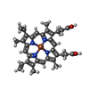 ChemComp-HEM: |
-Macromolecule #3: FLAVIN MONONUCLEOTIDE
| Macromolecule | Name: FLAVIN MONONUCLEOTIDE / type: ligand / ID: 3 / Number of copies: 2 / Formula: FMN |
|---|---|
| Molecular weight | Theoretical: 456.344 Da |
| Chemical component information | 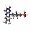 ChemComp-FMN: |
-Macromolecule #4: FLAVIN-ADENINE DINUCLEOTIDE
| Macromolecule | Name: FLAVIN-ADENINE DINUCLEOTIDE / type: ligand / ID: 4 / Number of copies: 2 / Formula: FAD |
|---|---|
| Molecular weight | Theoretical: 785.55 Da |
| Chemical component information | 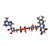 ChemComp-FAD: |
-Macromolecule #5: water
| Macromolecule | Name: water / type: ligand / ID: 5 / Number of copies: 2 / Formula: HOH |
|---|---|
| Molecular weight | Theoretical: 18.015 Da |
| Chemical component information |  ChemComp-HOH: |
-Experimental details
-Structure determination
| Method | cryo EM |
|---|---|
 Processing Processing | single particle reconstruction |
| Aggregation state | particle |
-Sample preparation
| Buffer | pH: 7.5 / Details: 25 mM HEPES,150 mM NaCl, pH 7.5 |
|---|---|
| Vitrification | Cryogen name: ETHANE |
- Electron microscopy
| Microscope | TFS KRIOS |
|---|---|
| Image recording | Film or detector model: FEI FALCON IV (4k x 4k) / Average electron dose: 52.0 e/Å2 |
| Electron beam | Acceleration voltage: 300 kV / Electron source:  FIELD EMISSION GUN FIELD EMISSION GUN |
| Electron optics | Illumination mode: FLOOD BEAM / Imaging mode: BRIGHT FIELD / Nominal defocus max: 2.0 µm / Nominal defocus min: 1.0 µm |
| Experimental equipment |  Model: Titan Krios / Image courtesy: FEI Company |

